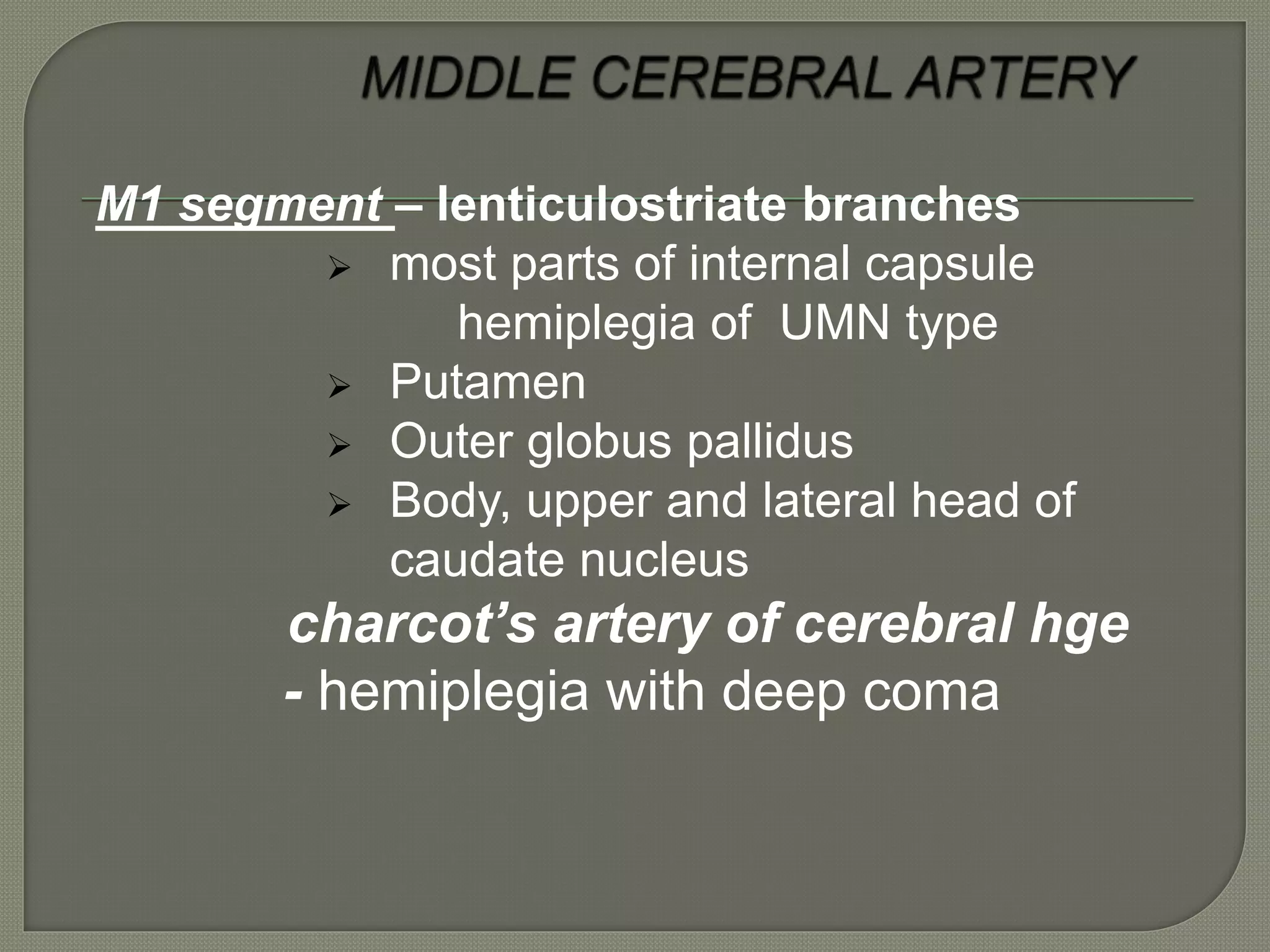
PRESENTER: DR. JITHIN GEORGE
BLOOD SUPPLY OF BRAIN
ANTERIOR CIRCULATION
Internal carotid artery and its
terminal branches
POSTERIOR CIRCULATION
Vertebral artery , basilar artery and
posterior cerebral artery
Branches
 Ophthalmic
 Anterior cerebral
 Middle cerebral
 Posterior communicating
 Anterior choroidal
 M1 segment
 M2 segment
M1 segment – lenticulostriate branches
 most parts of internal capsule
hemiplegia of UMN type
 Putamen
 Outer globus pallidus
 Body, upper and lateral head of
caudate nucleus
charcot’s artery of cerebral hge
- hemiplegia with deep coma
Superior division
 motor cortex
 sensory cortex
 broca’s area
 frontal eye field
Inferior division
 wernicke’s area
 auditory area
 optic radiation
2 segments – A1 segment ( pre communal )
A2 segment ( post communal )
A1 segment
 anterior limb of internal capsule
 anterior perforate substance
 anterior hypothalamus
 amygdala
 inferior part of head of caudate
nucleus
` Recurrent artery of Heubner –
faciobrachial monoplegia
 motor area of leg
 sensory area of leg
 paracentral lobule
 corpus callosum
 superior frontal gyrus
ACA
PCA
MCA
POSTERIOR CEREBRAL ARTERY
 In 75% cases both PCA arises
from the bifurcation of basilar
artery
 20% one from ipsilateral ICA
 5% both from respective
ipsilateral ICA
Two segments
P1 segment
P2 segment
The artery of Percheron – inferomedial and
anterior thalamus and subthalamus
Thalamogeniculate branches
Medial posterior choroidal artery
Lateral posterior choroidal artery
Other areas – middle cerebral peduncle
substantia nigra
red nucleus
medial lemniscus
MLF
Lateral part of cerebral peduncle
Medial lemniscus
Tegmentum of midbrain
Superior colliculi
Lateral geniculate body
4 segments
V1, V2, V3, V4
Branches(cranial)
 Anterior spinal
 Posterior spinal
PICA
Meningeal
Medullary
Branches
 Pontine
 Labyrinthine
 Anterior inferior cerebellar
 Superior cerebellar
 Posterior cerebral
Supplies
base of pons and
superior cerebellum
third most frequent cause of stroke,
following cerebral embolism and
thrombotic disease.
 Vascular malformations
 Intracranial tumors
 Bleeding disorders, anticoagulant and
fibrinolytic treatment
 Cerebral amyloid angiopathy
 Granulomatous angiitis of the central nervous
system and other vasculitides, such as
polyarteritis nodosa
 Sympathomimetic agents (including
amphetamine and cocaine)
 Hemorrhagic infarction
 Head trauma
the most common sites of a cerebral
hemorrhage are
 (1) the putamen and adjacent internal capsule
 (2) the central white matter of the temporal,
parietal, or frontal lobes (lobar hemorrhages,
not strictly associated with hypertension);
 (3) the thalamus;
 (4) one or the other cerebellar hemisphere;
and
 (5) the pons.
Multiple, nearly simultaneous intracerebral
hemorrhages raise the possibility of
amyloid angiopathy or a bleeding diathesis
age <65 years,
 female sex,
nonsmoker,
lobar ICH,
Intraventricular extension,
absence of a history of hypertension or
coagulopathy
In the first hours and days following the
hemorrhage, varying degrees of edema
evolve around the clot and add to the
mass effect.
Hydrocephalus may occur as a result of
bleeding into the ventricular system or
from compression of the third ventricle.
The extravasated blood undergoes a
predictable series of changes. At first fluid,
the collection becomes a clot.
Before the clot forms, red cells settle in the
dependent part of the hematoma and form
a meniscus with the plasma above;
particularly in cases of anticoagulant-
induced hemorrhage. The resultant fluid-
fluid level ("hematocrit effect") .
Within a few days, hemoglobin products,
mainly hemosiderin and hematoidin, begin
to appear.
The hemosiderin forms within histiocytes
that have phagocytized red blood cells
(RBCs) and takes the form of ferritin
granules that stain positively for iron.
As oxyhemoglobin is liberated from the
RBCs and becomes deoxygenated,
methemoglobin appears. This begins
within a few days and imparts a brownish
hue to the periphery of the clot.
Phagocytosis of red cells begins within 24
h, and hemosiderin is first observed
around the margins of the clot in 5 to 6
days
 The clot changes color gradually over a few weeks
from dark red to pale red, and the border of
golden-brown hemosiderin widens.
 The edema disappears over many days or weeks.
 In 2 to 3 months, larger clots are filled with a
chrome-colored thick fluid, which is slowly
absorbed, leaving a smooth-walled cavity or a
yellow-brown scar.
 The iron pigment (hematin) becomes dispersed
and studs adjacent astrocytes and neurons and
may persist well beyond the border of the
hemorrhage for years.
On CT, fresh blood is visualized as a white
mass as soon as it is shed. The "spot
sign," the appearance of contrast within
the hemorrhage during CT angiography.
After 2 to 3 weeks, the surrounding edema
begins to recede and the density of the
hematoma decreases, first at the
periphery.
 At one point several weeks after the bleed,
the appearance may transiently simulate a
tumor or abscess.
 By :M.RI, either in conventional Tl- or T2-
weighted images, the hemorrhage is not
easily visible in the 2 or 3 days after bleeding,
as oxyhemoglobin is diamagnetic or, at most,
is slightly hypointense, so that only the mass
effect is evident.
 MR gradient echo or equivalent sequences
that display areas of magnetic susceptibility
show hemorrhages earlier and detect
remnants of deposited hemosiderin even
years afterward.
As deoxyhemoglobin and methemoglobin
form, the hematoma signal becomes bright
on Tl-weighted images and dark on T2.
The hematoma is then subacute and the
dark signal gradually brightens.
Massive refers to hemorrhages several
centimeters in diameter, usually larger than
50 mL; small applies to those 1 to 2 cm in
diameter and less than 20 mL in volume.
The hypertensive vascular lesion that
leads to arteriolar rupture in most cases
appears to arise from an arterial wall
altered by the effects of hypertension,
i.e., the change referred to in a preceding
section as segmental lipohyalinosis and
the false aneurysm (microaneurysm)
named for Charcot and Bouchard
Acute reactive hypertension
Vomiting
Nuchal rigidity
Seizures usually focal, occur in the first
few days in only 10 percent of cases of
supratentorial hemorrhage.
fundus hypertensive changes in the
arterioles.
 In putaminal hemorrhage, the eyes are
deviated to the side opposite the paralysis;
 in thalamic hemorrhage, ocular abnormality is
downward deviation of the eyes and the
pupils may be unreactive;
 in pontine hemorrhage, the eyeballs are fixed
and the pupils are tiny but reactive;
 in large cerebellar hemorrhage, the eyes may
be deviated laterally to the side opposite the
lesion and ocular bobbing may occur (as
often cerebellar hemorrhage in awake
patients there are no eye signs).
most common syndrome is the one caused
by putaminal hemorrhage with extension to
the adjacent internal capsule.
hemiplegia from interruption of the capsule
is a consistent feature of medium-sized
and large clots.
Vomiting occurs in about half the patients.
Headache is frequent
More advanced stages are characterized by
signs of upper brainstem compression ;
bilateral Babinski signs;
irregular or intermittent respiration;
dilated, fixed pupils, first on the side of the
clot; and
decerebrate rigidity.
 With hemorrhages confined to the anterior
segment of the putamen, the hemiplegia and
hyperreflexia tend to be less severe and to
clear more rapidly. also prominent abulia,
motor impersistence, temporary unilateral
neglect, and with left-sided lesions, nonfluent
aphasia, and dysgraphia.
 With small posterior lesions, weakness is also
mild and is attended by sensory loss,
hemianopia, impaired visual pursuit to the
opposite side, Wernicke-type aphasia (left-
sided lesions), and anosognosia (right-sided)
.
Those extending laterally and posteriorly
into the internal capsule behave much like
large putaminal hemorrhages.
Those extending medially into the lateral
ventricle give rise to drowsmess, stupor,
and either confusion and underactivity or
restlessness and agitation.
severe sensory loss on the entire
contralateral body.
If large or moderate in size, thalamic
hemorrhage also produces a hemiplegia or
hemiparesis by compression or destruction
of the adjacent internal capsule.
The sensory deficit involves all of the
opposite side including the trunk and may
exceed the motor weakness.
A fluent aphasia may be present with
lesions of the dominant side
contralateral neglect, with lesions of the
nondominant side.
Thalamic hemorrhage, by virtue of its
extension into the subthalamus and high
midbrain, may also cause a series of
ocular disturbances- pseudoabducens
palsies with one or both eyes turned
asymmetrically inward and slightly
downward, palsies of vertical and lateral
gaze,
forced deviation of the eyes downward,
inequality of pupils
absence of light reaction,
skew deviation with the eye ipsilateral to
the hemorrhage assuming a higher
position than the contralateral eye,
ipsilateral ptosis and miosis (Horner
syndrome),
Compression of the adjacent third ventricle
leads to enlargement of the lateral
ventricles that may require temporary
drainage.
early hydrocephalus is common
deep coma within a few minutes;
total paralysis with bilateral Babinski signs,
decerebrate rigidity,
small (1-mm) pupils that react to light.
Lateral eye movements, evoked by head
turning or caloric testing, are impaired or
absent.
 loss of consciousness at the onset is unusual.
 Repeated vomiting is a prominent feature,
 Occipital headache, vertigo, and inability to
sit, stand, or walk.
 A mild ipsilateral facial weakness, diminished
corneal reflex, paresis of conjugate lateral
gaze to the side of the hemorrhage, or an
ipsilateral sixth-nerve weakness occur with
larger hemorrhages
 or extend into the cerebellar peduncle.
 Dysarthria and dysphagia may be prominent
in some cases but usually are absent.
 infrequent ocular signs :blepharospasm,
involuntary closure of one eye, skew
deviation, "ocular bobbing," and small, often
unequal pupils that continue to react.
 Contralateral hemiplegia and ipsilateral facial
weakness occur if there is marked
displacement and compression of the medulla
against the clivus.
The plantar reflexes are flexor in the early
stages but extensor later. When these
signs occur, hydrocephalus is usually
found and may require drainage.
cerebellar hemorrhage is the most
amenable to surgical evacuation with good
results.
Bleeding in areas specifically in the
subcortical white matter of one of the lobes
of the cerebral hemisphere
not associated strictly with hypertension.
Causes: anticoagulation or thrombolytic
therapy; acquired coagulopathies, cranial
trauma, arteriovenous, trauma, and, in the
elderly, amyloidosis of the cerebral
vessels.
Most lobar hemorrhages are spherical or
ovoid, but a few follow the contour of the
subcortical white matter tracts and take the
form of a slit (subcortical slit hemorrhage).
many of these are the result of a bleeding
diathesis, such as thrombocytopenia.
 The spot sign on CT angiography in relation
to hematoma expansion.
 coexisting hydrocephalus, tumor, cerebral
swelling, and displacement of the intracranial
contents are readily appreciated.
 MRI is particularly useful for demonstrating
brainstem hemorrhages and residual
hemorrhages, which remain visible long after
they are no longer detectable on the CT (after
4 to 5 weeks).
The white cell count in the peripheral blood
may rise transiently to 15,000/mm3, a
higher figure than in thrombosis, but it is
most often normal.
The sedimentation rate may be mildly
elevated in some patients.
Determination of the INR, partial
thromboplastin time, and platelet count is
advisable.
A volume of 30 mL or less, calculated by
various methods from the CT predicted a
generally favorable outcome.
If the patient has a depressed level of
consciousness and a Glasgow Coma
Scale score of 8 or less, endotracheal
intubation should follow.
This is best performed with the
administration of short-acting IV agents
such as thiopental (1-5 mg/kg) or lidocaine
(1 mg/kg) to block the increases in ICP that
result from tracheal stimulation.
 the maintenance of adequate ventilation,
selective acute use of controlled
hyperventilation to a Pco2 of 25 to 30 mm Hg,
 monitoring of intracranial pressure
 tissue-dehydrating agents such as mannitol
(0.25-1 g/kg), (osmolality kept at 295 to 305
mOsm/L and Na at 145 to 150 mEq).
 limit intravenous infusions to normal saline.
Although dexamethasone is frequently
given with the purpose of decreasing
intracranial hypertension by reducing
cerebral edema, its use is not supported
by data
ICH due to heparin anticoagulation should
be treated with protamine sulfate, 1 mg per
100 units of heparin estimated in plasma,
those on warfarin should receive 5 to 25
mg of IV vitamin K1 and, most important,
fresh frozen plasma (10-20 mL/kg) or
prothrombin complex concentrate.
 FEIBA (factor VIII inhibitor bypassing activity)
or rFVIIa may be better for the direct thrombin
inhibitor dabigatran, whereas other PCCs
may be better for the factor Xa inhibitors
rivaroxaban and apixaban.
 Activated charcoal can be used if the most
recent dose of dabigatran, apixaban, or
rivaroxaban was taken within the previous
couple of hours.
 Hemodialysis has been noted as an option for
dabigatran, but less so for rivaroxaban or
apixaban because these are more highly
protein bound
PCCs are plasma-derived factor
concentrates originally developed to treat
factor IX deficiency (hemophilia B).
Instances of ICH after thrombolytic therapy
are best treated with 4 to 6 units of
cryoprecipitate or fresh frozen plasma, as
well as single-donor platelets.
Although rFVIIa can limit the extent of
hematoma expansion in noncoagulopathic
ICH patients, there is an increase in
thromboembolic risk with rFVIIa and no
clear clinical benefit in unselected patients.
Thus, rFVIIa is not recommended
 intermittent pneumatic compression for prevention
of venous thromboembolism beginning the day of
hospital admission
 Graduated compression stockings are not
beneficial to reduce DVT or improve outcome)
 After documentation of cessation of bleeding,
lowdose subcutaneous low-molecular-weight
heparin or unfractionated heparin may be
considered for prevention of venous
thromboembolism in patients with lack of mobility
after 1 to 4 days from onset
Systemic anticoagulation or IVC filter
placement is probably indicated in ICH
patients with symptomatic DVT or PE
 Rapid reduction of moderately elevated blood
pressure (between 140 and 160 mm Hg systolic),
is not recommended,
 sustained mean blood pressures of greater than
110 mm Hg (generally above 160 mm Hg systolic)
use of beta-blocking drugs (esmolol, labetalol) or
angiotensin-converting enzyme (ACE) inhibitors is
recommended.
 The major calcium channel blocking drugs are
used less often for this purpose because of reports
of adverse effects on intracranial pressure,
although this information derives mainly from
patients with brain tumors
 Pharmacological correction of severe hypertension (blood pressure
>180/105 mm Hg) is recommended in the acute phases of ICH,
 the goal being maintenance of normal cerebral perfusion pressure levels on
the order of 60 to 70 mm Hg, aiming at a blood pressure of 160/90 mm Hg
 The antihypertensive agent of choice in this setting is the IV beta- and alpha-
blocking agent, labetalol, often used in combination with loop diuretics.
 The use of the IV calcium channel blocker, nicardipine, is an equally
appropriate choice in this setting in view of its lack of cerebral vasodilatory
effect. These IV agents have the advantage of being rapidly effective and
easy to titrate.
For ICH patients presenting with SBP
between 150 and 220 mm Hg and without
contraindication to acute BP treatment,
acute lowering of SBP to 140 mm Hg is
safe and can be effective for improving
functional outcome.
 For ICH patients presenting with SBP
>220 mm Hg, it may be reasonable to
consider aggressive reduction of BP with a
continuous intravenous infusion and
frequent BP monitoring
Seizures, a feature of the lobar rather than
deep ganglionic varieties of ICH, typically
occur at onset.
 In patients who did not have early
seizures, there is a negligible risk of late
epilepsy.
routine prophylactic use of anticonvulsants
in patients with ICH is not justified.
 A direct surgical approach is considered
frequently in patients with superficial (lobar)
hematomas of the cerebral hemispheres or
with cerebellar hemorrhage,
 patients with deep hemorrhages (caudate,
thalamic, pontine, mesencephalic, and
medullary in location) are rarely surgical
candidates.
 Putaminal hemorrhage occupies an
intermediate position and is most
controversial.
 In addition to direct evacuation of a
hematoma, there is the option of ventricular
drainage for the relief of hydrocephalus and
increased ICP in cases of cerebellar,
thalamic, and caudate ICH.
 In cerebellar hemorrhage, massive
hydrocephalus can be a major cause of
clinical deterioration, and ventriculostomy
may provide dramatic improvement
 the surgical evacuation of cerebellar
hematomas is a generally accepted treatment
and is a more urgent matter because of the
proximity of the mass to the brainstem and
the risk of abrupt progression to coma and
respiratory failure.
 Also, hydrocephalus from compression of the
fourth ventricle more often complicates the
clinical picture and further raises intracranial
pressure
cerebellar hematoma less than 2 cm in
diameter generally not requiring surgery.
Hematomas that are 4 Cm or more in
largest diameter, especially if located in the
vermis, pose the greatest risk,
recommended evacuation of lesions of this
size no matter what the clinical status of
the patient.
Lifestyle modifications, including
avoidance of alcohol use greater than 2
drinks per day, tobacco use, and illicit drug
use, as well as treatment of obstructive
sleep apnea, are probably beneficial.
There are insufficient data to recommend
restrictions on the use of statins in ICH
patients
Avoidance of long-term anticoagulation
with warfarin as a treatment for
nonvalvular atrial fibrillation is probably
recommended after warfarin-associated
spontaneous lobar ICH because of the
relatively high risk of recurrence
Anticoagulation after nonlobar ICH and
antiplatelet monotherapy after any ICH
might be considered, particularly when
there are strong indications for these
agents
The optimal timing to resume oral
anticoagulation after anticoagulant-related
ICH is uncertain.
Avoidance of oral anticoagulation for at
least 4 weeks, in patients without
mechanical heart valves, might decrease
the risk of ICH recurrence
If indicated, aspirin monotherapy can
probably be restarted in the days after ICH
Intracerebral hemorrhage

Intracerebral hemorrhage

  • 1.
  • 2.
  • 3.
    ANTERIOR CIRCULATION Internal carotidartery and its terminal branches POSTERIOR CIRCULATION Vertebral artery , basilar artery and posterior cerebral artery
  • 5.
    Branches  Ophthalmic  Anteriorcerebral  Middle cerebral  Posterior communicating  Anterior choroidal
  • 6.
  • 7.
    M1 segment –lenticulostriate branches  most parts of internal capsule hemiplegia of UMN type  Putamen  Outer globus pallidus  Body, upper and lateral head of caudate nucleus charcot’s artery of cerebral hge - hemiplegia with deep coma
  • 8.
    Superior division  motorcortex  sensory cortex  broca’s area  frontal eye field Inferior division  wernicke’s area  auditory area  optic radiation
  • 9.
    2 segments –A1 segment ( pre communal ) A2 segment ( post communal )
  • 10.
    A1 segment  anteriorlimb of internal capsule  anterior perforate substance  anterior hypothalamus  amygdala  inferior part of head of caudate nucleus ` Recurrent artery of Heubner – faciobrachial monoplegia
  • 11.
     motor areaof leg  sensory area of leg  paracentral lobule  corpus callosum  superior frontal gyrus ACA PCA MCA
  • 12.
    POSTERIOR CEREBRAL ARTERY In 75% cases both PCA arises from the bifurcation of basilar artery  20% one from ipsilateral ICA  5% both from respective ipsilateral ICA Two segments P1 segment P2 segment
  • 13.
    The artery ofPercheron – inferomedial and anterior thalamus and subthalamus Thalamogeniculate branches Medial posterior choroidal artery Lateral posterior choroidal artery Other areas – middle cerebral peduncle substantia nigra red nucleus medial lemniscus MLF
  • 14.
    Lateral part ofcerebral peduncle Medial lemniscus Tegmentum of midbrain Superior colliculi Lateral geniculate body
  • 16.
    4 segments V1, V2,V3, V4 Branches(cranial)  Anterior spinal  Posterior spinal PICA Meningeal Medullary
  • 17.
    Branches  Pontine  Labyrinthine Anterior inferior cerebellar  Superior cerebellar  Posterior cerebral Supplies base of pons and superior cerebellum
  • 22.
    third most frequentcause of stroke, following cerebral embolism and thrombotic disease.
  • 23.
     Vascular malformations Intracranial tumors  Bleeding disorders, anticoagulant and fibrinolytic treatment  Cerebral amyloid angiopathy  Granulomatous angiitis of the central nervous system and other vasculitides, such as polyarteritis nodosa  Sympathomimetic agents (including amphetamine and cocaine)  Hemorrhagic infarction  Head trauma
  • 25.
    the most commonsites of a cerebral hemorrhage are  (1) the putamen and adjacent internal capsule  (2) the central white matter of the temporal, parietal, or frontal lobes (lobar hemorrhages, not strictly associated with hypertension);  (3) the thalamus;  (4) one or the other cerebellar hemisphere; and  (5) the pons.
  • 26.
    Multiple, nearly simultaneousintracerebral hemorrhages raise the possibility of amyloid angiopathy or a bleeding diathesis
  • 29.
    age <65 years, female sex, nonsmoker, lobar ICH, Intraventricular extension, absence of a history of hypertension or coagulopathy
  • 31.
    In the firsthours and days following the hemorrhage, varying degrees of edema evolve around the clot and add to the mass effect. Hydrocephalus may occur as a result of bleeding into the ventricular system or from compression of the third ventricle.
  • 32.
    The extravasated bloodundergoes a predictable series of changes. At first fluid, the collection becomes a clot. Before the clot forms, red cells settle in the dependent part of the hematoma and form a meniscus with the plasma above; particularly in cases of anticoagulant- induced hemorrhage. The resultant fluid- fluid level ("hematocrit effect") .
  • 33.
    Within a fewdays, hemoglobin products, mainly hemosiderin and hematoidin, begin to appear. The hemosiderin forms within histiocytes that have phagocytized red blood cells (RBCs) and takes the form of ferritin granules that stain positively for iron.
  • 34.
    As oxyhemoglobin isliberated from the RBCs and becomes deoxygenated, methemoglobin appears. This begins within a few days and imparts a brownish hue to the periphery of the clot. Phagocytosis of red cells begins within 24 h, and hemosiderin is first observed around the margins of the clot in 5 to 6 days
  • 35.
     The clotchanges color gradually over a few weeks from dark red to pale red, and the border of golden-brown hemosiderin widens.  The edema disappears over many days or weeks.  In 2 to 3 months, larger clots are filled with a chrome-colored thick fluid, which is slowly absorbed, leaving a smooth-walled cavity or a yellow-brown scar.  The iron pigment (hematin) becomes dispersed and studs adjacent astrocytes and neurons and may persist well beyond the border of the hemorrhage for years.
  • 36.
    On CT, freshblood is visualized as a white mass as soon as it is shed. The "spot sign," the appearance of contrast within the hemorrhage during CT angiography. After 2 to 3 weeks, the surrounding edema begins to recede and the density of the hematoma decreases, first at the periphery.
  • 38.
     At onepoint several weeks after the bleed, the appearance may transiently simulate a tumor or abscess.  By :M.RI, either in conventional Tl- or T2- weighted images, the hemorrhage is not easily visible in the 2 or 3 days after bleeding, as oxyhemoglobin is diamagnetic or, at most, is slightly hypointense, so that only the mass effect is evident.  MR gradient echo or equivalent sequences that display areas of magnetic susceptibility show hemorrhages earlier and detect remnants of deposited hemosiderin even years afterward.
  • 39.
    As deoxyhemoglobin andmethemoglobin form, the hematoma signal becomes bright on Tl-weighted images and dark on T2. The hematoma is then subacute and the dark signal gradually brightens. Massive refers to hemorrhages several centimeters in diameter, usually larger than 50 mL; small applies to those 1 to 2 cm in diameter and less than 20 mL in volume.
  • 42.
    The hypertensive vascularlesion that leads to arteriolar rupture in most cases appears to arise from an arterial wall altered by the effects of hypertension, i.e., the change referred to in a preceding section as segmental lipohyalinosis and the false aneurysm (microaneurysm) named for Charcot and Bouchard
  • 43.
    Acute reactive hypertension Vomiting Nuchalrigidity Seizures usually focal, occur in the first few days in only 10 percent of cases of supratentorial hemorrhage. fundus hypertensive changes in the arterioles.
  • 44.
     In putaminalhemorrhage, the eyes are deviated to the side opposite the paralysis;  in thalamic hemorrhage, ocular abnormality is downward deviation of the eyes and the pupils may be unreactive;  in pontine hemorrhage, the eyeballs are fixed and the pupils are tiny but reactive;  in large cerebellar hemorrhage, the eyes may be deviated laterally to the side opposite the lesion and ocular bobbing may occur (as often cerebellar hemorrhage in awake patients there are no eye signs).
  • 45.
    most common syndromeis the one caused by putaminal hemorrhage with extension to the adjacent internal capsule. hemiplegia from interruption of the capsule is a consistent feature of medium-sized and large clots. Vomiting occurs in about half the patients. Headache is frequent
  • 46.
    More advanced stagesare characterized by signs of upper brainstem compression ; bilateral Babinski signs; irregular or intermittent respiration; dilated, fixed pupils, first on the side of the clot; and decerebrate rigidity.
  • 47.
     With hemorrhagesconfined to the anterior segment of the putamen, the hemiplegia and hyperreflexia tend to be less severe and to clear more rapidly. also prominent abulia, motor impersistence, temporary unilateral neglect, and with left-sided lesions, nonfluent aphasia, and dysgraphia.  With small posterior lesions, weakness is also mild and is attended by sensory loss, hemianopia, impaired visual pursuit to the opposite side, Wernicke-type aphasia (left- sided lesions), and anosognosia (right-sided) .
  • 49.
    Those extending laterallyand posteriorly into the internal capsule behave much like large putaminal hemorrhages. Those extending medially into the lateral ventricle give rise to drowsmess, stupor, and either confusion and underactivity or restlessness and agitation.
  • 51.
    severe sensory losson the entire contralateral body. If large or moderate in size, thalamic hemorrhage also produces a hemiplegia or hemiparesis by compression or destruction of the adjacent internal capsule. The sensory deficit involves all of the opposite side including the trunk and may exceed the motor weakness.
  • 52.
    A fluent aphasiamay be present with lesions of the dominant side contralateral neglect, with lesions of the nondominant side. Thalamic hemorrhage, by virtue of its extension into the subthalamus and high midbrain, may also cause a series of ocular disturbances- pseudoabducens palsies with one or both eyes turned asymmetrically inward and slightly downward, palsies of vertical and lateral gaze,
  • 53.
    forced deviation ofthe eyes downward, inequality of pupils absence of light reaction, skew deviation with the eye ipsilateral to the hemorrhage assuming a higher position than the contralateral eye, ipsilateral ptosis and miosis (Horner syndrome),
  • 54.
    Compression of theadjacent third ventricle leads to enlargement of the lateral ventricles that may require temporary drainage. early hydrocephalus is common
  • 56.
    deep coma withina few minutes; total paralysis with bilateral Babinski signs, decerebrate rigidity, small (1-mm) pupils that react to light. Lateral eye movements, evoked by head turning or caloric testing, are impaired or absent.
  • 58.
     loss ofconsciousness at the onset is unusual.  Repeated vomiting is a prominent feature,  Occipital headache, vertigo, and inability to sit, stand, or walk.  A mild ipsilateral facial weakness, diminished corneal reflex, paresis of conjugate lateral gaze to the side of the hemorrhage, or an ipsilateral sixth-nerve weakness occur with larger hemorrhages
  • 59.
     or extendinto the cerebellar peduncle.  Dysarthria and dysphagia may be prominent in some cases but usually are absent.  infrequent ocular signs :blepharospasm, involuntary closure of one eye, skew deviation, "ocular bobbing," and small, often unequal pupils that continue to react.  Contralateral hemiplegia and ipsilateral facial weakness occur if there is marked displacement and compression of the medulla against the clivus.
  • 60.
    The plantar reflexesare flexor in the early stages but extensor later. When these signs occur, hydrocephalus is usually found and may require drainage. cerebellar hemorrhage is the most amenable to surgical evacuation with good results.
  • 61.
    Bleeding in areasspecifically in the subcortical white matter of one of the lobes of the cerebral hemisphere not associated strictly with hypertension. Causes: anticoagulation or thrombolytic therapy; acquired coagulopathies, cranial trauma, arteriovenous, trauma, and, in the elderly, amyloidosis of the cerebral vessels.
  • 62.
    Most lobar hemorrhagesare spherical or ovoid, but a few follow the contour of the subcortical white matter tracts and take the form of a slit (subcortical slit hemorrhage). many of these are the result of a bleeding diathesis, such as thrombocytopenia.
  • 65.
     The spotsign on CT angiography in relation to hematoma expansion.  coexisting hydrocephalus, tumor, cerebral swelling, and displacement of the intracranial contents are readily appreciated.  MRI is particularly useful for demonstrating brainstem hemorrhages and residual hemorrhages, which remain visible long after they are no longer detectable on the CT (after 4 to 5 weeks).
  • 66.
    The white cellcount in the peripheral blood may rise transiently to 15,000/mm3, a higher figure than in thrombosis, but it is most often normal. The sedimentation rate may be mildly elevated in some patients. Determination of the INR, partial thromboplastin time, and platelet count is advisable.
  • 67.
    A volume of30 mL or less, calculated by various methods from the CT predicted a generally favorable outcome.
  • 71.
    If the patienthas a depressed level of consciousness and a Glasgow Coma Scale score of 8 or less, endotracheal intubation should follow. This is best performed with the administration of short-acting IV agents such as thiopental (1-5 mg/kg) or lidocaine (1 mg/kg) to block the increases in ICP that result from tracheal stimulation.
  • 72.
     the maintenanceof adequate ventilation, selective acute use of controlled hyperventilation to a Pco2 of 25 to 30 mm Hg,  monitoring of intracranial pressure  tissue-dehydrating agents such as mannitol (0.25-1 g/kg), (osmolality kept at 295 to 305 mOsm/L and Na at 145 to 150 mEq).  limit intravenous infusions to normal saline.
  • 73.
    Although dexamethasone isfrequently given with the purpose of decreasing intracranial hypertension by reducing cerebral edema, its use is not supported by data
  • 74.
    ICH due toheparin anticoagulation should be treated with protamine sulfate, 1 mg per 100 units of heparin estimated in plasma, those on warfarin should receive 5 to 25 mg of IV vitamin K1 and, most important, fresh frozen plasma (10-20 mL/kg) or prothrombin complex concentrate.
  • 75.
     FEIBA (factorVIII inhibitor bypassing activity) or rFVIIa may be better for the direct thrombin inhibitor dabigatran, whereas other PCCs may be better for the factor Xa inhibitors rivaroxaban and apixaban.  Activated charcoal can be used if the most recent dose of dabigatran, apixaban, or rivaroxaban was taken within the previous couple of hours.  Hemodialysis has been noted as an option for dabigatran, but less so for rivaroxaban or apixaban because these are more highly protein bound
  • 76.
    PCCs are plasma-derivedfactor concentrates originally developed to treat factor IX deficiency (hemophilia B). Instances of ICH after thrombolytic therapy are best treated with 4 to 6 units of cryoprecipitate or fresh frozen plasma, as well as single-donor platelets.
  • 77.
    Although rFVIIa canlimit the extent of hematoma expansion in noncoagulopathic ICH patients, there is an increase in thromboembolic risk with rFVIIa and no clear clinical benefit in unselected patients. Thus, rFVIIa is not recommended
  • 78.
     intermittent pneumaticcompression for prevention of venous thromboembolism beginning the day of hospital admission  Graduated compression stockings are not beneficial to reduce DVT or improve outcome)  After documentation of cessation of bleeding, lowdose subcutaneous low-molecular-weight heparin or unfractionated heparin may be considered for prevention of venous thromboembolism in patients with lack of mobility after 1 to 4 days from onset
  • 79.
    Systemic anticoagulation orIVC filter placement is probably indicated in ICH patients with symptomatic DVT or PE
  • 80.
     Rapid reductionof moderately elevated blood pressure (between 140 and 160 mm Hg systolic), is not recommended,  sustained mean blood pressures of greater than 110 mm Hg (generally above 160 mm Hg systolic) use of beta-blocking drugs (esmolol, labetalol) or angiotensin-converting enzyme (ACE) inhibitors is recommended.  The major calcium channel blocking drugs are used less often for this purpose because of reports of adverse effects on intracranial pressure, although this information derives mainly from patients with brain tumors
  • 81.
     Pharmacological correctionof severe hypertension (blood pressure >180/105 mm Hg) is recommended in the acute phases of ICH,  the goal being maintenance of normal cerebral perfusion pressure levels on the order of 60 to 70 mm Hg, aiming at a blood pressure of 160/90 mm Hg  The antihypertensive agent of choice in this setting is the IV beta- and alpha- blocking agent, labetalol, often used in combination with loop diuretics.  The use of the IV calcium channel blocker, nicardipine, is an equally appropriate choice in this setting in view of its lack of cerebral vasodilatory effect. These IV agents have the advantage of being rapidly effective and easy to titrate.
  • 82.
    For ICH patientspresenting with SBP between 150 and 220 mm Hg and without contraindication to acute BP treatment, acute lowering of SBP to 140 mm Hg is safe and can be effective for improving functional outcome.  For ICH patients presenting with SBP >220 mm Hg, it may be reasonable to consider aggressive reduction of BP with a continuous intravenous infusion and frequent BP monitoring
  • 83.
    Seizures, a featureof the lobar rather than deep ganglionic varieties of ICH, typically occur at onset.  In patients who did not have early seizures, there is a negligible risk of late epilepsy. routine prophylactic use of anticonvulsants in patients with ICH is not justified.
  • 84.
     A directsurgical approach is considered frequently in patients with superficial (lobar) hematomas of the cerebral hemispheres or with cerebellar hemorrhage,  patients with deep hemorrhages (caudate, thalamic, pontine, mesencephalic, and medullary in location) are rarely surgical candidates.  Putaminal hemorrhage occupies an intermediate position and is most controversial.
  • 85.
     In additionto direct evacuation of a hematoma, there is the option of ventricular drainage for the relief of hydrocephalus and increased ICP in cases of cerebellar, thalamic, and caudate ICH.  In cerebellar hemorrhage, massive hydrocephalus can be a major cause of clinical deterioration, and ventriculostomy may provide dramatic improvement
  • 86.
     the surgicalevacuation of cerebellar hematomas is a generally accepted treatment and is a more urgent matter because of the proximity of the mass to the brainstem and the risk of abrupt progression to coma and respiratory failure.  Also, hydrocephalus from compression of the fourth ventricle more often complicates the clinical picture and further raises intracranial pressure
  • 87.
    cerebellar hematoma lessthan 2 cm in diameter generally not requiring surgery. Hematomas that are 4 Cm or more in largest diameter, especially if located in the vermis, pose the greatest risk, recommended evacuation of lesions of this size no matter what the clinical status of the patient.
  • 88.
    Lifestyle modifications, including avoidanceof alcohol use greater than 2 drinks per day, tobacco use, and illicit drug use, as well as treatment of obstructive sleep apnea, are probably beneficial. There are insufficient data to recommend restrictions on the use of statins in ICH patients
  • 89.
    Avoidance of long-termanticoagulation with warfarin as a treatment for nonvalvular atrial fibrillation is probably recommended after warfarin-associated spontaneous lobar ICH because of the relatively high risk of recurrence Anticoagulation after nonlobar ICH and antiplatelet monotherapy after any ICH might be considered, particularly when there are strong indications for these agents
  • 90.
    The optimal timingto resume oral anticoagulation after anticoagulant-related ICH is uncertain. Avoidance of oral anticoagulation for at least 4 weeks, in patients without mechanical heart valves, might decrease the risk of ICH recurrence If indicated, aspirin monotherapy can probably be restarted in the days after ICH