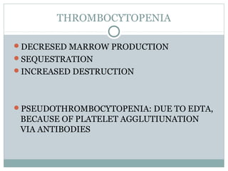
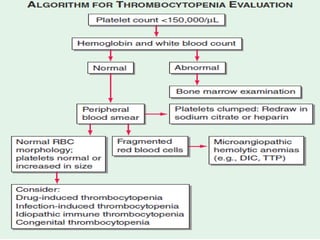
DR. JITHIN GEORGE
DISORDERS OF PLATELETS
Normal endothelium prevents thrombosis by
inhibiting platelet function. When vascular
endothelium injured these inhibitions overcome.
Platelets adhere through VWF.
PLATELET ADHESION leads to activation of GpIIb
IIIa receptor. Result in platelet aggregation.
Platelet plug is stabilised by fibrin mesh
Major regulator of platelet production –
thrombopoietin (TPO) is synthesized in ?
Harrison’s 18th Ed. 965
A. Kidney
B. Liver
C. Muscle
D. Adipose tissue
QUANTITATIVE DISORDERS
QUALITATIVE DIOSRDERS
QUANTITATIVE DISORDERS
THROMBOCYTOPENIA
DECRESED MARROW PRODUCTION
SEQUESTRATION
INCREASED DESTRUCTION
PSEUDOTHROMBOCYTOPENIA: DUE TO EDTA,
BECAUSE OF PLATELET AGGLUTIUNATION
VIA ANTIBODIES
Petechiae first appears where more venous pressure.
Ankles and feet.
Drug induced thrombocytopenia
Drug induced thrombocytopenia usually resolves
how many days after drug withdrawal ?
Harrison’s 18th Ed. 967
A. 1 - 3 days
B. 3 - 7 days
C. 7 - 10 days
D. 14 - 21 days
Heparin-Induced Thrombocytopenia
Drug-induced thrombocytopenia due to heparin differs
from that seen with other drugs in two major ways.
 (1) The thrombocytopenia is not usually severe, with nadir counts
rarely <20,000/L.
 (2) Heparin-induced thrombocytopenia (HIT) is not associated
with bleeding and, in fact, markedly increases the risk of
thrombosis.
HIT results from antibody formation to a complex of
the platelet-specific protein platelet factor 4 (PF4) and
heparin.
The antiheparin/PF4 antibody can activate platelets
through the FcRIIa receptor and also activate
monocytes and endothelial cells.
HIT can occur after exposure to low-molecular-
weight heparin (LMWH) as well as unfractionated
heparin (UFH), although it is about 10 times more
common with the latter.
Most patients develop HIT after exposure to heparin
for 5–14 days.
Laboratory Testing for HIT
HIT (anti-heparin/PF4) antibodies can be detected
using two types of assays. The most widely
available is an enzyme-linked immunoassay
(ELISA) with PF4/polyanion complex as the
antigen
The other assay is a platelet activation assay, which
measures the ability of the patient's serum to
activate platelets in the presence of heparin in a
concentration-dependent manner.
Treatment: Heparin-Induced Thrombocytopenia
discontinuation of heparin and use of alternative
anticoagulants.
 The direct thrombin inhibitors (DTIs) argatroban and
lepirudin are effective in HIT.
 The DTI bivalirudin and the antithrombin-binding
pentasaccharide fondaparinux are also effective but not
yet approved .
Danaparoid also can use
Warfarin therapy, if started, should be overlapped with a
DTI or fondaparinux, and started after resolution of the
thrombocytopenia and lessening of the prothrombotic
state.
Pseudothrombocytopenia is due to ?
 Harrison’s 18th Ed. 965
A. Exercise
B. Laboratory artifact
C. Cold weather
D. Fasting
Pseudothrombocytopenia is most likely when blood
is collected with which of the following
anticoagulants ?
Harrison’s 18th Ed. 965
A. EDTA
B. Sodium citrate
C. Heparin
D. All of the above
IMMUNE THROMBOCYTOPENIC PURPURA
also termed idiopathic thrombocytopenic purpura
acquired disorder in which there is immune-
mediated destruction of platelets and possibly
inhibition of platelet release from the
megakaryocyte.
*Children are affected by an acute type of illness
following URT infection (usually viral).
* = incidence.♂ ♀
*Adults are affected by a more gradual onset of
disease with chronic course & without preceding
illness.
*Age < 40 years
* : = 3-4:1♀ ♂
 ITP is termed secondary if it is associated with an
underlying disorder; autoimmune disorders,
particularly systemic lupus erythematosus (SLE),
and infections, such as HIV and hepatitis C, are
common causes.
Skin shows petechiae (pinpoint red hemorrhagic
spots) & ecchymoses.
Mucosal bleeding --- bleeding gum, epistaxis,
menorrhagia, metrorrhagia, GI bleeding &
hematuria
Intracranial hemorrhage is seen in 1%. Risk of
death 5%
No splenomegaly.
Laboratory Testing in ITP
Laboratory testing for antibodies (serologic testing)
is usually not helpful due to the low sensitivity and
specificity.
Bone marrow examination can be reserved for older
adults (usually >60 years) or those who have other
signs or laboratory abnormalities not explained by
ITP, or in patients who do not respond to initial
therapy. The peripheral blood smear may show large
platelets, with otherwise normal morphology.
Depending on the bleeding history, iron deficiency
anemia may be present
Laboratory testing is performed to evaluate for secondary
causes of ITP.
It should include testing for HIV infection and hepatitis C
(and other infections if indicated);
serologic testing for SLE, serum protein electrophoresis,
and immunoglobulin levels to potentially detect
hypogammaglobulinemia;
selective testing for IgA deficiency or monoclonal
gammopathies ,
 if anemia is present, direct antiglobulin testing (Coombs
test) to rule out combined autoimmune hemolytic
anemia with ITP (Evans syndrome).
Treatment: Immune Thrombocytopenic Purpura
prednisone at 1 mg/kg usually
Rh0(D) immune globulin therapy (WinRho SDF), at
50–75 g/kg, is also being used in this setting.
Rh0(D) immune globulin must be used only in Rh-
positive patients.
Intravenous gamma globulin (IVIgG),
 IVIgG has more efficacy than anti-Rh0(D) in
postsplenectomized patients. IVIgG is dosed at 2
g/kg total, given in divided doses over 2–5 days.
Rituximab, an anti-CD20 (B cell) antibody, has
shown efficacy in the treatment of refractory ITP.
Splenectomy has been used for treatment of patients
who relapse after glucocorticoids are tapered
Vaccination against encapsulated organisms
(especially pneumococcus, but also meningococcus
and Haemophilus influenzae, depending on patient
age and potential exposure) is recommended before
splenectomy
Thrombopoietin receptor agonists are now available
for the treatment of ITP.
Two agents, one administered subcutaneously
(romiplostim) and another orally (eltrombopag).
Evans’s syndrome refers to ? Harrison’s 18th Ed. 968
A. Autoimmune hemolytic anemia with ITP
B. SLE with ITP
C. HIV with ITP
D. Hepatitis C with ITP
Which of the following drugs is useful in refractory
ITP ? Harrison’s 18th Ed. 969
A. Rituximab
B. Romiplostim
C. Eltrombopag
D. All of the above
Inherited Thrombocytopenia
inherited in an autosomal dominant, autosomal
recessive, or X-linked pattern.
autosomal dominant thrombocytopenia are now
known to be associated with mutations in the
nonmuscle myosin heavy chain MYH9 gene.
Interestingly, these include the May-Hegglin
anomaly, and Sebastian, Epstein's, and Fechtner
syndromes, all of which have distinct distinguishing
features. A common feature of these disorders is
large platelets
Autosomal recessive disorders include congenital
amegakaryocytic thrombocytopenia,
thrombocytopenia with absent radii, and Bernard
Soulier syndrome. The latter is primarily a functional
platelet disorder due to absence of GPIb-IX-V, the
VWF adhesion receptor.
X-linked disorders include Wiskott-Aldrich
syndrome and a dyshematopoietic syndrome
resulting from a mutation in GATA-1, an important
transcriptional regulator of hematopoiesis
Thrombotic thrombocytopenic
microangiopathies
characterized by thrombocytopenia, a
microangiopathic hemolytic anemia evident by
fragmented RBCs and laboratory evidence of
hemolysis, and microvascular thrombosis.
thrombotic thrombocytopenic purpura (TTP) and
hemolytic uremic syndrome (HUS), as well as
syndromes complicating bone marrow
transplantation, certain medications and infections,
pregnancy and vasculitis.
 In DIC, while thrombocytopenia and
microangiopathy are seen, a coagulopathy
predominates, with consumption of clotting factors
and fibrinogen resulting in an elevated prothrombin
time (PT), and often activated partial thromboplastin
time (aPTT).
The PT and aPTT are characteristically normal in
TTP or HUS.
Thrombotic Thrombocytopenic Purpura (TTP)
Medication-related microangiopathic hemolytic
anemia may be
secondary to antibody formation (ticlopidine and
possibly clopidogrel) or
direct endothelial toxicity (cyclosporine, mitomycin
C, tacrolimus, quinine
Rx: application of plasma exchange.
Treatment: Thrombotic Thrombocytopenic
Purpura
Plasma exchange remains the mainstay of treatment
Glucocorticoids
other immunomodulatory therapies have been
reported to be successful in refractory or relapsing
TTP, including rituximab, vincristine,
cyclophosphamide, and splenectomy.
Hemolytic Uremic Syndrome
Treatment: Hemolytic Uremic Syndrome
Treatment of HUS is primarily supportive. In
D+HUS, many (~40%) children require at least some
period of support with dialysis
Plasma infusion or plasma exchange
Which of the following is false regarding thrombotic
thrombocytopenic microangiopathies ? Harrison’s
18th Ed. 969
A. Thrombocytopenia
B. Laboratory evidence of hemolysis
C. Abnormal PT and aPTT
D. Microvascular thrombosis
TTP is due to deficiency in the activity of ?
Harrison’s 18th Ed. 969
A. ADAMTS 11
B. ADAMTS 12
C. ADAMTS 13
D. ADAMTS 14
Features of Hemolytic Uremic Syndrome include all
except ? Harrison’s 18th Ed. 970
A. Chronic renal failure
B. Microangiopathic hemolytic anemia
C. Thrombocytopenia
D. Frequently preceded by an episode of diarrhea
Which of the following is not used in the treatment
of TTP ?
A. Platelet concentrates
B. Plasma exchange
C. Fresh-frozen plasma (FFP)
D. Solvent/detergent-treated plasma (SD-FFP)
answer
Plasma exchange remains the mainstay of treatment of
TTP.
Use of platelet concentrates and Desmopressin, or
DDAVP in the treatment of TTP is contraindicated as
it leads to extensive platelet aggregation in the CNS.
Thromobcytosis
Due to
(1) iron deficiency;
(2) inflammation, cancer, or infection (reactive
thrombocytosis); or
(3) an underlying myeloproliferative process
[essential thrombocythemia or polycythemia vera)
or, rarely, the 5q- myelodysplastic process.
Thrombocytosis is due to deficiency of ? Harrison’s
18th Ed. 970
A. Vitamin B12
 B. Folic acid
C. Iron
 D. Calcium
Qualitative Disorders of Platelet Function
autosomal recessive disorders Glanzmann's
thrombasthenia (absence of the platelet GpIIbIIIa
receptor) and Bernard Soulier syndrome (absence of
the platelet GpIb-IX-V receptor).
 Both are inherited in an autosomal recessive fashion
and present with bleeding symptoms in childhood.
Platelet storage pool disorder (SPD) is the classic
autosomal dominant qualitative platelet disorder.
This results from abnormalities of platelet granule
formation. It is also seen as a part of inherited
disorders of granule formation, such as Hermansky-
Pudlak syndrome
In which of the following disorders, there is absence
of the platelet GpIIbIIIa receptor ?
Harrison’s 18th Ed. 970
A. Glanzmann’s thrombasthenia
B. Bernard Soulier syndrome
C. Hermansky-Pudlak syndrome
D. Heyde’s syndrome
VWD
three major types and four subtypes of type 2
Not all patients with low VWF levels have bleeding
symptoms. Whether patients bleed or not will
depend on the overall hemostatic balance they
have inherited, along with environmental
influences and the type of hemostatic challenges
they experience.
Although the inheritance of VWD is autosomal,
many factors modulate both VWF levels and
bleeding symptoms. These have not all been
defined, but include blood type, thyroid hormone
status, race, stress, exercise, and hormonal (both
endogenous and exogenous) influences.
Patients with type O blood have VWF protein levels
of approximately one-half that of patients with AB
blood type; and, in fact, the normal range for
patients with type O blood overlaps that which has
been considered diagnostic for VWD. A mildly
decreased VWF level should perhaps be viewed more
as a risk factor for bleeding than as an actual disease
Patients with Type 2 VWD have functional defects;
thus, the VWF antigen measurement is
significantly higher than the test of function.
 For types 2A, 2B and 2M, VWF activity is
decreased, measured as ristocetin cofactor or
collagen-binding activity.
 In type 2A VWD, the impaired function is due
either to increased susceptibility to cleavage by
ADAMTS13, resulting in loss of intermediate and
high-molecular-weight multimers, or to decreased
secretion of these multimers by the cell.
 Type 2B VWD results from gain of function
mutations that result in increased spontaneous
binding of VWF to platelets in circulation, with
subsequent clearance of this complex by the
reticuloendothelial system.
Type 2M occurs as a consequence of a group of
mutations that cause dysfunction of the molecule but
do not affect multimer structure.
Type 2N VWD is due to mutations in VWF that
preclude binding of FVIII. As FVIII is stabilized by
binding to VWF, the FVIII in patients with type 2N
VWD has a very short half-life, and the FVIII level is
markedly decreased. This is sometimes termed
autosomal hemophilia.
Type 3 VWD, or severe VWD, describes patients with
virtually no VWF protein and FVIII levels <10%.
Treatment: von Willebrand Disease
For type 1 VWD is 1-deamino-8-d-arginine
vasopressin (DDAVP, or desmopressin), which
results in release of VWF and FVIII from endothelial
stores.
 DDAVP can be given intravenously or by a high
concentration intranasal spray (1.5 mg/mL).
The peak activity when given intravenously is
approximately 30 minutes, while it is 2 h when given
intranasally. The usual dose is 0.3 g/kg
intravenously or 2 squirts (1 in each nostril) for
patients >50 kg (1 squirt for those <50 kg).
Some patients with types 2A and 2M VWD respond
to DDAVP such that it can be used for minor
procedures.
For the other subtypes, for type 3 disease, and for
major procedures requiring longer periods of normal
hemostasis, VWF replacement can be given. Virally
inactivated VWF-containing factor concentrates are
thought to be safer than cryoprecipitate as the
replacement product.
Antifibrinolytic therapy using either -aminocaproic
acid or tranexamic acid is an important therapy,
either alone or in an adjunctive capacity, particularly
for the prevention or treatment of mucosal bleeding.
These agents are particularly useful in prophylaxis
for dental procedures, with DDAVP for dental
extractions and tonsillectomy, menorrhagia, and
prostate procedures.
 It is contraindicated in the setting of upper urinary
tract bleeding, due to the risk of ureteral obstruction
Which of the following vWD type is most severe ?
Harrison’s 16th Ed. 676
 A. Type I
B. Type IIA
C. Type IIB
D. Type III
Disorders of platelets

Disorders of platelets

  • 1.
  • 3.
    Normal endothelium preventsthrombosis by inhibiting platelet function. When vascular endothelium injured these inhibitions overcome. Platelets adhere through VWF. PLATELET ADHESION leads to activation of GpIIb IIIa receptor. Result in platelet aggregation. Platelet plug is stabilised by fibrin mesh
  • 4.
    Major regulator ofplatelet production – thrombopoietin (TPO) is synthesized in ? Harrison’s 18th Ed. 965 A. Kidney B. Liver C. Muscle D. Adipose tissue
  • 5.
  • 6.
  • 7.
    THROMBOCYTOPENIA DECRESED MARROW PRODUCTION SEQUESTRATION INCREASEDDESTRUCTION PSEUDOTHROMBOCYTOPENIA: DUE TO EDTA, BECAUSE OF PLATELET AGGLUTIUNATION VIA ANTIBODIES
  • 8.
    Petechiae first appearswhere more venous pressure. Ankles and feet.
  • 13.
  • 14.
    Drug induced thrombocytopeniausually resolves how many days after drug withdrawal ? Harrison’s 18th Ed. 967 A. 1 - 3 days B. 3 - 7 days C. 7 - 10 days D. 14 - 21 days
  • 15.
    Heparin-Induced Thrombocytopenia Drug-induced thrombocytopeniadue to heparin differs from that seen with other drugs in two major ways.  (1) The thrombocytopenia is not usually severe, with nadir counts rarely <20,000/L.  (2) Heparin-induced thrombocytopenia (HIT) is not associated with bleeding and, in fact, markedly increases the risk of thrombosis. HIT results from antibody formation to a complex of the platelet-specific protein platelet factor 4 (PF4) and heparin. The antiheparin/PF4 antibody can activate platelets through the FcRIIa receptor and also activate monocytes and endothelial cells.
  • 16.
    HIT can occurafter exposure to low-molecular- weight heparin (LMWH) as well as unfractionated heparin (UFH), although it is about 10 times more common with the latter. Most patients develop HIT after exposure to heparin for 5–14 days.
  • 18.
    Laboratory Testing forHIT HIT (anti-heparin/PF4) antibodies can be detected using two types of assays. The most widely available is an enzyme-linked immunoassay (ELISA) with PF4/polyanion complex as the antigen The other assay is a platelet activation assay, which measures the ability of the patient's serum to activate platelets in the presence of heparin in a concentration-dependent manner.
  • 19.
    Treatment: Heparin-Induced Thrombocytopenia discontinuationof heparin and use of alternative anticoagulants.  The direct thrombin inhibitors (DTIs) argatroban and lepirudin are effective in HIT.  The DTI bivalirudin and the antithrombin-binding pentasaccharide fondaparinux are also effective but not yet approved . Danaparoid also can use Warfarin therapy, if started, should be overlapped with a DTI or fondaparinux, and started after resolution of the thrombocytopenia and lessening of the prothrombotic state.
  • 20.
    Pseudothrombocytopenia is dueto ?  Harrison’s 18th Ed. 965 A. Exercise B. Laboratory artifact C. Cold weather D. Fasting
  • 21.
    Pseudothrombocytopenia is mostlikely when blood is collected with which of the following anticoagulants ? Harrison’s 18th Ed. 965 A. EDTA B. Sodium citrate C. Heparin D. All of the above
  • 22.
    IMMUNE THROMBOCYTOPENIC PURPURA alsotermed idiopathic thrombocytopenic purpura acquired disorder in which there is immune- mediated destruction of platelets and possibly inhibition of platelet release from the megakaryocyte.
  • 23.
    *Children are affectedby an acute type of illness following URT infection (usually viral). * = incidence.♂ ♀ *Adults are affected by a more gradual onset of disease with chronic course & without preceding illness. *Age < 40 years * : = 3-4:1♀ ♂
  • 24.
     ITP istermed secondary if it is associated with an underlying disorder; autoimmune disorders, particularly systemic lupus erythematosus (SLE), and infections, such as HIV and hepatitis C, are common causes.
  • 25.
    Skin shows petechiae(pinpoint red hemorrhagic spots) & ecchymoses. Mucosal bleeding --- bleeding gum, epistaxis, menorrhagia, metrorrhagia, GI bleeding & hematuria Intracranial hemorrhage is seen in 1%. Risk of death 5% No splenomegaly.
  • 26.
    Laboratory Testing inITP Laboratory testing for antibodies (serologic testing) is usually not helpful due to the low sensitivity and specificity. Bone marrow examination can be reserved for older adults (usually >60 years) or those who have other signs or laboratory abnormalities not explained by ITP, or in patients who do not respond to initial therapy. The peripheral blood smear may show large platelets, with otherwise normal morphology. Depending on the bleeding history, iron deficiency anemia may be present
  • 27.
    Laboratory testing isperformed to evaluate for secondary causes of ITP. It should include testing for HIV infection and hepatitis C (and other infections if indicated); serologic testing for SLE, serum protein electrophoresis, and immunoglobulin levels to potentially detect hypogammaglobulinemia; selective testing for IgA deficiency or monoclonal gammopathies ,  if anemia is present, direct antiglobulin testing (Coombs test) to rule out combined autoimmune hemolytic anemia with ITP (Evans syndrome).
  • 28.
    Treatment: Immune ThrombocytopenicPurpura prednisone at 1 mg/kg usually Rh0(D) immune globulin therapy (WinRho SDF), at 50–75 g/kg, is also being used in this setting. Rh0(D) immune globulin must be used only in Rh- positive patients. Intravenous gamma globulin (IVIgG),
  • 29.
     IVIgG hasmore efficacy than anti-Rh0(D) in postsplenectomized patients. IVIgG is dosed at 2 g/kg total, given in divided doses over 2–5 days. Rituximab, an anti-CD20 (B cell) antibody, has shown efficacy in the treatment of refractory ITP. Splenectomy has been used for treatment of patients who relapse after glucocorticoids are tapered
  • 30.
    Vaccination against encapsulatedorganisms (especially pneumococcus, but also meningococcus and Haemophilus influenzae, depending on patient age and potential exposure) is recommended before splenectomy
  • 31.
    Thrombopoietin receptor agonistsare now available for the treatment of ITP. Two agents, one administered subcutaneously (romiplostim) and another orally (eltrombopag).
  • 32.
    Evans’s syndrome refersto ? Harrison’s 18th Ed. 968 A. Autoimmune hemolytic anemia with ITP B. SLE with ITP C. HIV with ITP D. Hepatitis C with ITP
  • 33.
    Which of thefollowing drugs is useful in refractory ITP ? Harrison’s 18th Ed. 969 A. Rituximab B. Romiplostim C. Eltrombopag D. All of the above
  • 34.
    Inherited Thrombocytopenia inherited inan autosomal dominant, autosomal recessive, or X-linked pattern. autosomal dominant thrombocytopenia are now known to be associated with mutations in the nonmuscle myosin heavy chain MYH9 gene. Interestingly, these include the May-Hegglin anomaly, and Sebastian, Epstein's, and Fechtner syndromes, all of which have distinct distinguishing features. A common feature of these disorders is large platelets
  • 35.
    Autosomal recessive disordersinclude congenital amegakaryocytic thrombocytopenia, thrombocytopenia with absent radii, and Bernard Soulier syndrome. The latter is primarily a functional platelet disorder due to absence of GPIb-IX-V, the VWF adhesion receptor.
  • 36.
    X-linked disorders includeWiskott-Aldrich syndrome and a dyshematopoietic syndrome resulting from a mutation in GATA-1, an important transcriptional regulator of hematopoiesis
  • 37.
  • 38.
    characterized by thrombocytopenia,a microangiopathic hemolytic anemia evident by fragmented RBCs and laboratory evidence of hemolysis, and microvascular thrombosis. thrombotic thrombocytopenic purpura (TTP) and hemolytic uremic syndrome (HUS), as well as syndromes complicating bone marrow transplantation, certain medications and infections, pregnancy and vasculitis.
  • 39.
     In DIC,while thrombocytopenia and microangiopathy are seen, a coagulopathy predominates, with consumption of clotting factors and fibrinogen resulting in an elevated prothrombin time (PT), and often activated partial thromboplastin time (aPTT). The PT and aPTT are characteristically normal in TTP or HUS.
  • 40.
  • 42.
    Medication-related microangiopathic hemolytic anemiamay be secondary to antibody formation (ticlopidine and possibly clopidogrel) or direct endothelial toxicity (cyclosporine, mitomycin C, tacrolimus, quinine Rx: application of plasma exchange.
  • 43.
    Treatment: Thrombotic Thrombocytopenic Purpura Plasmaexchange remains the mainstay of treatment Glucocorticoids other immunomodulatory therapies have been reported to be successful in refractory or relapsing TTP, including rituximab, vincristine, cyclophosphamide, and splenectomy.
  • 44.
  • 45.
    Treatment: Hemolytic UremicSyndrome Treatment of HUS is primarily supportive. In D+HUS, many (~40%) children require at least some period of support with dialysis Plasma infusion or plasma exchange
  • 46.
    Which of thefollowing is false regarding thrombotic thrombocytopenic microangiopathies ? Harrison’s 18th Ed. 969 A. Thrombocytopenia B. Laboratory evidence of hemolysis C. Abnormal PT and aPTT D. Microvascular thrombosis
  • 47.
    TTP is dueto deficiency in the activity of ? Harrison’s 18th Ed. 969 A. ADAMTS 11 B. ADAMTS 12 C. ADAMTS 13 D. ADAMTS 14
  • 48.
    Features of HemolyticUremic Syndrome include all except ? Harrison’s 18th Ed. 970 A. Chronic renal failure B. Microangiopathic hemolytic anemia C. Thrombocytopenia D. Frequently preceded by an episode of diarrhea
  • 49.
    Which of thefollowing is not used in the treatment of TTP ? A. Platelet concentrates B. Plasma exchange C. Fresh-frozen plasma (FFP) D. Solvent/detergent-treated plasma (SD-FFP)
  • 50.
    answer Plasma exchange remainsthe mainstay of treatment of TTP. Use of platelet concentrates and Desmopressin, or DDAVP in the treatment of TTP is contraindicated as it leads to extensive platelet aggregation in the CNS.
  • 51.
    Thromobcytosis Due to (1) irondeficiency; (2) inflammation, cancer, or infection (reactive thrombocytosis); or (3) an underlying myeloproliferative process [essential thrombocythemia or polycythemia vera) or, rarely, the 5q- myelodysplastic process.
  • 52.
    Thrombocytosis is dueto deficiency of ? Harrison’s 18th Ed. 970 A. Vitamin B12  B. Folic acid C. Iron  D. Calcium
  • 53.
    Qualitative Disorders ofPlatelet Function autosomal recessive disorders Glanzmann's thrombasthenia (absence of the platelet GpIIbIIIa receptor) and Bernard Soulier syndrome (absence of the platelet GpIb-IX-V receptor).  Both are inherited in an autosomal recessive fashion and present with bleeding symptoms in childhood.
  • 55.
    Platelet storage pooldisorder (SPD) is the classic autosomal dominant qualitative platelet disorder. This results from abnormalities of platelet granule formation. It is also seen as a part of inherited disorders of granule formation, such as Hermansky- Pudlak syndrome
  • 56.
    In which ofthe following disorders, there is absence of the platelet GpIIbIIIa receptor ? Harrison’s 18th Ed. 970 A. Glanzmann’s thrombasthenia B. Bernard Soulier syndrome C. Hermansky-Pudlak syndrome D. Heyde’s syndrome
  • 57.
  • 58.
    three major typesand four subtypes of type 2 Not all patients with low VWF levels have bleeding symptoms. Whether patients bleed or not will depend on the overall hemostatic balance they have inherited, along with environmental influences and the type of hemostatic challenges they experience. Although the inheritance of VWD is autosomal, many factors modulate both VWF levels and bleeding symptoms. These have not all been defined, but include blood type, thyroid hormone status, race, stress, exercise, and hormonal (both endogenous and exogenous) influences.
  • 59.
    Patients with typeO blood have VWF protein levels of approximately one-half that of patients with AB blood type; and, in fact, the normal range for patients with type O blood overlaps that which has been considered diagnostic for VWD. A mildly decreased VWF level should perhaps be viewed more as a risk factor for bleeding than as an actual disease
  • 60.
    Patients with Type2 VWD have functional defects; thus, the VWF antigen measurement is significantly higher than the test of function.  For types 2A, 2B and 2M, VWF activity is decreased, measured as ristocetin cofactor or collagen-binding activity.  In type 2A VWD, the impaired function is due either to increased susceptibility to cleavage by ADAMTS13, resulting in loss of intermediate and high-molecular-weight multimers, or to decreased secretion of these multimers by the cell.
  • 61.
     Type 2BVWD results from gain of function mutations that result in increased spontaneous binding of VWF to platelets in circulation, with subsequent clearance of this complex by the reticuloendothelial system. Type 2M occurs as a consequence of a group of mutations that cause dysfunction of the molecule but do not affect multimer structure.
  • 62.
    Type 2N VWDis due to mutations in VWF that preclude binding of FVIII. As FVIII is stabilized by binding to VWF, the FVIII in patients with type 2N VWD has a very short half-life, and the FVIII level is markedly decreased. This is sometimes termed autosomal hemophilia. Type 3 VWD, or severe VWD, describes patients with virtually no VWF protein and FVIII levels <10%.
  • 63.
    Treatment: von WillebrandDisease For type 1 VWD is 1-deamino-8-d-arginine vasopressin (DDAVP, or desmopressin), which results in release of VWF and FVIII from endothelial stores.  DDAVP can be given intravenously or by a high concentration intranasal spray (1.5 mg/mL). The peak activity when given intravenously is approximately 30 minutes, while it is 2 h when given intranasally. The usual dose is 0.3 g/kg intravenously or 2 squirts (1 in each nostril) for patients >50 kg (1 squirt for those <50 kg).
  • 64.
    Some patients withtypes 2A and 2M VWD respond to DDAVP such that it can be used for minor procedures. For the other subtypes, for type 3 disease, and for major procedures requiring longer periods of normal hemostasis, VWF replacement can be given. Virally inactivated VWF-containing factor concentrates are thought to be safer than cryoprecipitate as the replacement product.
  • 65.
    Antifibrinolytic therapy usingeither -aminocaproic acid or tranexamic acid is an important therapy, either alone or in an adjunctive capacity, particularly for the prevention or treatment of mucosal bleeding. These agents are particularly useful in prophylaxis for dental procedures, with DDAVP for dental extractions and tonsillectomy, menorrhagia, and prostate procedures.  It is contraindicated in the setting of upper urinary tract bleeding, due to the risk of ureteral obstruction
  • 66.
    Which of thefollowing vWD type is most severe ? Harrison’s 16th Ed. 676  A. Type I B. Type IIA C. Type IIB D. Type III