PRIMORDIAL GERM CELLS
M. VharshiniM. Vharshini
MSc 1MSc 1stst
yearyear
Roll No.: 35Roll No.: 35
Introduction
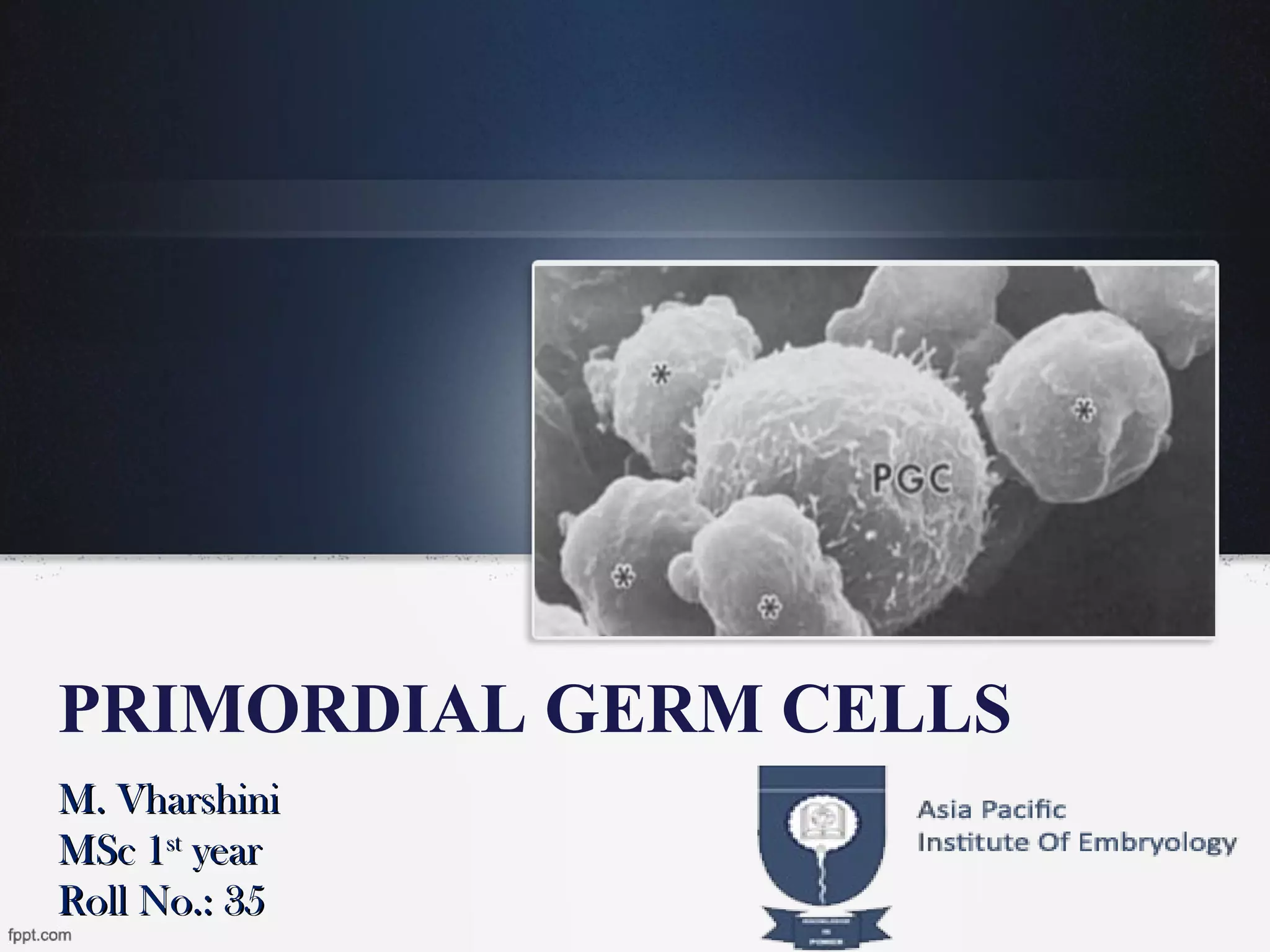
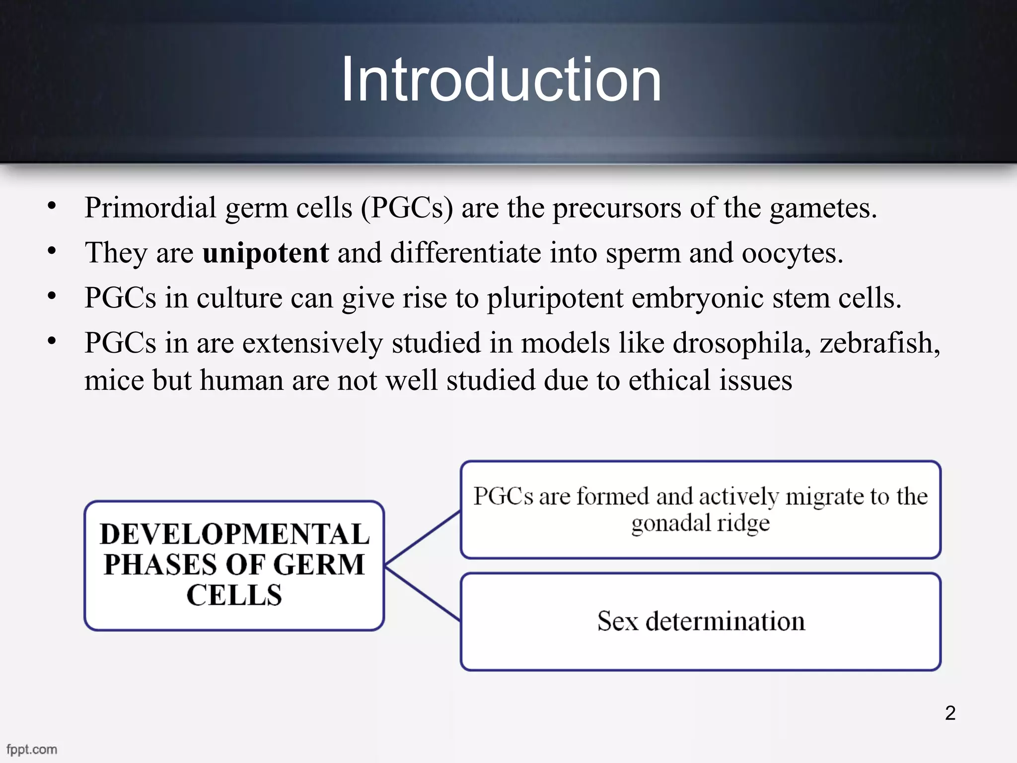
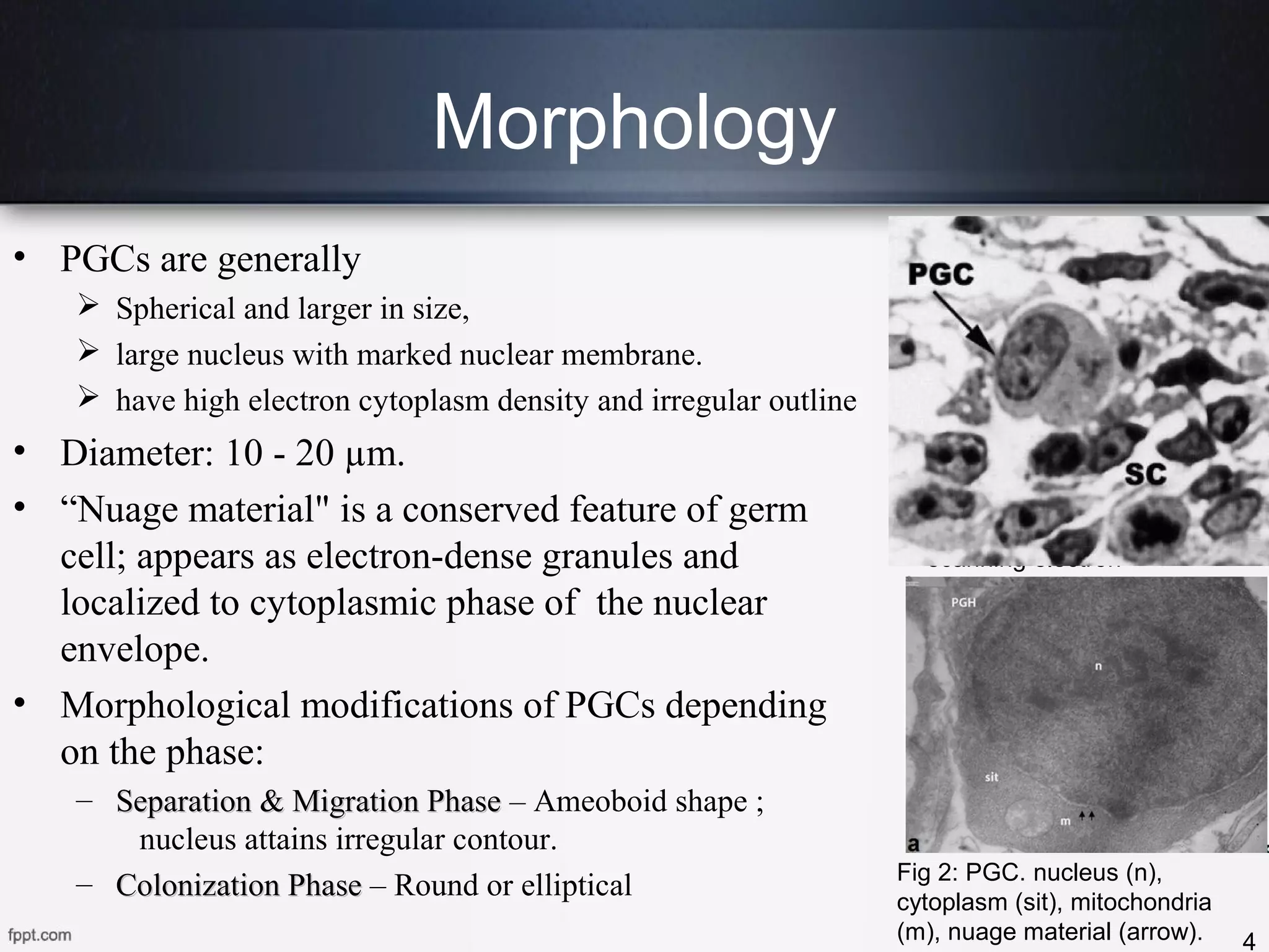
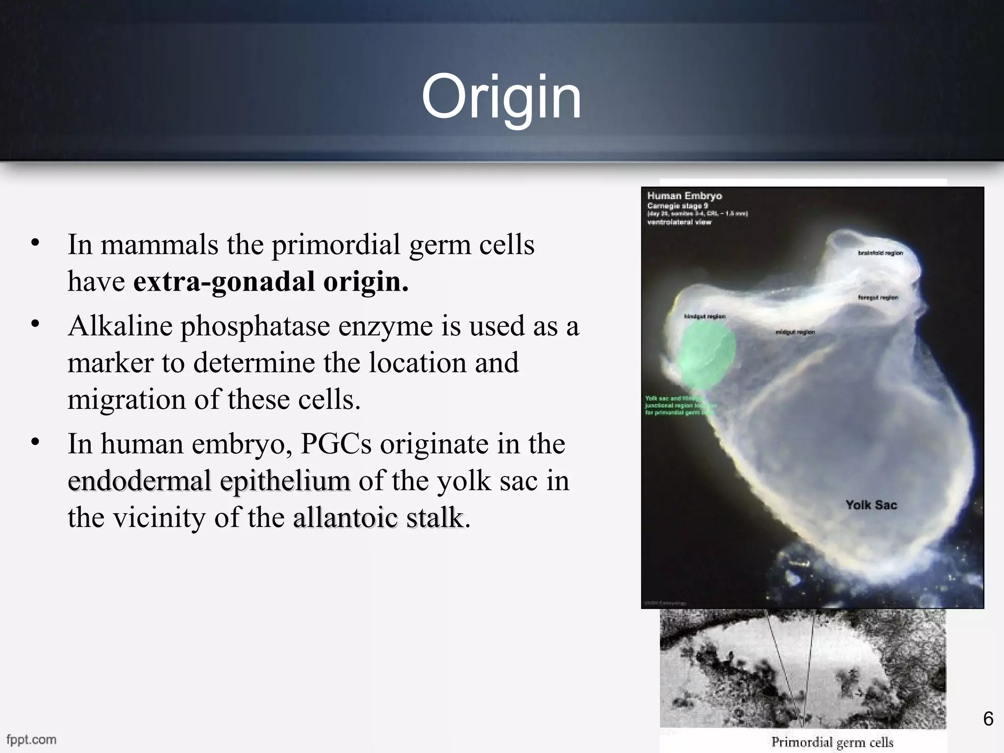
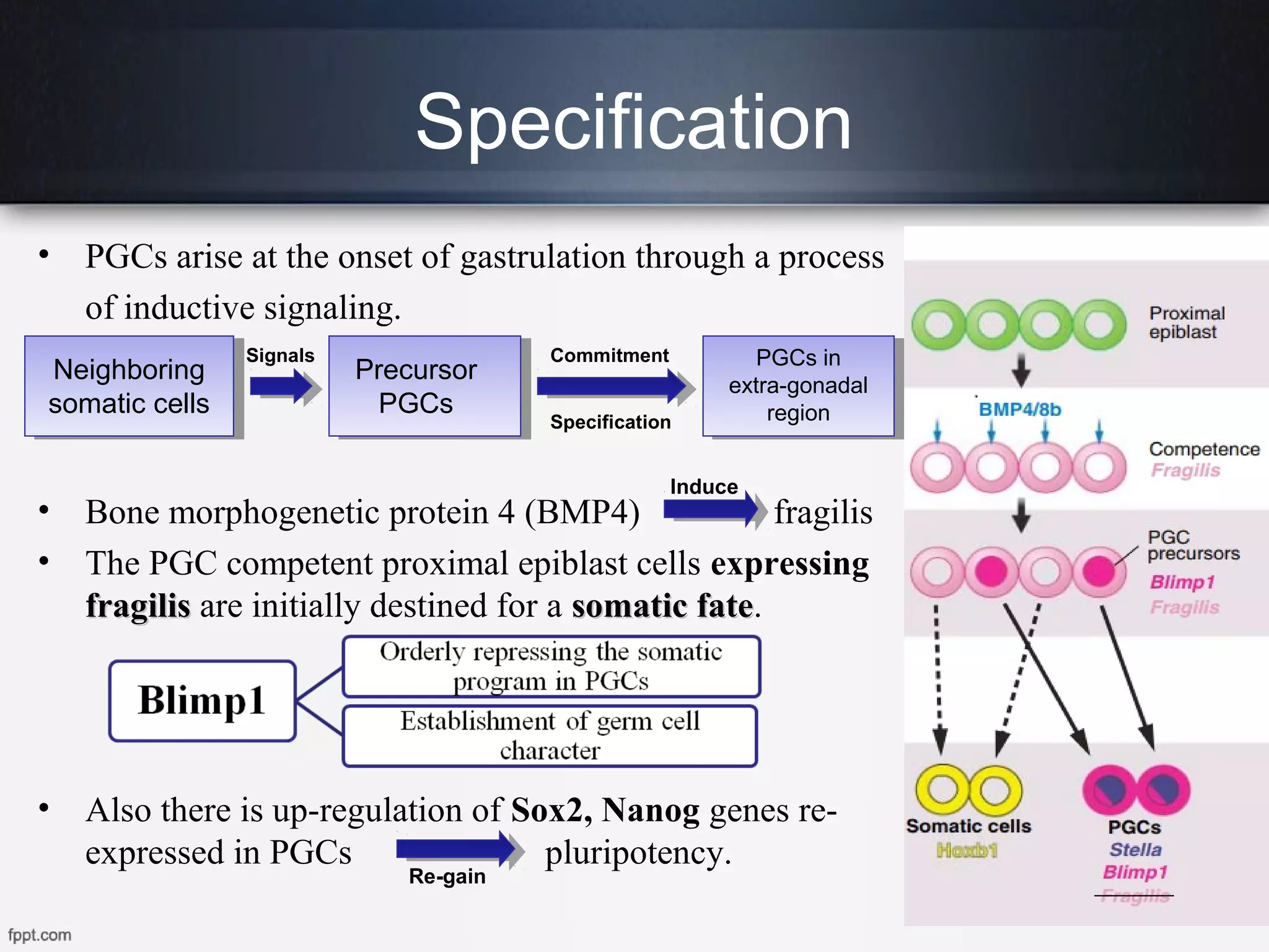
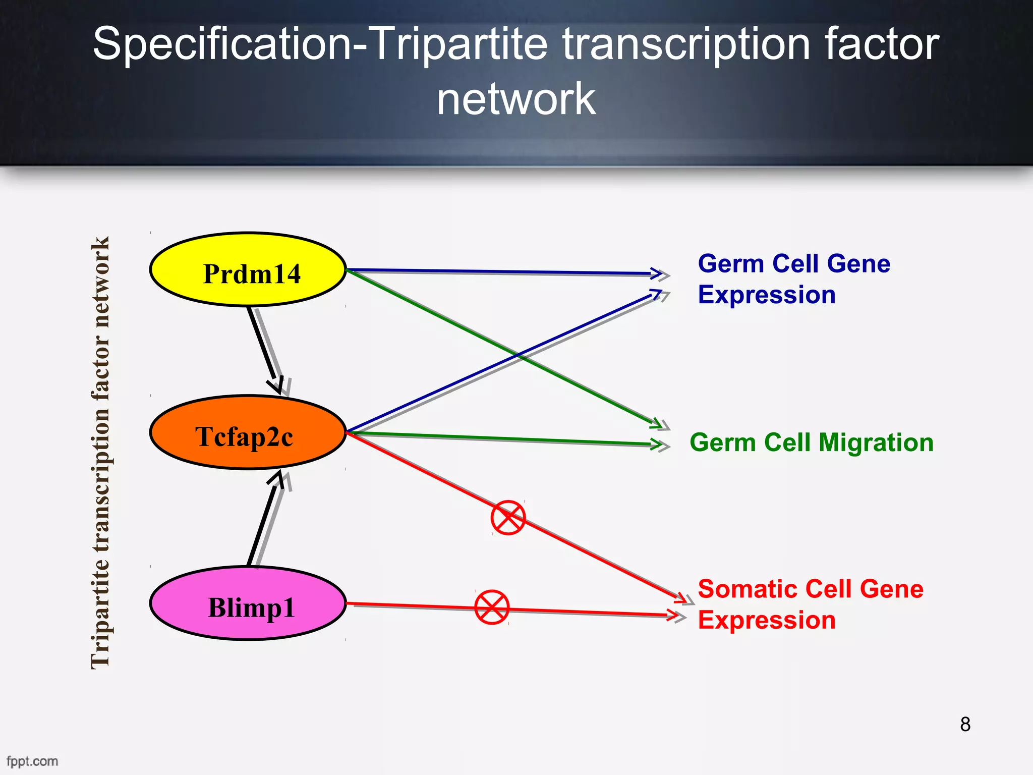
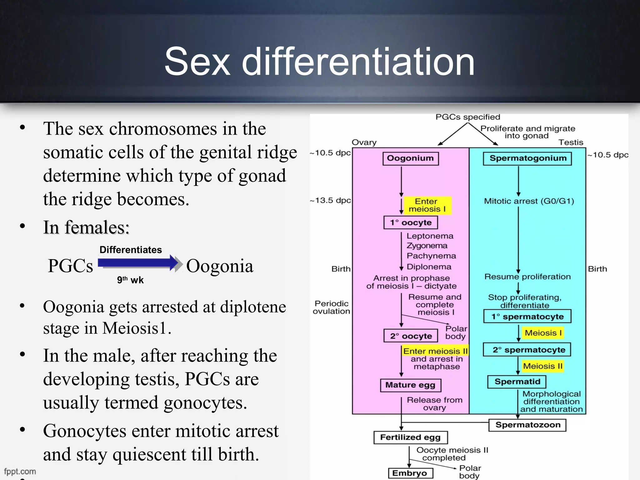
• Primordial germ cells (PGCs) are the precursors of the gametes.
• They are unipotent and differentiate into sperm and oocytes.
• PGCs in culture can give rise to pluripotent embryonic stem cells.
• PGCs in are extensively studied in models like drosophila, zebrafish,
mice but human are not well studied due to ethical issues
2
Time line of PGCs in Mouse and
Human
3
• PGCs are generally
 Spherical and larger in size,
 large nucleus with marked nuclear membrane.
 have high electron cytoplasm density and irregular outline
• Diameter: 10 - 20 µm.
• “Nuage material" is a conserved feature of germ
cell; appears as electron-dense granules and
localized to cytoplasmic phase of the nuclear
envelope.
• Morphological modifications of PGCs depending
on the phase:
– Separation & Migration PhaseSeparation & Migration Phase – Ameoboid shape ;
nucleus attains irregular contour.
– Colonization PhaseColonization Phase – Round or elliptical
4
Fig1: PGC (chicken)
scanning electron
micrograph.
Morphology
Fig 2: PGC. nucleus (n),
cytoplasm (sit), mitochondria
(m), nuage material (arrow).
• PGCs are stained more densely than somatic cells.
• Peripheral cytoplasm shows high alkaline phosphatase
(AP) activity – Marker
• The "nuage material" has electron-dense granules
give positivepositive reactionreaction with H&E staining.
• VasaVasa and nanosnanos genes can be used as molecular marker.
• Currently PGCs are most identified by using the monoclonal
antibodies such as stage-specific embryonic antigens α-SSEA-
1, α-SSEA-3, SSEA-4.
• α-SSEA-1 is the most widely used marker for PGCs.
5
Histological and Immunohistochemical
Identification
Fig3: α-SSEA-1
positive PGCs
Origin
• In mammals the primordial germ cells
have extra-gonadal origin.
• Alkaline phosphatase enzyme is used as a
marker to determine the location and
migration of these cells.
• In human embryo, PGCs originate in the
endodermal epitheliumendodermal epithelium of the yolk sac in
the vicinity of the allantoic stalkallantoic stalk.
6
Specification
• PGCs arise at the onset of gastrulation through a process
of inductive signaling.
• Bone morphogenetic protein 4 (BMP4) fragilis
• The PGC competent proximal epiblast cells expressing
fragilisfragilis are initially destined for a somatic fatesomatic fate.
• Also there is up-regulation of Sox2, Nanog genes re-
expressed in PGCs pluripotency.
7
Neighboring
somatic cells
Precursor
PGCs
PGCs in
extra-gonadal
region
Commitment
Specification
Signals
Induce
Re-gain
Specification-Tripartite transcription factor
network
8
Prdm14
Blimp1
Tcfap2c
Germ Cell Gene
Expression
Germ Cell Migration
Somatic Cell Gene
Expression
Tripartitetranscriptionfactornetwork
Migration
9
•Begins to migrate from yolk sac in the 4th
week and reached gonads by 5th
week
in mammals.
•Controlled through two separate ligand-receptor chemo-attractant signaling
interactions.
Migration
• The GR secretes stromal cell-derived
factor1 (SDF1) provides directional cue for
PGC.
• Germ cells lacking the SDF1 receptor fail to
colonise the gonads efficiently
• Alkaline phosphatase enzyme is used as a
marker to identify the migration path.
• Genetic reprogramming, DNA methylation,
erasure of gene imprinting occurs during
migration.
• By default, bipotenital gonad produces
female reproductive organs, unless male-
specific factors are upregulated.
10
11
• The sex chromosomes in the
somatic cells of the genital ridge
determine which type of gonad
the ridge becomes.
• In females:In females:
PGCs Oogonia
• Oogonia gets arrested at diplotene
stage in Meiosis1.
• In the male, after reaching the
developing testis, PGCs are
usually termed gonocytes.
• Gonocytes enter mitotic arrest
and stay quiescent till birth.
Sex differentiation
Differentiates
9th
wk
SRY gene
12
THANK YOU
13

Primordial Germ Cells

  • 1.
    PRIMORDIAL GERM CELLS M.VharshiniM. Vharshini MSc 1MSc 1stst yearyear Roll No.: 35Roll No.: 35
  • 2.
    Introduction • Primordial germcells (PGCs) are the precursors of the gametes. • They are unipotent and differentiate into sperm and oocytes. • PGCs in culture can give rise to pluripotent embryonic stem cells. • PGCs in are extensively studied in models like drosophila, zebrafish, mice but human are not well studied due to ethical issues 2
  • 3.
    Time line ofPGCs in Mouse and Human 3
  • 4.
    • PGCs aregenerally  Spherical and larger in size,  large nucleus with marked nuclear membrane.  have high electron cytoplasm density and irregular outline • Diameter: 10 - 20 µm. • “Nuage material" is a conserved feature of germ cell; appears as electron-dense granules and localized to cytoplasmic phase of the nuclear envelope. • Morphological modifications of PGCs depending on the phase: – Separation & Migration PhaseSeparation & Migration Phase – Ameoboid shape ; nucleus attains irregular contour. – Colonization PhaseColonization Phase – Round or elliptical 4 Fig1: PGC (chicken) scanning electron micrograph. Morphology Fig 2: PGC. nucleus (n), cytoplasm (sit), mitochondria (m), nuage material (arrow).
  • 5.
    • PGCs arestained more densely than somatic cells. • Peripheral cytoplasm shows high alkaline phosphatase (AP) activity – Marker • The "nuage material" has electron-dense granules give positivepositive reactionreaction with H&E staining. • VasaVasa and nanosnanos genes can be used as molecular marker. • Currently PGCs are most identified by using the monoclonal antibodies such as stage-specific embryonic antigens α-SSEA- 1, α-SSEA-3, SSEA-4. • α-SSEA-1 is the most widely used marker for PGCs. 5 Histological and Immunohistochemical Identification Fig3: α-SSEA-1 positive PGCs
  • 6.
    Origin • In mammalsthe primordial germ cells have extra-gonadal origin. • Alkaline phosphatase enzyme is used as a marker to determine the location and migration of these cells. • In human embryo, PGCs originate in the endodermal epitheliumendodermal epithelium of the yolk sac in the vicinity of the allantoic stalkallantoic stalk. 6
  • 7.
    Specification • PGCs ariseat the onset of gastrulation through a process of inductive signaling. • Bone morphogenetic protein 4 (BMP4) fragilis • The PGC competent proximal epiblast cells expressing fragilisfragilis are initially destined for a somatic fatesomatic fate. • Also there is up-regulation of Sox2, Nanog genes re- expressed in PGCs pluripotency. 7 Neighboring somatic cells Precursor PGCs PGCs in extra-gonadal region Commitment Specification Signals Induce Re-gain
  • 8.
    Specification-Tripartite transcription factor network 8 Prdm14 Blimp1 Tcfap2c GermCell Gene Expression Germ Cell Migration Somatic Cell Gene Expression Tripartitetranscriptionfactornetwork
  • 9.
    Migration 9 •Begins to migratefrom yolk sac in the 4th week and reached gonads by 5th week in mammals. •Controlled through two separate ligand-receptor chemo-attractant signaling interactions.
  • 10.
    Migration • The GRsecretes stromal cell-derived factor1 (SDF1) provides directional cue for PGC. • Germ cells lacking the SDF1 receptor fail to colonise the gonads efficiently • Alkaline phosphatase enzyme is used as a marker to identify the migration path. • Genetic reprogramming, DNA methylation, erasure of gene imprinting occurs during migration. • By default, bipotenital gonad produces female reproductive organs, unless male- specific factors are upregulated. 10
  • 11.
    11 • The sexchromosomes in the somatic cells of the genital ridge determine which type of gonad the ridge becomes. • In females:In females: PGCs Oogonia • Oogonia gets arrested at diplotene stage in Meiosis1. • In the male, after reaching the developing testis, PGCs are usually termed gonocytes. • Gonocytes enter mitotic arrest and stay quiescent till birth. Sex differentiation Differentiates 9th wk
  • 12.
  • 13.

Editor's Notes

  • #11 (Epigenetic mechanism that occurs by addition of methyl group to DNA, modifying the function of gene),