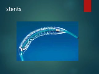
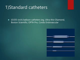
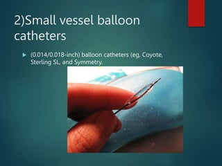
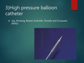
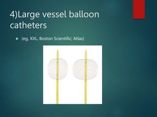
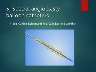
Percutaneous
Transluminal
Coronary
Angioplasty
PERCUTANEOUS
TRANSLUMINAL CORONARY
ANGIOPLASTY (PTCA)
Is performed to open blocked
coronary arteries caused by
coronary artery disease and
to restore arterial blood flow
to the heart tissue without
open heart surgery.
MEANING
PTCA is a minimally invasive
procedure to open up
blocked coronary arteries
allowing blood to circulate
unobstructed to the heart
muscle.
Intracoronary stents are
used to treat abrupt or
threatened abrupt
closure and re-stenosis
following balloon
angioplasty.
STENTS:-
A stent is an expandable
mesh like structure
designed to maintain
vessel patency by
compressing the arterial
walls and resisting
vasoconstriction
stents
INDICATIONS:-
 Acute ST-elevation myocardial infarction (STEMI)
 Non–ST-elevation acute coronary syndrome (NSTE-
ACS)
 Stable angina
 Anginal equivalent (eg, dyspnea, arrhythmia, or
dizziness or syncope)
 Asymptomatic or mildly symptomatic patient with
moderate to severe ischemia on noninvasive testing
 Angiographic indications that include
hemodynamically significant lesions in vessels.
 Atherosclerosis
USES OF STENTS:-
 Short lesions in large native coronary arteries not
previously treated with PTCA
 Focal lesions in saphenous vein grafts
 Treatment of abrupt closure during PTCA
CONTRAINDICATIONS:-
ABSOLUTE CONTRAINDICATIONS:-
 Lack of cardiac surgical support
 Significant obstruction of the left main coronary artery
without a non-obstructed bypass graft to the left
anterior descending or left circumflex arteries
RELATIVE CONTRAINDICATIONS:-
 Coagulopathy
 Hypercoagulable states
 Diffusely diseased vessels without focal stenosis
 A single diseased vessel providing all perfusion to the
myocardium
 Total occlusion of a coronary artery
 Stenosis < 50%
 Bare metal stents (BMS)
Made of nickal titanium alloy
 Drug-eluting stents (DES)
Traditional stents that are coated with drugs which release
certain drugs over time-
Zotarolimus, sirolimus, everolimus
These types of stents help prevent re-stenosis of the
artery
Bare metal stent
DES STENT
TYPES OF ANGIOPLASTY
BALLOONS:-
 Standard
 Small vessel balloon catheters
 High pressure balloon catheters
 Large vessel balloon catheters
 Special angioplasty balloon catheters
1)Standard catheters
 (0.035-inch) balloon catheters (eg, Ultra-thin Diamond,
Boston Scientific; OPTA Pro, Cordis Endovascular
2)Small vessel balloon
catheters
 (0.014/0.018-inch) balloon catheters (eg, Coyote,
Sterling SL, and Symmetry.
3)High pressure balloon
catheter
 (eg, Mustang, Boston Scientific; Dorado and Conquest,
BARD)
4)Large vessel balloon
catheters
 (eg, XXL, Boston Scientific; Atlas)
5) Special angioplasty
balloon catheters
 (eg, Cutting Balloon and PolarCath, Boston Scientific)
Before procedure:-
 a consent form must be signed after explaining the
procedure
 Notify the physician if patient is allergic to iodine or
seafood or contrast material in the past
 NPO from the previous night .
 If patient is pregnant or suspected to be pregnant,
notify the physician.
 Notify the physician of all medications (prescription
and over-the-counter) and herbal supplements that
patient is taking.
 Notify the physician if patient has had heart valve
disease, as antibiotics may be needed prior to the
procedure.
 Notify the physician if patient has a history of bleeding
disorders or is on any anticoagulant medications,
aspirin, or other medications that affect blood clotting.
 Notify the physician if patient has a pacemaker.
 Part preparation must be done
 Remove any jewelry or other objects that may interfere
with the procedure but dentures or hearing aid can be
worn.
 Provide gown to wear.
 Ask patient to empty the bladder prior to the
procedure.
 An intravenous (IV) line must be started prior to the
procedure
 Explain that patient may feel some effects when the
contrast dye is injected into the IV line. These effects
include a flushing sensation, a salty or metallic taste in
the mouth, and/or a brief headache. The effects usually
last for a few moments.
TECHNIQUE
AFTER CARE
 The removal of the sheath, is done after approximately
four to six hours, to allow the effects of blood-
thinning medication given during the procedure to
wear off.
 Instruct patient to lie flat until sheath is removed
 NOTE: If the insertion was in the groin, do not allow to
bend the leg for several hours. IF in the arm , it must
be kept elevated on pillows and kept straight by
placing the arm in an arm guard or arm board
PRESSURE DRESSING
Anticoagulation :-
 to reduce the incidence of thrombosis at the site of
balloon dilation
 .Clopidogrel( in combination with aspirin) is continued
for 9 to 12 months after PCI.
 Ca channel blockers and nitrates may also reduce risk
of coronary spasm.
Complications:-
a)Abrupt block of the artery: It may result in a heart attack
during the procedure and require an emergency
bypass surgery.
b)Perforation of the artery may occur rarely
c)Restenosis of the artery could occur, requiring a repeat
procedure.
d)others:-stroke, damage to the kidneys caused by the
dye used during angiography, and infection at the site
of the catheter
e)Thrombosis or hematoma formation
f)Bleeding from the site of insertion
g)Death could occur due to heart failure or occlusion of
the coronary artery
Radiofrequency
ablation
Definition
 is a medical procedure where part of
the electrical conduction system of the
heart, tumor or other
dysfunctional tissue is ablated using the heat
generated from the high frequency
alternating current to treat a medical disorder
 Cryoablation, which damages tissue by
freezing rather than heating, may be used
rather than radiofrequency energy
RFA
RFA
 Special wires or catheters are threaded into
the heart and radiofrequency energy (low-
voltage, high-frequency electricity) are
targeted towards the areas that are causing
the abnormal heart rhythm, and permanently
damages those small areas of tissue with
heat.
 The damaged tissue will no longer be capable
of generating or conducting electrical
impulses.
 If the procedure is successful, this prevents
the arrhythmia from being generated, curing
the patient.
 RFA procedures are performed under image
guidance (such as X-ray screening, CT
scan or ultrasound) by an interventional pain
specialist (such as an
anesthesiologist),interventional radiologist or
a cardiac electrophysiologist, a subspecialty
of cardiologists.
Indications:-
 Paroxysmal supraventricular tachycardia
(PSVT), which includes:
 Wolff-Parkinson-White syndrome
 AV nodal reentrant tachycardia
 Atrial tachycardia
 Atrial flutter
 Ventricular tachycardia and premature
ventricular contractions
 Atrial fibrillation
PREPARATION:-
 Patients may be asked to discontinue certain
medications in the days before the procedure.
 Most patients are advised to stop eating and
drinking at midnight the night before the
procedure.
 A pregnancy test is usually done immediately
before the procedure in women of child
bearing capacity because of the radiation
exposure.
PROCEDURE:-
 Before the procedure ,a sedative medication
is given.
 Small catheters are threaded into the heart.
The catheters are usually inserted into the
vein or artery in the right and left femoral
site and are positioned within the chambers
of the heart using fluoroscopy . Other sites:-
subclavian,internal jugular brachial or radial
arm.
 The doctor tests various parts of the heart
and usually will try to provoke the arrhythmia
 The sections of the heart that are causing
arrhythmia can be identified. The doctor will
then use the radiofrequency energy or
cryoablation to treat the problem area.
 During the procedure, heart rate and
rhythm, oxygen level, and blood pressure are
monitored.
 When the procedure is completed, the doctor
will again try to provoke the arrhythmia. If it
cannot be initiated, the procedure is
considered to be a success. However, if the
arrhythmia can still be initiated, additional
radiofrequency energy is delivered.
 Typically, the procedure lasts two hours or
more.
POST ABLATION CARE
 The catheter site will be monitored for
bleeding and the heart rhythm is observed
closely during this period.
 Several hours of bed rest is advised to reduce
the risk of bleeding from the catheter site.
Analgesics are needed for only a short time,
 Some patients are admitted to the hospital after the
procedure while others go home later the same day.
 Certain activities are not recommended for a brief
period to avoid straining the catheter site example
flexing the arm or leg at insertion site.
 Aspirin every day for several weeks after the procedure
to prevent blood clots is prescribed
COMPLICATIONS:-
 Problems related to threading the catheters
through the blood vessels such as bleeding,
infection, blood clots, bruising, and injury to
the vessel(s)
 Injury to the heart as a result of the catheters;
this includes a perforation through the
muscle or damage to one of the valves within
the heart
 pulmonary embolism or brain (stroke)
 Heart block-This complication, if permanent,
requires implantation of a pacemaker.
 New arrhythmias
 Exposure to radiation during the
procedure,causes small risk of cancer or
genetic defects.
 Death

ptca rfa ppt.pptx, PTCA RFA, Percutaneous translu

  • 1.
  • 2.
    PERCUTANEOUS TRANSLUMINAL CORONARY ANGIOPLASTY (PTCA) Isperformed to open blocked coronary arteries caused by coronary artery disease and to restore arterial blood flow to the heart tissue without open heart surgery.
  • 4.
    MEANING PTCA is aminimally invasive procedure to open up blocked coronary arteries allowing blood to circulate unobstructed to the heart muscle.
  • 5.
    Intracoronary stents are usedto treat abrupt or threatened abrupt closure and re-stenosis following balloon angioplasty.
  • 6.
    STENTS:- A stent isan expandable mesh like structure designed to maintain vessel patency by compressing the arterial walls and resisting vasoconstriction
  • 7.
  • 8.
    INDICATIONS:-  Acute ST-elevationmyocardial infarction (STEMI)  Non–ST-elevation acute coronary syndrome (NSTE- ACS)  Stable angina  Anginal equivalent (eg, dyspnea, arrhythmia, or dizziness or syncope)
  • 9.
     Asymptomatic ormildly symptomatic patient with moderate to severe ischemia on noninvasive testing  Angiographic indications that include hemodynamically significant lesions in vessels.  Atherosclerosis
  • 10.
    USES OF STENTS:- Short lesions in large native coronary arteries not previously treated with PTCA  Focal lesions in saphenous vein grafts  Treatment of abrupt closure during PTCA
  • 11.
    CONTRAINDICATIONS:- ABSOLUTE CONTRAINDICATIONS:-  Lackof cardiac surgical support  Significant obstruction of the left main coronary artery without a non-obstructed bypass graft to the left anterior descending or left circumflex arteries
  • 12.
    RELATIVE CONTRAINDICATIONS:-  Coagulopathy Hypercoagulable states  Diffusely diseased vessels without focal stenosis  A single diseased vessel providing all perfusion to the myocardium  Total occlusion of a coronary artery  Stenosis < 50%
  • 13.
     Bare metalstents (BMS) Made of nickal titanium alloy  Drug-eluting stents (DES) Traditional stents that are coated with drugs which release certain drugs over time- Zotarolimus, sirolimus, everolimus These types of stents help prevent re-stenosis of the artery
  • 14.
  • 15.
  • 16.
    TYPES OF ANGIOPLASTY BALLOONS:- Standard  Small vessel balloon catheters  High pressure balloon catheters  Large vessel balloon catheters  Special angioplasty balloon catheters
  • 17.
    1)Standard catheters  (0.035-inch)balloon catheters (eg, Ultra-thin Diamond, Boston Scientific; OPTA Pro, Cordis Endovascular
  • 18.
    2)Small vessel balloon catheters (0.014/0.018-inch) balloon catheters (eg, Coyote, Sterling SL, and Symmetry.
  • 19.
    3)High pressure balloon catheter (eg, Mustang, Boston Scientific; Dorado and Conquest, BARD)
  • 20.
    4)Large vessel balloon catheters (eg, XXL, Boston Scientific; Atlas)
  • 21.
    5) Special angioplasty ballooncatheters  (eg, Cutting Balloon and PolarCath, Boston Scientific)
  • 22.
    Before procedure:-  aconsent form must be signed after explaining the procedure  Notify the physician if patient is allergic to iodine or seafood or contrast material in the past  NPO from the previous night .  If patient is pregnant or suspected to be pregnant, notify the physician.  Notify the physician of all medications (prescription and over-the-counter) and herbal supplements that patient is taking.
  • 23.
     Notify thephysician if patient has had heart valve disease, as antibiotics may be needed prior to the procedure.  Notify the physician if patient has a history of bleeding disorders or is on any anticoagulant medications, aspirin, or other medications that affect blood clotting.
  • 24.
     Notify thephysician if patient has a pacemaker.  Part preparation must be done  Remove any jewelry or other objects that may interfere with the procedure but dentures or hearing aid can be worn.  Provide gown to wear.  Ask patient to empty the bladder prior to the procedure.
  • 25.
     An intravenous(IV) line must be started prior to the procedure  Explain that patient may feel some effects when the contrast dye is injected into the IV line. These effects include a flushing sensation, a salty or metallic taste in the mouth, and/or a brief headache. The effects usually last for a few moments.
  • 26.
  • 27.
    AFTER CARE  Theremoval of the sheath, is done after approximately four to six hours, to allow the effects of blood- thinning medication given during the procedure to wear off.  Instruct patient to lie flat until sheath is removed  NOTE: If the insertion was in the groin, do not allow to bend the leg for several hours. IF in the arm , it must be kept elevated on pillows and kept straight by placing the arm in an arm guard or arm board
  • 28.
  • 29.
    Anticoagulation :-  toreduce the incidence of thrombosis at the site of balloon dilation  .Clopidogrel( in combination with aspirin) is continued for 9 to 12 months after PCI.  Ca channel blockers and nitrates may also reduce risk of coronary spasm.
  • 30.
    Complications:- a)Abrupt block ofthe artery: It may result in a heart attack during the procedure and require an emergency bypass surgery. b)Perforation of the artery may occur rarely c)Restenosis of the artery could occur, requiring a repeat procedure.
  • 31.
    d)others:-stroke, damage tothe kidneys caused by the dye used during angiography, and infection at the site of the catheter e)Thrombosis or hematoma formation f)Bleeding from the site of insertion g)Death could occur due to heart failure or occlusion of the coronary artery
  • 32.
  • 33.
    Definition  is amedical procedure where part of the electrical conduction system of the heart, tumor or other dysfunctional tissue is ablated using the heat generated from the high frequency alternating current to treat a medical disorder  Cryoablation, which damages tissue by freezing rather than heating, may be used rather than radiofrequency energy
  • 34.
  • 35.
    RFA  Special wiresor catheters are threaded into the heart and radiofrequency energy (low- voltage, high-frequency electricity) are targeted towards the areas that are causing the abnormal heart rhythm, and permanently damages those small areas of tissue with heat.
  • 36.
     The damagedtissue will no longer be capable of generating or conducting electrical impulses.  If the procedure is successful, this prevents the arrhythmia from being generated, curing the patient.
  • 37.
     RFA proceduresare performed under image guidance (such as X-ray screening, CT scan or ultrasound) by an interventional pain specialist (such as an anesthesiologist),interventional radiologist or a cardiac electrophysiologist, a subspecialty of cardiologists.
  • 38.
    Indications:-  Paroxysmal supraventriculartachycardia (PSVT), which includes:  Wolff-Parkinson-White syndrome  AV nodal reentrant tachycardia  Atrial tachycardia  Atrial flutter  Ventricular tachycardia and premature ventricular contractions  Atrial fibrillation
  • 39.
    PREPARATION:-  Patients maybe asked to discontinue certain medications in the days before the procedure.  Most patients are advised to stop eating and drinking at midnight the night before the procedure.  A pregnancy test is usually done immediately before the procedure in women of child bearing capacity because of the radiation exposure.
  • 40.
    PROCEDURE:-  Before theprocedure ,a sedative medication is given.  Small catheters are threaded into the heart. The catheters are usually inserted into the vein or artery in the right and left femoral site and are positioned within the chambers of the heart using fluoroscopy . Other sites:- subclavian,internal jugular brachial or radial arm.  The doctor tests various parts of the heart and usually will try to provoke the arrhythmia
  • 41.
     The sectionsof the heart that are causing arrhythmia can be identified. The doctor will then use the radiofrequency energy or cryoablation to treat the problem area.  During the procedure, heart rate and rhythm, oxygen level, and blood pressure are monitored.
  • 42.
     When theprocedure is completed, the doctor will again try to provoke the arrhythmia. If it cannot be initiated, the procedure is considered to be a success. However, if the arrhythmia can still be initiated, additional radiofrequency energy is delivered.  Typically, the procedure lasts two hours or more.
  • 43.
    POST ABLATION CARE The catheter site will be monitored for bleeding and the heart rhythm is observed closely during this period.  Several hours of bed rest is advised to reduce the risk of bleeding from the catheter site. Analgesics are needed for only a short time,
  • 44.
     Some patientsare admitted to the hospital after the procedure while others go home later the same day.  Certain activities are not recommended for a brief period to avoid straining the catheter site example flexing the arm or leg at insertion site.  Aspirin every day for several weeks after the procedure to prevent blood clots is prescribed
  • 45.
    COMPLICATIONS:-  Problems relatedto threading the catheters through the blood vessels such as bleeding, infection, blood clots, bruising, and injury to the vessel(s)  Injury to the heart as a result of the catheters; this includes a perforation through the muscle or damage to one of the valves within the heart
  • 46.
     pulmonary embolismor brain (stroke)  Heart block-This complication, if permanent, requires implantation of a pacemaker.  New arrhythmias  Exposure to radiation during the procedure,causes small risk of cancer or genetic defects.  Death