This document discusses different views and techniques for chest x-rays. It covers positioning for PA, AP, lateral, lordotic and other views. It describes interpreting the trachea, heart, mediastinum, diaphragm, lungs, fissures and hila. Signs discussed include the silhouette sign for localizing lesions, air bronchograms for consolidation, and signs of volume loss like fissure displacement.
![DIFFERENT VIEWS
BASIC - PA VIEW-ERECT.
ALTERNATE - AP VIEW -ERECT.
-SUPINE.
-SEMIERECT
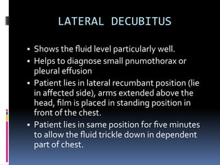
SUPPLYMENTARY- LATERAL VIEW [erect & decubitus].
- LORDOTIC.
- PA VEIW EXPIRTION
Surface dose for standard patient is 0.3mGy](https://image.slidesharecdn.com/chest-220916124430-d1135239/85/chest-examination-pptx-pdf-3-320.jpg)