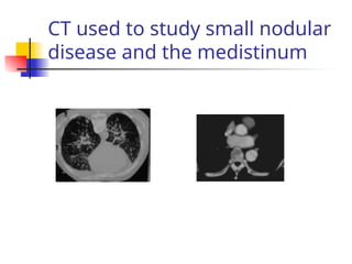
IMAGING THE THORAX–
PART 1
DR. MOSES ACAN
DEPARTMENT OF RADIOLOGY
 Chest x-ray
 Computerised tomography
 Ultrasound
 Magnetic resonance imaging
 New advances
Thoracic Imaging
Chest x-ray adults:
Technique
 A Chest X-ray (CXR) is normally taken
erect and PA (posterior anterior)
 at a distance 150 or 200cm.
 anterior chest wall is against the film
cassette
 the X-ray tube behind the patient aimed
towards the film.
Qualities of a good PA film
(1)
 The patient should not be rotated. Look
at the anterior ends of the clavicles. They
should be equidistant from the spinous
processes.
 If the patient is rotated one side of the chest
will look paler than the other & the
mediastinum will appear abnormal
Qualities of a good film (2)
 The film should be taken on full inspiration.
 The top of the R diaphragm should lie below the
anterior end of the 6th
right rib.
 The left diaphragm is usually lower.
 The ribs should be sloping and not horizontal.
 If they lie horizontally, it may be that the patient was
leaning backwards and the diaphragms will
obliterate the lung bases.
 The anterior end of the first rib should lie just below
the clavicle.
Film should not be rotated
The distance x should be equal on the 2 sides
Where is the 6th
anterior rib?
Is this a good film?
Qualities of a good film (3)
 The scapulae should not overlay the lung fields.
 The vertebrae should be faintly visible through
the heart shadow: lesions behind or in front of
the heart will not be missed.
 The bony detail should not be visible: lungs will then
appear too dark.
 The blood vessels in the lower lobe should just be
seen through the heart.
 The whole of the lung fields should be fully
included on the film including costophrenic
angles & apices.
When do we need an
expiratory film
 This is taken when the patient has
breathed out.
 This may help to show bronchial
obstruction with air trapping (e.g. inhaled
foreign body in a child):
When do we need a lateral
film
 A lateral film should never be part of a standard chest
examination especially for medical purposes or for
follow up of a known lesion.
 The PA film should be examined. If there is an
abnormality, a lateral film may then be useful in the
following:
 If the film is too light & a lesion is suspected clinically,
 Further assessment & localisation of abnormalities seen or
suspected on the PA film.
 Earlier detection of small pleural effusions if
ultrasound is not available
When do we need oblique
views
 These are helpful in assessing rib lesions
& some pneumothoraces.
Normal chest: soft tissues
and bones
SOFT TISSUES AND BONES.
 Look at the breast shadows, ribs,
clavicles, spine and shoulders for signs
of disease
Diaphragms and costophrenic angles
 Diaphragms have smooth
outline and are convex
upwards.
 The right dome lies 1-2cm
higher than the left: at or
below the level of the 10th
posterior rib or 6th
anterior
rib
 The posterior ribs
differentiated from the
anterior:horizontal
 There may be a fat pad at
heart border in obese
patients.
 diaphragmatic hump
(localised bulge) is a normal
variant.
Clear C-P angles
Fissures: Which fissures are
shown?
Which fissures are shown?
Normal lung hilae
 The left hilum normally lies 0.5 – 1.5cm
higher than the right.
 They are of similar density and size with
concave borders.
 They are composed of the pulmonary
arteries and veins.
 Normal lymph nodes are not seen.
Normal mediastinum
 the trachea should be central in position.
 the subcarinal angle, (under the division of the
trachea), is normally 60-70 degrees.
 the heart size should not measure greater than
14.5/15 cm in diameter in women, or 15.5/16cm in
men. cardio-thoracic ratio (CT ratio) should not be
greater than 50%.
 In other words, the transverse diameter of the heart
should not exceed half the transverse diameter of the
thorax at the level of the diaphragms
 the heart borders should be clearly visible, the right
border should be seen to the right of the spine.
 The aorta descends just to the left of the spine and is
more or less parallel with it.
Normal lungs and lung
markings
 Normal lung markings (lines or shadows seen across the
lungs) are due to, pulmonary vessels or fissures
 Bronchi are normally only visible near the hilae :bronchus
should only be 1mm or less in thickness.
 Bronchi seen end-on near the hilae should roughly be the
same diameter as a vessel.
 Bronchi are hollow ring shadows & vessels are solid grey discs.
 In the erect film lower zone vessels are larger than those in the
upper lobe.
 In the supine chest X-ray, upper lobe vessels are the same size
as the lower lobe vessels.
 The lungs should be of equal transradiancy.
Tricky or review areas
 lesions lying in some areas on chest X-ray
are more likely to be overlooked than
others.
 These are called the “review areas” and
are: the apices, mediastinum, hilae,
retrocardiac & retrodiaphragmatic
areas & the bones
Lung Anatomy
(1) aortic arch
(2) pulmonary trunk
(3) left atrial appendage
(4) left ventricle
(5) right atrium
(6) superior vena cava
(7 & 8) diaphragm
(9) transverse fissure
Lung Anatomy
(1) oblique fissure
(2) transverse fissure
(3) retrocardiac space
(4) retrosternal space
Correct CXR
 Name
 Date of birth
 Date
 Left and right, marker/stomach
Chest x-ray viewing guide
 PA, AP, lateral or decubitus view
 Rotation – Sternal end clavicles equal
from vertebral body
 If AP what position
Patient Position
 How dark or light a film is
 Should faintly see vertebral bodies
through heart
Exposure
 Breast shadows
 Air in tissues
 Tissue folds in obese
 Medical equipment
Soft Tissues
Breast
shadows
 Ribs
 Scapulae
 Clavicles
 Vertebrae
Bony Structures
 Deviated
 Carina
 Artificial airway
Trachea
 Deviated
 Hilar shadows
 Aortic arch
Mediastinum
Size
 No larger than half width of chest
Position
 Two thirds on the left
Borders
 Clear
Mediastinum - Heart
 Shape
 Height: right –6rib ant, left – 7 ant
 Cardiophrenic angle
 Costophrenic angle
Diaphragm
 Black with lung markings
 Other opacity indicated pathology
 Fissures
 Zones
 Air bronchograms
 Consolidation
Lung Fields
normal
OTHER IMAGING
MODALITIES FOR THE CHEST
Radionuclide scanning
 This is of most value in the diagnosis of pulmonary embolus.
If the scan is perfectly normal this virtually excludes it. The
major drawback is the lack of specificity and a report can only
be made when a recent chest X-ray and a ventilation scan are
available. The main indications are:
 1. Pulmonary embolus
 2. Evaluation of emphysema
 3. To determine the extent of parenchymal disease
 Monitoring therapy
Perfusion scan & ventilation
scan
A lung perfusion scan. There are multiple
wedge-shaped defects in the lungs. A
ventilation scan should also be performed to
see if the defects match or are seen only on the
perfusion scan. If so, they are due to
pulmonary emboli
A ventilation lung scan performed
on the same patient. This shows
no defects, the lungs showing
even uptake of the isotope.
This confirms the diagnosis of pulmonary
emboli.
Computed tomography of the thorax
 Computed tomography is being
increasingly used for examining the
chest despite its high radiation dose.
 The newer spiral CT machines allow
coverage of the entire thorax with
one breath hold.
The main indications for computed
tomography of the thorax:
 1. Staging of lung cancer and other
malignant tumours
 2. Diffuse lung disease – high resolution CT
 3. Pleural disease e.g. fluid versus malignant
tumour; empyema versus lung abscess
 4. Mediastinal disease: masses, aortic
dissection
 5. Conditions which are may be incoclusive at
CXR eg subtle signs of a pneumonia
CT used to study small nodular
disease and the medistinum
Magnetic resonance imaging of the
chest
 The role of MRI is not yet clearly defined. It
is good for vessels and lesions in the
mediastinal and hilar regions.
 A major disadvantage is respiratory and
cardiac motion. It is unable to visualise the
small pulmonary vessels and bronchi and
lung parenchyma.
 It is unable to provide the same anatomic
detail as high resolution computed
tomography and CT is the procedure of
choice for most diseases of the thorax.
Ultrasound imaging in the chest
 The acoustic mismatch between the chest wall
and the aerated lung results in almost total
reflection of the ultrasound beam
 Therefore ultrasound is little used for lung
pathology except for diagnosis and localisation
of effusions or other collections.
 Pleural fluid appears as an echofree area outlining
the pleural space.
 Internal echoes may be due to blood or pus, with
septa indicating loculation and a thick wall
suggesting an empyema.
 Pleural based masses are usually of low
echogenicity and pleural thickening is easily
identified.
LUNG ABNORMALITIES:
The abnormal chest
 overlying soft tissues or rib cage,
 lung disease
 disease of the pleura
 heart
 Other Mediastinal structures
 hilar areas
 diaphragm
OPACITY DUE TO OVERLYING SOFT
TISSUES OR RIB ABNORMALITIES
 Masses in the breast or other soft tissues of the thorax: can
be mistaken for pulmonary disease.
 structures superimposed on each other can mimic lung
disease e.g scapula overlying a posterior rib.
 Buttons or other fasteners on patients clothes may cast
confusing shadows & buttons may look like pulmonary
deposits.
 it is important not to assume that an opacity is on the
clothing. This should be confirmed by repeat PA film without
clothes.
 A lateral film is sometimes necessary.
 Rib fractures or deposits in the ribs commonly show a
peripheral opacity related to the rib.
LUNG SHADOWING/OPACITIES
 The fine network of the lungs is made up of: a) the alveoli or
air spaces b) the interstitium (i.e. the soft tissues between
the alveoli in which the bronchi & blood vessels lie)
 Disease processes may involve the alveoli, the interstitium,
or both.
 It is important but difficult to differentiate alveolar from
interstitial shadowing.
 There are 2 important signs, which help to interpret lung
shadowing.
 The silhouette sign
 The air bronchogram
In these two conditions, bronchi are
visible, which are the two conditions?
What conditions cause bronchial walls to thickening?, what is the
Explanation for the bronchus to be seen clearly when there is
Alveolar consolidation?
ALVEOLAR SHADOWING
(AIRSPACE
OPACITIES/DENSITIES)
ALVEOLAR SHADOWING (AIRSPACE
OPACITIES/DENSITIES)
 If the alveoli are involved in a disease process,
they fill with fluid or solid tissue.
 In the very early stages it may be that the alveoli
show as small separate rounded 6mm opacities
 small opacities rapidly become confluent
shadowing with fluffy ill defined margins.
 The vessels are obscured and there may
sometimes be an air bronchogram.
 There may also be a silhouette sign.
 When the alveoli fill with fluid or other
substances, it is called consolidation.
Signs of alveolar shadowing
 In the early stages it may present as a faint homogenous
haze or “ground glass” appearance
 Shadowing may be homogenous, patchy or blotchy in
appearance
 Fluffy, ill defined areas of opacification
 Areas of shadowing tend to coalesce and become
consolidation
 2 patterns of distribution occur: 1. Segmental or lobar 2.
“batswing”.
 Bats wing occurs typically in pulmonary oedema. It is non-
segmental.
 Air bronchograms may be visible.
Describe what is shown on these
pts with alveolar shadowing!
Pathogenesis of alveolar
shadowing (1)
 Note that all produce the same appearance on the CXR:
only differentiated by the distribution and the clinical
features.
 TRANSUDATE:
 the alveoli become filled with a clear transudate of low
protein content and this occurs in e.g. pulmonary
oedema, which may be of cardiogenic or non cardiogenic
origin.
 EXUDATE
 alveoli become filled with fluid of a high protein content and
the commonest cause is infection. Other less common
causes are acute Respiratory Distress Syndrome (ARDS),
pulmonary oesinophilia, and alveolar proteinosis.
 HAEMORRHAGE:
 alveoli fill with blood and this may occur in trauma, lung
Causes of alveoar
shadowing (2)
 INFILTRATION:
 the alveoli become filled with solid tissue
such as lymphoma, alveolar cell
carcinoma or carcinomatosis
 MISCELLANEOUS:
 Aspiration of gastric contents is an
important cause. Other causes are
inhaled foreign body, oesophageal leak,
oesophageal stricture due to carcinoma
or corrosive, radiation therapy and in
babies hyaline membrane disease.
Common causes of alveolar
shadowing: Lobar/segmental pattern
 Infection
 Pulmonary infarct
 Alveolar cell carcinoma
 Contusion
 Note: Alveolar shadowing, which is purely lobar will be
bounded by the fissures and depending on the segments
may produce a sharp outline of one border next to a
fissure.
Segmental/alveolar shadows:
In what part of the lung is this located, what radiological signs can you see? Why is the
® hemi diaphragm still clear? Name the lung lobes and segments! What do you see on
a PA and lat CXR when you have consolidation in each of the lobes/ segments
Alveolar consolidation of the
“bats –wing pattern”
 a) acute:
 - Pulmonary oedema
 b) chronic:
 - Lymphoma/leukaemia
 - Sarcoidosis but the interstitial form
is much commoner
 - Pulmonary alveolar proteinosis
 - Alveolar cell carcinoma but the
localised form is much commoner
Alveolar consolidation: bats
wing
Alveolar pneumonia not fitting in with
segmental or bats wing patterns.
 Alveolar shadowing may not fit into any
of the above patterns e.g.
 - Bronchopneumonia as in
staphylococcal septicaemia or pulmonary
tuberculosis.
 - Fat embolism (occurs 1-2 days
following major trauma, particularly
fractures of the large bones of the lower
limbs)
 Eosinophilic pneumonia
INTERSTITIAL
OPACITIES/SHADOWING
INTERSTITIAL SHADOWING:
 This is due to disease in the interstitium
i.e. the tissue in which the blood vessels
and bronchi lie within the lungs.
 This leads to a non homogenous pattern
of shadowing which may take many
forms.
 The alveoli are still aerated: no air
bronchogram or silhouette sign.
 blood vessels become ill defined or
obscured.
Causes of interstitial
shadowing.
 These diseases often look identical on CXR
regardless of the cause but distribution is
important.
 Some are characteristically more predominant in
the lower zones and others in the upper zones.
 Interstitial disease is better imaged by high
resolution CT for DDX, but some may need lung
biopsy for DX.
 clinical features help in DDX
 Diseases may also be classified on the basis of
whether acute or chronic as well as the
distribution in the lungs.
Interstitial disease:
common patterns
 Linear
 Miliary
 Reticulo-nodular
 Honeycomb
 Nodular
Linear Pattern
 abnormal network of fine lines running through the lungs
due to thickened connective tissue septae. The most
common are:
 Kerley A lines: long thin lines in the upper lobes, seen in
both PA & lateral views(under the sternum in the latter)
 Kerley B lines: short thin lines predominantly in the
periphery of the lower zones extending 1-2cm horizontally
inwards from the lung surface.
 Kerley C lines: diffuse linear pattern through the entire lung:
these can be difficult to recognise.
 Causes of Kerly lines are interstitial pulmonary oedema and
lymphangitis carcinomatosis.
Interstitial shadows: linear
pattern
Interstitial shadows: nodular
pattern
 interstitial nodules are small (1-
5mm), well defined, and not
associated with air bronchograms.
 often very numerous and
distributed evenly through the
lungs.
 if low in density may be difficult to
appreciate in the early stages
Interstitial shadows: causes
of nodullar pattern
 ACUTE:
 Infections such as tuberculosis, chickenpox
 Pulmonary oedema
 Acute extrinsic allergic alveolitis
 Fat emboli
 LESS ACUTE
 Carcinomatosis – especially thyroid carcinoma,
chorionepithelioma, breast.
 CHRONIC
 Sarcoidosis – predominantly mid zones.
 Coal miners pneumoconiosis, silicosis
 Fibrosing alveolitis
Interstitial shadows: nodular
pattern
Interstitial shadows: reticulo-
nodular pattern:
 this due to very fine nodular opacities
superimposed on a fine network of
short lines.
 This may be seen in TB, silicosis,
rheumatoid lung disease and
fibrosing alveolitis as well as drug
induced lung changes.
 It is usually more obvious at the
bases.
Interstitial shadows:
reticulonodular pattern.
Interstitial shadows: the
honey-comb pattern
 represents the end stage of many interstitial
processes and implies extensive destruction of
pulmonary tissue.
 lung parenchyma replaced by cysts, which range
in size from tiny up to 2 cm diameter. These are
separated by coarse linear interstitial markings.
 cysts have very thin walls
 normal pulmonary vasculature cannot be seen
 may be complicated by pneumothorax.
 Examples are: Chronic heart failure, Rheumatoid,
Scleroderma, Fibrosing alveolitis, & Histiocytosis.
BRONCHIECTASIS
 Usually occurs secondary to infection.
 postnasal drop in chronic sinusitis, pertussis, TB,
pneumonia of any kind and any cause of segmental
atelectasis.
 The bronchi become infected, dilate peripherally and
develop thick walls visible on chest X-rays.
 Commonly seen in the lower zones, the bronchi appear as
thick lines enclosing the aerated lumen.
 If seen in cross section the bronchi look dilated surrounded
by a white cuff, which represents the thickened wall.
 super-added inflammatory changes appearing as alveolar
shadowing may co-exist.
 involve any lobe, segment or complete lung.
Interstitial shadows:
Bilateral lower zone
bronchiectasis
Interstitial shadowing: diseases
causing milliary shadows.
 multiple small opacities less than 5mm in diameter are:
1. Tuberculosis
2. Miliary metastases – any primary site but especially thyroid
& chorion epithelioma
3. Chickenpox
4. Pneumocystis Carinii pneumonia, Cytomegalic infection
5. Sarcoidosis
6. Dust inhalation – pneumoconioses
7. Haemosiderosis
8. Extrinsic allergic alveolitis, eosinophilia
 Fibrosing alveolitis
Interstitial shadows: acute diseases which
cause linear patterns.
 Pulmonary oedema – cardiac, non
cardiac causes
 Infections – particularly viruses,
pneumocystis, measles, malaria &
tuberculosis (less often)
Interstitial shadows: chronic
diseases that give a linear pattern
 Linear interstitial shadowing due to long standing
pulmonary oedema with Kerley B lines
 Lymphangitis Carcinomatosis – may be unilateral. Often
associated hilar adenopathy
 Sarcoidosis
 Early silicosis & asbestosis
 Allergic alveolitis & pulmonary oesinophilia
 Drugs & inhaled irritants such as chlorine or tear gas
 Connective disorders: Rheumatoid arthritis, scleroderma,
systemic lupus,graulomatosis
 Miscellaneous: histiocytosis, tuberous sclerosis
 Bronchiectasis: usually a mixed alveolar/interstitial pattern
PULMONARY COLAPSE
PULMONARY COLLAPSE:
• A cause of opacity on chest X-ray
• features to distinguish it from
alveolar/interstitial shadowing are:
-The loss of lung volume may affect the
position of adjacent structures such as the
diaphragm, hilum, mediastinum and fissures.
-There may be a silhouette sign but no air
bronchogram unless associated with
consolidation
Collapse: causes of collapse
• Obstruction of a bronchus by a foreign
body, tumour, mucus plug, aspiration,
or a stricture
• Compressive collapse due to air or fluid
in the pleural cavity.
• Cicatrization. This is commonly seen
following pulmonary tuberculosis
Collapse: what part of lung is
involved
• Collapse may involve the whole lung,
• lobe of the lung (lobar collapse) or it
may involve just
• part of a lobe such as a segment or
subsegment.
• Subsegmental collapse is often called
“plate atelectasis” because of its
appearance.
SIGNS OF COLLAPSE:
• Whole lung collapse:
• complete opacity of the hemithorax
• displacement of the mediastinum to
the side of the opacity
• silhouette sign with loss of
diaphragm and mediastinal outlines.
• herniation of the unaffected lung across
the midline
Collapse
Describe these changes, what other pathology causes homogenous Opacities?
What are the causes of collapse?, How does the pt. present and and what are the
CXR signs
Collapse: lobar collapse
• Lobar collapse
• The lobes collapse in characteristic
fashion:
• The upper lobes collapse upwards,
medially and anteriorly
• The middle lobe goes downwards and
medially
• The lower lobes collapse posteriorly,
medially and downwards.
Collapse: sign of lobar
collapse
  increased density due to loss of aeration
  crowding of the blood vessels
  displacement of the hilum – upwards or downwards
  displacement of a fissure which may be bowed
  shift of the mediastinum, trachea towards the collapsed part
of the lung
  elevation of the diaphragm
  silhouette sign
  the remainder of the lung may be overexpanded &
hypertranslucent with fewer vessels showing.
• rib crowding may occur in children or with long standing
collapse
Collapse: R upper lobe
• movement of the horizontal fissure
upwards,
• The collapsed lung assumes an
increased density at the R apex, it’s
lower border being sharply defined by
the horizontal fissure,
• Horizontal fissure may be bowed.
• The hilum is usually distorted & pulled
up.
Collapse lung A
Describe the changes in these CXRs and give a differential diagnosis. What are
the radiological features of: lung, lobar, segmental and subsegmental collapse?
What are the radiological changes in upper lobe, mid and lower lobe collapse?
What may cause collapse of these lobes?
Collapse middle lobe
• relatively small lobe so collapse
produces only minor changes on the PA
film.
• There is increased density lateral to the
R heart border & blurring of the outline.
• On the lateral view it shows as a
triangular opacity anteriorly.
Collapse lower lobe A
• The oblique fissure moves backwards
maintaining the same slope.
• On the PA view, left lower lobe collapse
shows as a triangular opacity projected
through the heart shadow.
• on the right, it may be accompanied by
collapse of the middle lobe leading to a larger
opacity
• Complete collapse of the lower lobes is often
difficult to detect as the lobe comes to lie
against the mediastinum on the PA view &
may be invisible.
Collapse of lobes
What do these diagrams represent?, what would one see on the
CXRs?
Collapse lower lobes
B
• the L hilum is pulled downwards & hidden by the
cardiac shadow normally should lie above the R
hilum.
• The mediastinum is displaced to the L & the L heart
border straightened.
• The L diaphragm is may lie higher than the R
though.
• As the collapse increases the collapsed lobe overlies
the spine on the lateral view and is no longer visible
in this projection. The only clue is that the spine
appears of even density throughout whereas
normally it looks darker in the lower part of the chest
Collapse lower lobe 3 (right
lower lobe)
• R lower lobe collapse produces shadowing adjacent to the R
heart border.
• There is usually a well -defined lateral boundary.
• The R hilum disappears as it is pulled downwards towards the
heart.
• In combined collapse of the R middle & lower lobe the
shadowing is greater and more obvious
• The lesser fissure may be bowed inferiorly.
• There is a silhouette sign of the R heart border & diaphragm.
• The upper border is bounded by the lesser fissure indicating
that the middle lobe is involved.
• The heart may be displaced a little to the R.
Patterns of collapse cont.
What part of the lung has collapsed in these pictures? How
would this appear on CXR?
Collapse L upper lobe
• This behaves differently than collapse of
the R upper lobe because of the lingular
segment,
• The lingular is equivalent to the middle
lobe on the R, but is part of the L upper
lobe.
• The oblique fissure moves forwards & the
collapsed lobe lies anteriorly against the
chest wall.
• This produces hazy ill -defined opacity in
the L upper & mid zones on the PA film
with a
• silhouette sign affecting the L
collapse
Describe the changes on the CXRs. Give differential diagnosis/
How do the CXR changes in lobar collapse of the right lung differ
From the left? What cause slund collapse on the left side?
Collapse left lower lobe B
• Lateral view may showing bowing
backwards of the oblique fissure.
• The anterior boundary of the collapsed
lobe may be difficult to identify
because the lobe lies against the
mediastinum.
• The posterior edge is usually visible
because it is bounded by the oblique
fissure.
Segmental & subsegmental
collapse
• Only a segment or subsegment of a
lobe may collapse.
• Small linear subsegmental atelectatic
shadows at the lung bases are called
“plate atelectasis” and are due to poor
aeration or perfusion.
Causes of segmental or
subsegmental collapse
• post abdominal surgery
• abdominal distension due to tumour or
ascites
• post MI (myocardial infarction)
• mucous plugs (asthma)
• pulmonary infarct
• segmental collapse may occur secondary to
• infection or distal to a small tumour.
• common in children with bronchiolitis
Subsegmental collapse
Describe the changes. Give a differential diagnosis. What are the
Causes of subsegmental collapse? What are myocardial infarction,
pulmonary embolism, Pulmonary infarction, asthma and bronchiolitis
and how do these conditions present clinically and at CXR?

LECTURE 3 chest vjftfyftufsidgsyudftygiugf

  • 1.
    IMAGING THE THORAX– PART1 DR. MOSES ACAN DEPARTMENT OF RADIOLOGY
  • 2.
     Chest x-ray Computerised tomography  Ultrasound  Magnetic resonance imaging  New advances Thoracic Imaging
  • 3.
    Chest x-ray adults: Technique A Chest X-ray (CXR) is normally taken erect and PA (posterior anterior)  at a distance 150 or 200cm.  anterior chest wall is against the film cassette  the X-ray tube behind the patient aimed towards the film.
  • 4.
    Qualities of agood PA film (1)  The patient should not be rotated. Look at the anterior ends of the clavicles. They should be equidistant from the spinous processes.  If the patient is rotated one side of the chest will look paler than the other & the mediastinum will appear abnormal
  • 5.
    Qualities of agood film (2)  The film should be taken on full inspiration.  The top of the R diaphragm should lie below the anterior end of the 6th right rib.  The left diaphragm is usually lower.  The ribs should be sloping and not horizontal.  If they lie horizontally, it may be that the patient was leaning backwards and the diaphragms will obliterate the lung bases.  The anterior end of the first rib should lie just below the clavicle.
  • 6.
    Film should notbe rotated The distance x should be equal on the 2 sides
  • 7.
    Where is the6th anterior rib? Is this a good film?
  • 8.
    Qualities of agood film (3)  The scapulae should not overlay the lung fields.  The vertebrae should be faintly visible through the heart shadow: lesions behind or in front of the heart will not be missed.  The bony detail should not be visible: lungs will then appear too dark.  The blood vessels in the lower lobe should just be seen through the heart.  The whole of the lung fields should be fully included on the film including costophrenic angles & apices.
  • 9.
    When do weneed an expiratory film  This is taken when the patient has breathed out.  This may help to show bronchial obstruction with air trapping (e.g. inhaled foreign body in a child):
  • 10.
    When do weneed a lateral film  A lateral film should never be part of a standard chest examination especially for medical purposes or for follow up of a known lesion.  The PA film should be examined. If there is an abnormality, a lateral film may then be useful in the following:  If the film is too light & a lesion is suspected clinically,  Further assessment & localisation of abnormalities seen or suspected on the PA film.  Earlier detection of small pleural effusions if ultrasound is not available
  • 11.
    When do weneed oblique views  These are helpful in assessing rib lesions & some pneumothoraces.
  • 12.
    Normal chest: softtissues and bones SOFT TISSUES AND BONES.  Look at the breast shadows, ribs, clavicles, spine and shoulders for signs of disease
  • 13.
    Diaphragms and costophrenicangles  Diaphragms have smooth outline and are convex upwards.  The right dome lies 1-2cm higher than the left: at or below the level of the 10th posterior rib or 6th anterior rib  The posterior ribs differentiated from the anterior:horizontal  There may be a fat pad at heart border in obese patients.  diaphragmatic hump (localised bulge) is a normal variant. Clear C-P angles
  • 14.
  • 15.
  • 16.
    Normal lung hilae The left hilum normally lies 0.5 – 1.5cm higher than the right.  They are of similar density and size with concave borders.  They are composed of the pulmonary arteries and veins.  Normal lymph nodes are not seen.
  • 17.
    Normal mediastinum  thetrachea should be central in position.  the subcarinal angle, (under the division of the trachea), is normally 60-70 degrees.  the heart size should not measure greater than 14.5/15 cm in diameter in women, or 15.5/16cm in men. cardio-thoracic ratio (CT ratio) should not be greater than 50%.  In other words, the transverse diameter of the heart should not exceed half the transverse diameter of the thorax at the level of the diaphragms  the heart borders should be clearly visible, the right border should be seen to the right of the spine.  The aorta descends just to the left of the spine and is more or less parallel with it.
  • 18.
    Normal lungs andlung markings  Normal lung markings (lines or shadows seen across the lungs) are due to, pulmonary vessels or fissures  Bronchi are normally only visible near the hilae :bronchus should only be 1mm or less in thickness.  Bronchi seen end-on near the hilae should roughly be the same diameter as a vessel.  Bronchi are hollow ring shadows & vessels are solid grey discs.  In the erect film lower zone vessels are larger than those in the upper lobe.  In the supine chest X-ray, upper lobe vessels are the same size as the lower lobe vessels.  The lungs should be of equal transradiancy.
  • 19.
    Tricky or reviewareas  lesions lying in some areas on chest X-ray are more likely to be overlooked than others.  These are called the “review areas” and are: the apices, mediastinum, hilae, retrocardiac & retrodiaphragmatic areas & the bones
  • 20.
    Lung Anatomy (1) aorticarch (2) pulmonary trunk (3) left atrial appendage (4) left ventricle (5) right atrium (6) superior vena cava (7 & 8) diaphragm (9) transverse fissure
  • 21.
    Lung Anatomy (1) obliquefissure (2) transverse fissure (3) retrocardiac space (4) retrosternal space
  • 22.
    Correct CXR  Name Date of birth  Date  Left and right, marker/stomach Chest x-ray viewing guide
  • 23.
     PA, AP,lateral or decubitus view  Rotation – Sternal end clavicles equal from vertebral body  If AP what position Patient Position
  • 24.
     How darkor light a film is  Should faintly see vertebral bodies through heart Exposure
  • 25.
     Breast shadows Air in tissues  Tissue folds in obese  Medical equipment Soft Tissues
  • 26.
  • 27.
     Ribs  Scapulae Clavicles  Vertebrae Bony Structures
  • 28.
     Deviated  Carina Artificial airway Trachea
  • 29.
     Deviated  Hilarshadows  Aortic arch Mediastinum
  • 30.
    Size  No largerthan half width of chest Position  Two thirds on the left Borders  Clear Mediastinum - Heart
  • 31.
     Shape  Height:right –6rib ant, left – 7 ant  Cardiophrenic angle  Costophrenic angle Diaphragm
  • 32.
     Black withlung markings  Other opacity indicated pathology  Fissures  Zones  Air bronchograms  Consolidation Lung Fields
  • 33.
  • 34.
  • 35.
    Radionuclide scanning  Thisis of most value in the diagnosis of pulmonary embolus. If the scan is perfectly normal this virtually excludes it. The major drawback is the lack of specificity and a report can only be made when a recent chest X-ray and a ventilation scan are available. The main indications are:  1. Pulmonary embolus  2. Evaluation of emphysema  3. To determine the extent of parenchymal disease  Monitoring therapy
  • 36.
    Perfusion scan &ventilation scan A lung perfusion scan. There are multiple wedge-shaped defects in the lungs. A ventilation scan should also be performed to see if the defects match or are seen only on the perfusion scan. If so, they are due to pulmonary emboli A ventilation lung scan performed on the same patient. This shows no defects, the lungs showing even uptake of the isotope. This confirms the diagnosis of pulmonary emboli.
  • 37.
    Computed tomography ofthe thorax  Computed tomography is being increasingly used for examining the chest despite its high radiation dose.  The newer spiral CT machines allow coverage of the entire thorax with one breath hold.
  • 38.
    The main indicationsfor computed tomography of the thorax:  1. Staging of lung cancer and other malignant tumours  2. Diffuse lung disease – high resolution CT  3. Pleural disease e.g. fluid versus malignant tumour; empyema versus lung abscess  4. Mediastinal disease: masses, aortic dissection  5. Conditions which are may be incoclusive at CXR eg subtle signs of a pneumonia
  • 39.
    CT used tostudy small nodular disease and the medistinum
  • 40.
    Magnetic resonance imagingof the chest  The role of MRI is not yet clearly defined. It is good for vessels and lesions in the mediastinal and hilar regions.  A major disadvantage is respiratory and cardiac motion. It is unable to visualise the small pulmonary vessels and bronchi and lung parenchyma.  It is unable to provide the same anatomic detail as high resolution computed tomography and CT is the procedure of choice for most diseases of the thorax.
  • 41.
    Ultrasound imaging inthe chest  The acoustic mismatch between the chest wall and the aerated lung results in almost total reflection of the ultrasound beam  Therefore ultrasound is little used for lung pathology except for diagnosis and localisation of effusions or other collections.  Pleural fluid appears as an echofree area outlining the pleural space.  Internal echoes may be due to blood or pus, with septa indicating loculation and a thick wall suggesting an empyema.  Pleural based masses are usually of low echogenicity and pleural thickening is easily identified.
  • 42.
  • 43.
    The abnormal chest overlying soft tissues or rib cage,  lung disease  disease of the pleura  heart  Other Mediastinal structures  hilar areas  diaphragm
  • 44.
    OPACITY DUE TOOVERLYING SOFT TISSUES OR RIB ABNORMALITIES  Masses in the breast or other soft tissues of the thorax: can be mistaken for pulmonary disease.  structures superimposed on each other can mimic lung disease e.g scapula overlying a posterior rib.  Buttons or other fasteners on patients clothes may cast confusing shadows & buttons may look like pulmonary deposits.  it is important not to assume that an opacity is on the clothing. This should be confirmed by repeat PA film without clothes.  A lateral film is sometimes necessary.  Rib fractures or deposits in the ribs commonly show a peripheral opacity related to the rib.
  • 45.
    LUNG SHADOWING/OPACITIES  Thefine network of the lungs is made up of: a) the alveoli or air spaces b) the interstitium (i.e. the soft tissues between the alveoli in which the bronchi & blood vessels lie)  Disease processes may involve the alveoli, the interstitium, or both.  It is important but difficult to differentiate alveolar from interstitial shadowing.  There are 2 important signs, which help to interpret lung shadowing.  The silhouette sign  The air bronchogram
  • 48.
    In these twoconditions, bronchi are visible, which are the two conditions? What conditions cause bronchial walls to thickening?, what is the Explanation for the bronchus to be seen clearly when there is Alveolar consolidation?
  • 49.
  • 50.
    ALVEOLAR SHADOWING (AIRSPACE OPACITIES/DENSITIES) If the alveoli are involved in a disease process, they fill with fluid or solid tissue.  In the very early stages it may be that the alveoli show as small separate rounded 6mm opacities  small opacities rapidly become confluent shadowing with fluffy ill defined margins.  The vessels are obscured and there may sometimes be an air bronchogram.  There may also be a silhouette sign.  When the alveoli fill with fluid or other substances, it is called consolidation.
  • 51.
    Signs of alveolarshadowing  In the early stages it may present as a faint homogenous haze or “ground glass” appearance  Shadowing may be homogenous, patchy or blotchy in appearance  Fluffy, ill defined areas of opacification  Areas of shadowing tend to coalesce and become consolidation  2 patterns of distribution occur: 1. Segmental or lobar 2. “batswing”.  Bats wing occurs typically in pulmonary oedema. It is non- segmental.  Air bronchograms may be visible.
  • 52.
    Describe what isshown on these pts with alveolar shadowing!
  • 53.
    Pathogenesis of alveolar shadowing(1)  Note that all produce the same appearance on the CXR: only differentiated by the distribution and the clinical features.  TRANSUDATE:  the alveoli become filled with a clear transudate of low protein content and this occurs in e.g. pulmonary oedema, which may be of cardiogenic or non cardiogenic origin.  EXUDATE  alveoli become filled with fluid of a high protein content and the commonest cause is infection. Other less common causes are acute Respiratory Distress Syndrome (ARDS), pulmonary oesinophilia, and alveolar proteinosis.  HAEMORRHAGE:  alveoli fill with blood and this may occur in trauma, lung
  • 54.
    Causes of alveoar shadowing(2)  INFILTRATION:  the alveoli become filled with solid tissue such as lymphoma, alveolar cell carcinoma or carcinomatosis  MISCELLANEOUS:  Aspiration of gastric contents is an important cause. Other causes are inhaled foreign body, oesophageal leak, oesophageal stricture due to carcinoma or corrosive, radiation therapy and in babies hyaline membrane disease.
  • 55.
    Common causes ofalveolar shadowing: Lobar/segmental pattern  Infection  Pulmonary infarct  Alveolar cell carcinoma  Contusion  Note: Alveolar shadowing, which is purely lobar will be bounded by the fissures and depending on the segments may produce a sharp outline of one border next to a fissure.
  • 56.
    Segmental/alveolar shadows: In whatpart of the lung is this located, what radiological signs can you see? Why is the ® hemi diaphragm still clear? Name the lung lobes and segments! What do you see on a PA and lat CXR when you have consolidation in each of the lobes/ segments
  • 57.
    Alveolar consolidation ofthe “bats –wing pattern”  a) acute:  - Pulmonary oedema  b) chronic:  - Lymphoma/leukaemia  - Sarcoidosis but the interstitial form is much commoner  - Pulmonary alveolar proteinosis  - Alveolar cell carcinoma but the localised form is much commoner
  • 59.
  • 60.
    Alveolar pneumonia notfitting in with segmental or bats wing patterns.  Alveolar shadowing may not fit into any of the above patterns e.g.  - Bronchopneumonia as in staphylococcal septicaemia or pulmonary tuberculosis.  - Fat embolism (occurs 1-2 days following major trauma, particularly fractures of the large bones of the lower limbs)  Eosinophilic pneumonia
  • 62.
  • 63.
    INTERSTITIAL SHADOWING:  Thisis due to disease in the interstitium i.e. the tissue in which the blood vessels and bronchi lie within the lungs.  This leads to a non homogenous pattern of shadowing which may take many forms.  The alveoli are still aerated: no air bronchogram or silhouette sign.  blood vessels become ill defined or obscured.
  • 64.
    Causes of interstitial shadowing. These diseases often look identical on CXR regardless of the cause but distribution is important.  Some are characteristically more predominant in the lower zones and others in the upper zones.  Interstitial disease is better imaged by high resolution CT for DDX, but some may need lung biopsy for DX.  clinical features help in DDX  Diseases may also be classified on the basis of whether acute or chronic as well as the distribution in the lungs.
  • 65.
    Interstitial disease: common patterns Linear  Miliary  Reticulo-nodular  Honeycomb  Nodular
  • 66.
    Linear Pattern  abnormalnetwork of fine lines running through the lungs due to thickened connective tissue septae. The most common are:  Kerley A lines: long thin lines in the upper lobes, seen in both PA & lateral views(under the sternum in the latter)  Kerley B lines: short thin lines predominantly in the periphery of the lower zones extending 1-2cm horizontally inwards from the lung surface.  Kerley C lines: diffuse linear pattern through the entire lung: these can be difficult to recognise.  Causes of Kerly lines are interstitial pulmonary oedema and lymphangitis carcinomatosis.
  • 67.
  • 68.
    Interstitial shadows: nodular pattern interstitial nodules are small (1- 5mm), well defined, and not associated with air bronchograms.  often very numerous and distributed evenly through the lungs.  if low in density may be difficult to appreciate in the early stages
  • 69.
    Interstitial shadows: causes ofnodullar pattern  ACUTE:  Infections such as tuberculosis, chickenpox  Pulmonary oedema  Acute extrinsic allergic alveolitis  Fat emboli  LESS ACUTE  Carcinomatosis – especially thyroid carcinoma, chorionepithelioma, breast.  CHRONIC  Sarcoidosis – predominantly mid zones.  Coal miners pneumoconiosis, silicosis  Fibrosing alveolitis
  • 70.
  • 71.
    Interstitial shadows: reticulo- nodularpattern:  this due to very fine nodular opacities superimposed on a fine network of short lines.  This may be seen in TB, silicosis, rheumatoid lung disease and fibrosing alveolitis as well as drug induced lung changes.  It is usually more obvious at the bases.
  • 72.
  • 73.
    Interstitial shadows: the honey-combpattern  represents the end stage of many interstitial processes and implies extensive destruction of pulmonary tissue.  lung parenchyma replaced by cysts, which range in size from tiny up to 2 cm diameter. These are separated by coarse linear interstitial markings.  cysts have very thin walls  normal pulmonary vasculature cannot be seen  may be complicated by pneumothorax.  Examples are: Chronic heart failure, Rheumatoid, Scleroderma, Fibrosing alveolitis, & Histiocytosis.
  • 74.
    BRONCHIECTASIS  Usually occurssecondary to infection.  postnasal drop in chronic sinusitis, pertussis, TB, pneumonia of any kind and any cause of segmental atelectasis.  The bronchi become infected, dilate peripherally and develop thick walls visible on chest X-rays.  Commonly seen in the lower zones, the bronchi appear as thick lines enclosing the aerated lumen.  If seen in cross section the bronchi look dilated surrounded by a white cuff, which represents the thickened wall.  super-added inflammatory changes appearing as alveolar shadowing may co-exist.  involve any lobe, segment or complete lung.
  • 75.
  • 76.
    Interstitial shadowing: diseases causingmilliary shadows.  multiple small opacities less than 5mm in diameter are: 1. Tuberculosis 2. Miliary metastases – any primary site but especially thyroid & chorion epithelioma 3. Chickenpox 4. Pneumocystis Carinii pneumonia, Cytomegalic infection 5. Sarcoidosis 6. Dust inhalation – pneumoconioses 7. Haemosiderosis 8. Extrinsic allergic alveolitis, eosinophilia  Fibrosing alveolitis
  • 77.
    Interstitial shadows: acutediseases which cause linear patterns.  Pulmonary oedema – cardiac, non cardiac causes  Infections – particularly viruses, pneumocystis, measles, malaria & tuberculosis (less often)
  • 78.
    Interstitial shadows: chronic diseasesthat give a linear pattern  Linear interstitial shadowing due to long standing pulmonary oedema with Kerley B lines  Lymphangitis Carcinomatosis – may be unilateral. Often associated hilar adenopathy  Sarcoidosis  Early silicosis & asbestosis  Allergic alveolitis & pulmonary oesinophilia  Drugs & inhaled irritants such as chlorine or tear gas  Connective disorders: Rheumatoid arthritis, scleroderma, systemic lupus,graulomatosis  Miscellaneous: histiocytosis, tuberous sclerosis  Bronchiectasis: usually a mixed alveolar/interstitial pattern
  • 79.
  • 80.
    PULMONARY COLLAPSE: • Acause of opacity on chest X-ray • features to distinguish it from alveolar/interstitial shadowing are: -The loss of lung volume may affect the position of adjacent structures such as the diaphragm, hilum, mediastinum and fissures. -There may be a silhouette sign but no air bronchogram unless associated with consolidation
  • 81.
    Collapse: causes ofcollapse • Obstruction of a bronchus by a foreign body, tumour, mucus plug, aspiration, or a stricture • Compressive collapse due to air or fluid in the pleural cavity. • Cicatrization. This is commonly seen following pulmonary tuberculosis
  • 82.
    Collapse: what partof lung is involved • Collapse may involve the whole lung, • lobe of the lung (lobar collapse) or it may involve just • part of a lobe such as a segment or subsegment. • Subsegmental collapse is often called “plate atelectasis” because of its appearance.
  • 83.
    SIGNS OF COLLAPSE: •Whole lung collapse: • complete opacity of the hemithorax • displacement of the mediastinum to the side of the opacity • silhouette sign with loss of diaphragm and mediastinal outlines. • herniation of the unaffected lung across the midline
  • 84.
    Collapse Describe these changes,what other pathology causes homogenous Opacities? What are the causes of collapse?, How does the pt. present and and what are the CXR signs
  • 85.
    Collapse: lobar collapse •Lobar collapse • The lobes collapse in characteristic fashion: • The upper lobes collapse upwards, medially and anteriorly • The middle lobe goes downwards and medially • The lower lobes collapse posteriorly, medially and downwards.
  • 86.
    Collapse: sign oflobar collapse   increased density due to loss of aeration   crowding of the blood vessels   displacement of the hilum – upwards or downwards   displacement of a fissure which may be bowed   shift of the mediastinum, trachea towards the collapsed part of the lung   elevation of the diaphragm   silhouette sign   the remainder of the lung may be overexpanded & hypertranslucent with fewer vessels showing. • rib crowding may occur in children or with long standing collapse
  • 87.
    Collapse: R upperlobe • movement of the horizontal fissure upwards, • The collapsed lung assumes an increased density at the R apex, it’s lower border being sharply defined by the horizontal fissure, • Horizontal fissure may be bowed. • The hilum is usually distorted & pulled up.
  • 88.
    Collapse lung A Describethe changes in these CXRs and give a differential diagnosis. What are the radiological features of: lung, lobar, segmental and subsegmental collapse? What are the radiological changes in upper lobe, mid and lower lobe collapse? What may cause collapse of these lobes?
  • 89.
    Collapse middle lobe •relatively small lobe so collapse produces only minor changes on the PA film. • There is increased density lateral to the R heart border & blurring of the outline. • On the lateral view it shows as a triangular opacity anteriorly.
  • 90.
    Collapse lower lobeA • The oblique fissure moves backwards maintaining the same slope. • On the PA view, left lower lobe collapse shows as a triangular opacity projected through the heart shadow. • on the right, it may be accompanied by collapse of the middle lobe leading to a larger opacity • Complete collapse of the lower lobes is often difficult to detect as the lobe comes to lie against the mediastinum on the PA view & may be invisible.
  • 91.
    Collapse of lobes Whatdo these diagrams represent?, what would one see on the CXRs?
  • 92.
    Collapse lower lobes B •the L hilum is pulled downwards & hidden by the cardiac shadow normally should lie above the R hilum. • The mediastinum is displaced to the L & the L heart border straightened. • The L diaphragm is may lie higher than the R though. • As the collapse increases the collapsed lobe overlies the spine on the lateral view and is no longer visible in this projection. The only clue is that the spine appears of even density throughout whereas normally it looks darker in the lower part of the chest
  • 93.
    Collapse lower lobe3 (right lower lobe) • R lower lobe collapse produces shadowing adjacent to the R heart border. • There is usually a well -defined lateral boundary. • The R hilum disappears as it is pulled downwards towards the heart. • In combined collapse of the R middle & lower lobe the shadowing is greater and more obvious • The lesser fissure may be bowed inferiorly. • There is a silhouette sign of the R heart border & diaphragm. • The upper border is bounded by the lesser fissure indicating that the middle lobe is involved. • The heart may be displaced a little to the R.
  • 94.
    Patterns of collapsecont. What part of the lung has collapsed in these pictures? How would this appear on CXR?
  • 95.
    Collapse L upperlobe • This behaves differently than collapse of the R upper lobe because of the lingular segment, • The lingular is equivalent to the middle lobe on the R, but is part of the L upper lobe. • The oblique fissure moves forwards & the collapsed lobe lies anteriorly against the chest wall. • This produces hazy ill -defined opacity in the L upper & mid zones on the PA film with a • silhouette sign affecting the L
  • 96.
    collapse Describe the changeson the CXRs. Give differential diagnosis/ How do the CXR changes in lobar collapse of the right lung differ From the left? What cause slund collapse on the left side?
  • 97.
    Collapse left lowerlobe B • Lateral view may showing bowing backwards of the oblique fissure. • The anterior boundary of the collapsed lobe may be difficult to identify because the lobe lies against the mediastinum. • The posterior edge is usually visible because it is bounded by the oblique fissure.
  • 98.
    Segmental & subsegmental collapse •Only a segment or subsegment of a lobe may collapse. • Small linear subsegmental atelectatic shadows at the lung bases are called “plate atelectasis” and are due to poor aeration or perfusion.
  • 99.
    Causes of segmentalor subsegmental collapse • post abdominal surgery • abdominal distension due to tumour or ascites • post MI (myocardial infarction) • mucous plugs (asthma) • pulmonary infarct • segmental collapse may occur secondary to • infection or distal to a small tumour. • common in children with bronchiolitis
  • 100.
    Subsegmental collapse Describe thechanges. Give a differential diagnosis. What are the Causes of subsegmental collapse? What are myocardial infarction, pulmonary embolism, Pulmonary infarction, asthma and bronchiolitis and how do these conditions present clinically and at CXR?

Editor's Notes

  • #2 MRI not widely used but can show malignancy especially We will look at actual films in the tutorials/practicals
  • #20 Transverse fissure – 6th rib laterally Does not estend beyond pulm artery medially Visible in 50%
  • #21 Oblique fissure from t4 posteriorly Propeller shaped Differentiation between sides- left is more vertical, has more posterior junction with the diaphragm= does not intersect transverse fissure Left diaphragm is lower and possesses stomach bubble by 2.5cm in 94% population
  • #22 How to view Check patient and x-ray details Left or right, markers placed on by radiographer, stomach on left. Heart not always on left
  • #23 Quality IF AP will have poorer inspiration and larger heart If patient supine will not see pleural effusions very well
  • #24 AP will show KV/MAS
  • #25 Breast shadows – mastectomy! Medical equipment, lines (CVP, ICD), endotracheal tube, NG tube, metal implants, pacemakers
  • #27 Ribs fractures, osteoporosis. Ribs even Scoliosis Scapulae need to be identified so do not confuse when looking at lung fields
  • #28 Trachea – can be pushed or pulled Air filled sacs keep trachea in middle
  • #29 Hilar shadows, pulmonary vasculature and lymph nodes, right side is slightly further out and the left is usually higher by 2 cm, with COPD will get upper lobe diversion Aortic arch may be calcified
  • #30 Heart Size, is usually half the width of the chest, is increased with AP picture, and in cardiac disease. Will look smaller if lungs hyperinflated and larger if very poor inspiration Positioned two thirds to the left unless have dextrocardia
  • #31 Shape Domed, flattened with hyperinflation more domed with poor inspiration or paralysis, gas in stomach Height Right 6 rib ant, left 7 rib anter in 95% of population Left lower because of weigh of heart Bear in mind structures below as stomach can push up occasionally liver can push up, ascites will push up Angles Clear if cardiophrenic poor collapse, if costophrenic blurred pleural effusion
  • #32 Lung markings of vessels, absent if a Pneumothorax Pulmonary oedema - bilateral Fissures Right horizontal, present in 80% of PA’s Third thoracic spine, goes down and anteriorly Fluid present? Moved? Oblique on lateral only Zones Upper, above 2nd rib ant Middle, 2-4 rib ant Lower, below forth rib Opacity increased with fluid, consolidation, malignancy