Downloaded 103 times
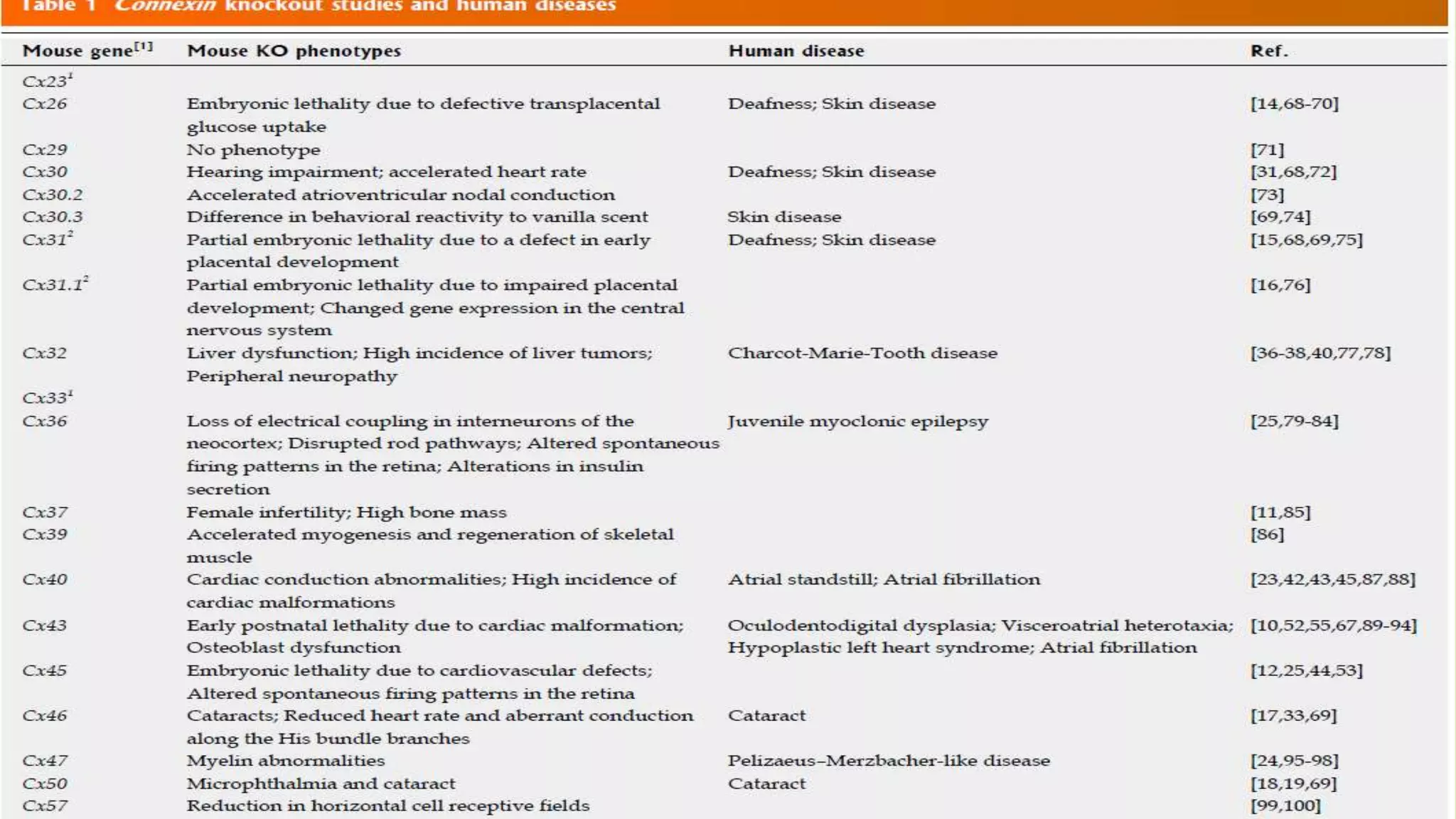
![Cx mutant associated
Cx gene knockout (KO) strategies in mice were first applied to Cx43 by
Reaume et al[10] in 1995.
Mutant mice embryos lacking Cx43 die at birth as a result of a failure in
pulmonary gas exchange caused by a swelling and blockage of the right
ventricular outflow tract from the heart, indicating that Cx43 plays an
essential role in heart development](https://image.slidesharecdn.com/cellularcommunicationinmulticellularorganisms-150902092308-lva1-app6891/75/Cellular-communication-in-multicellular-organisms-31-2048.jpg)


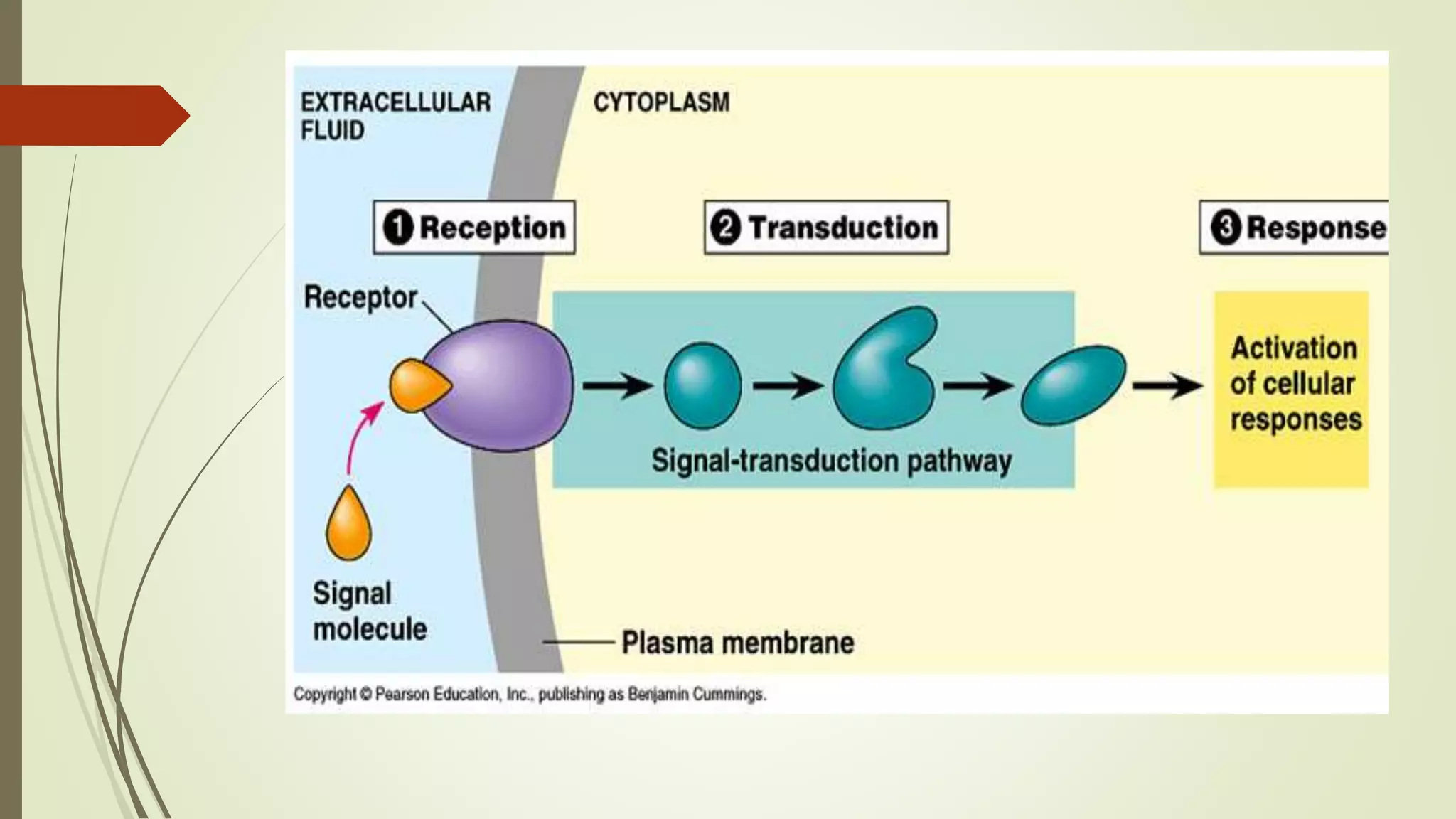
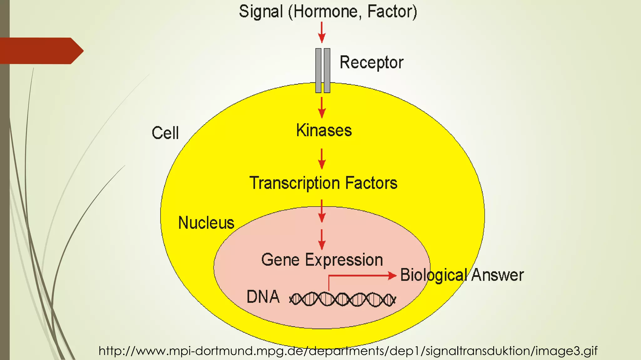
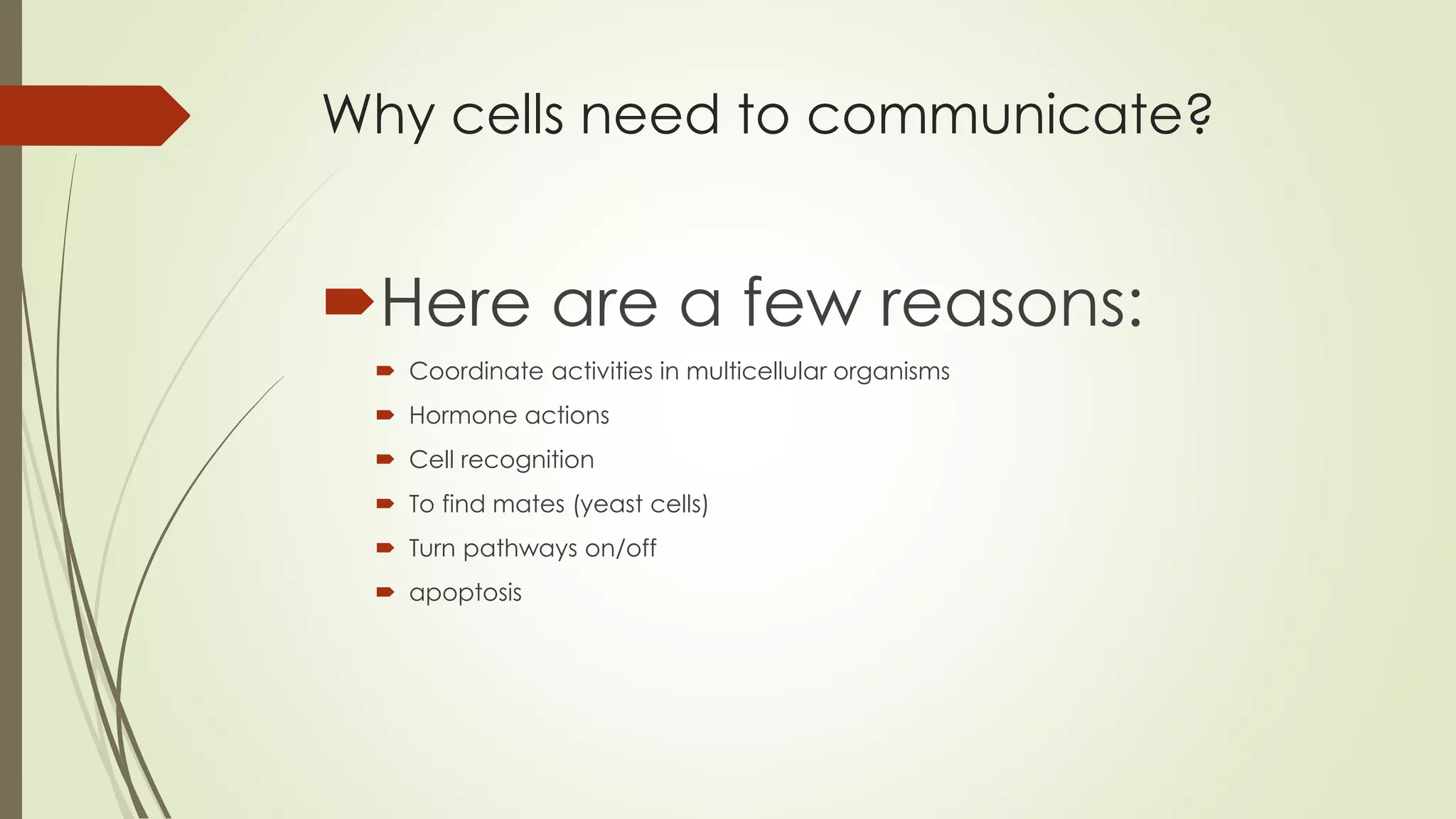
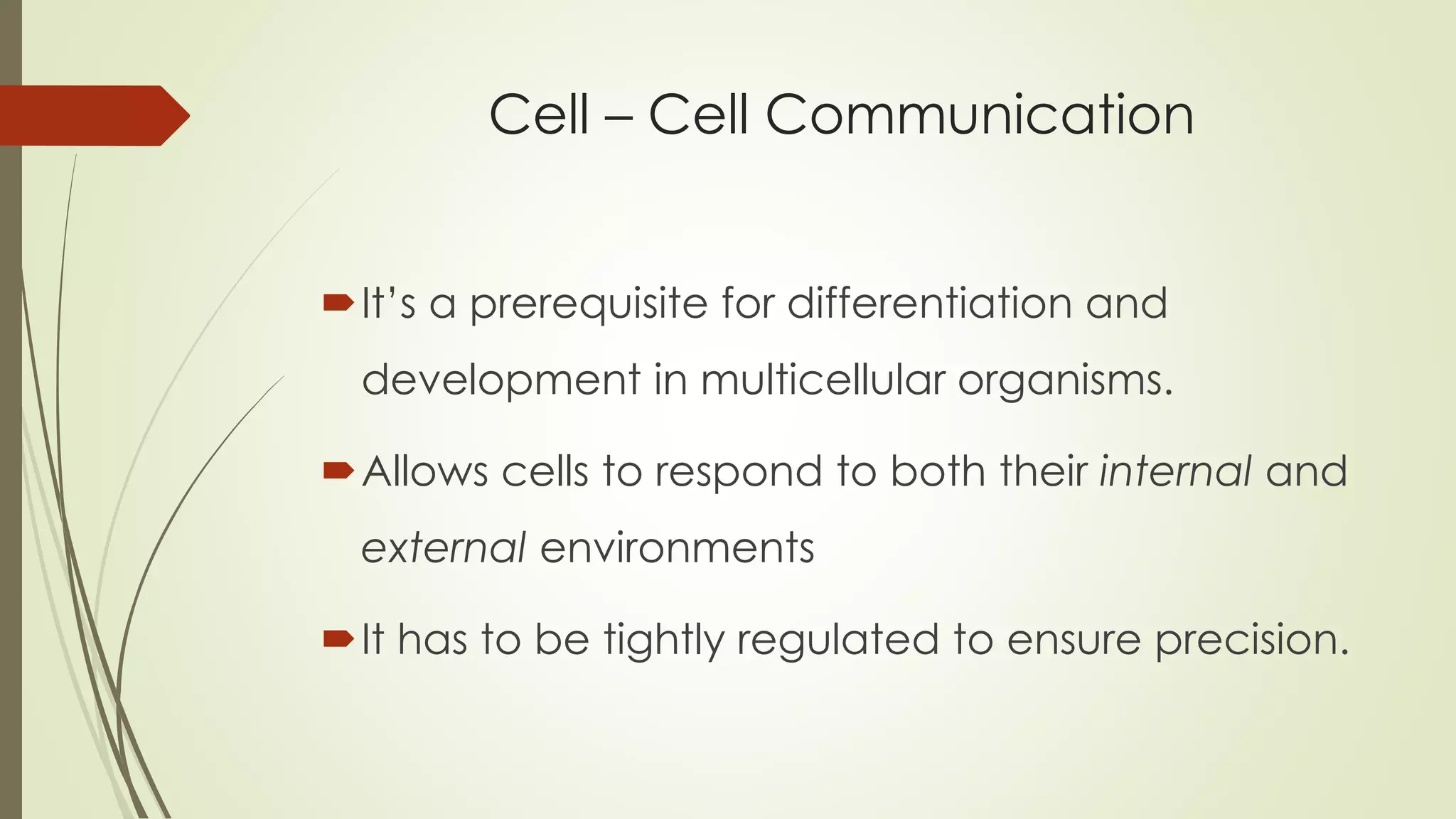
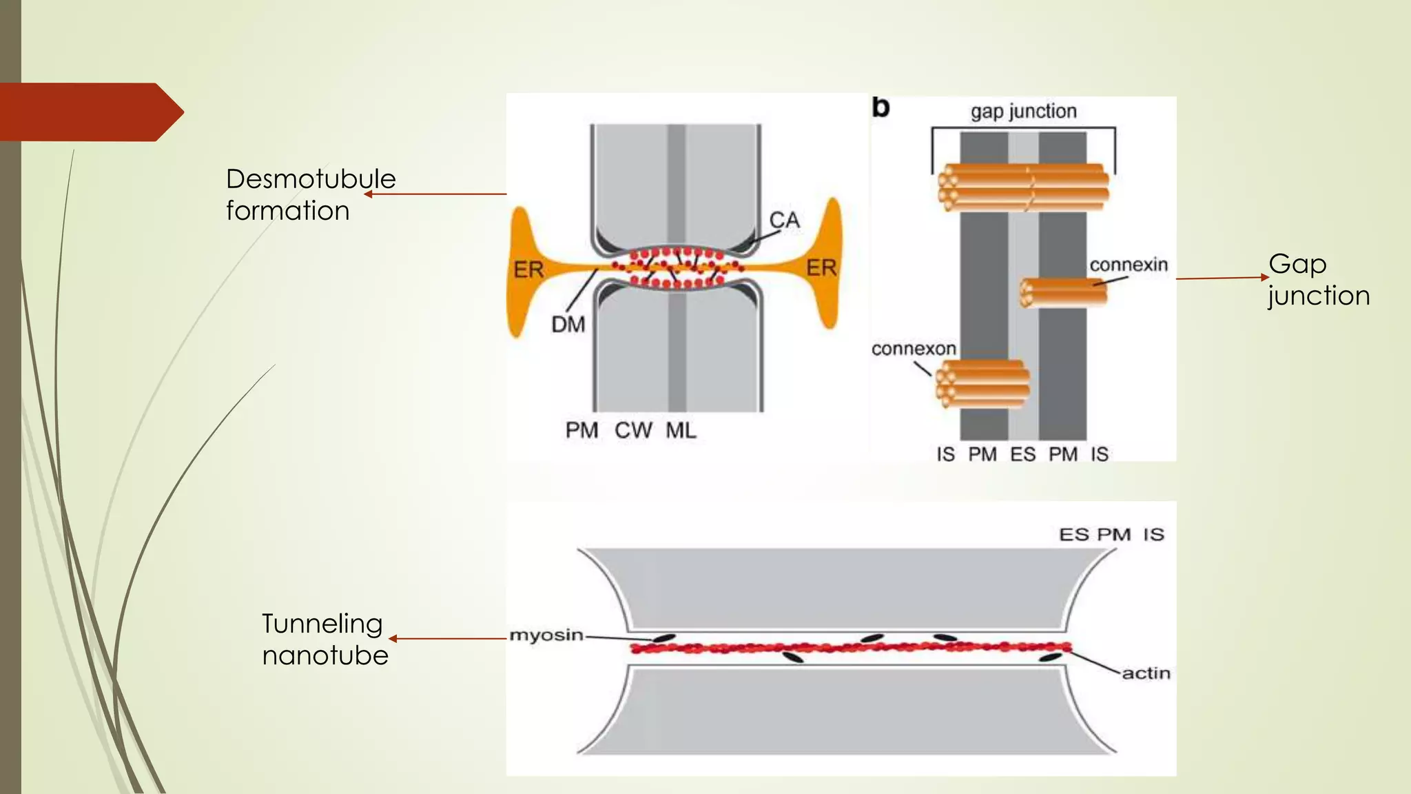
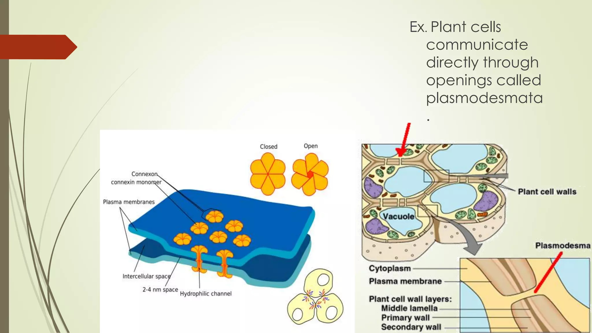
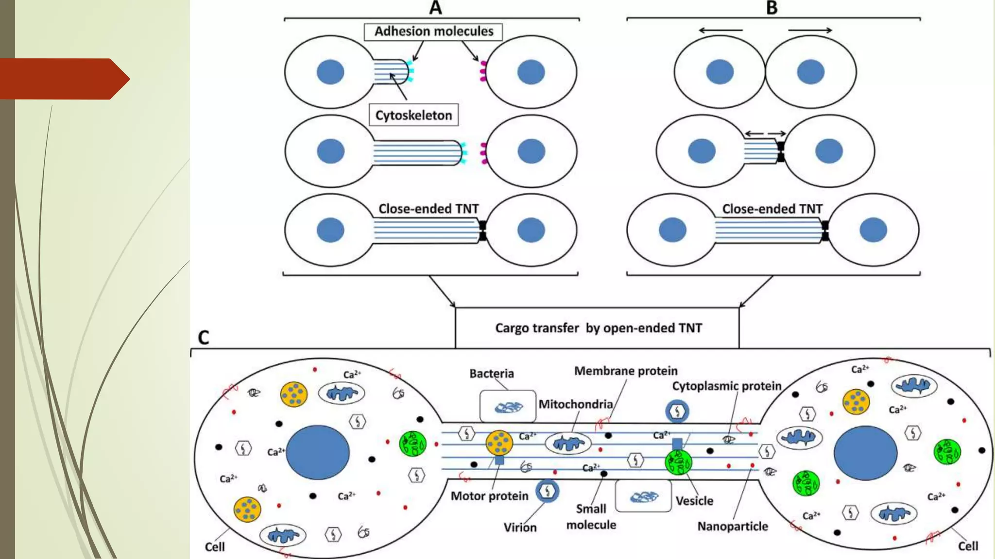
Cells communicate through chemical signaling and direct connections between cells. In multicellular organisms, cell communication allows for coordination between cells and tissues. Cells communicate through several mechanisms including gap junctions, plasmodesmata in plants, and tunneling nanotubes. Gap junctions and tunneling nanotubes allow direct cytoplasmic connections between cells through which small molecules can pass. Communication through these connections helps coordinate activities in tissues and is important for processes like development, but can also enable the spread of pathogens between cells.