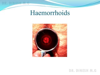
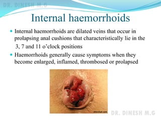
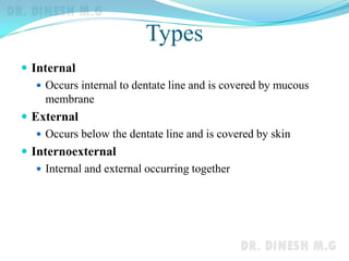
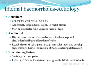
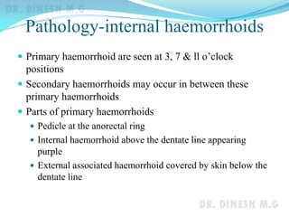
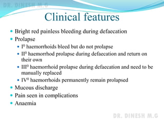
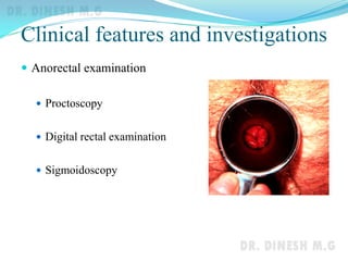
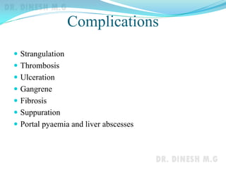
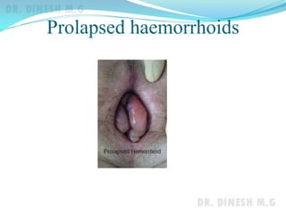
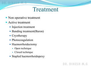
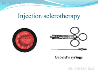
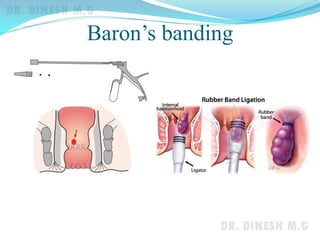
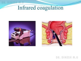
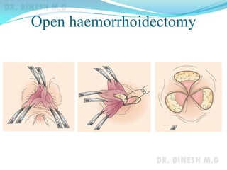
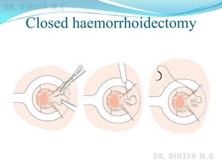
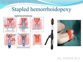
Dr. Dinesh. M.G
Professor of Surgery
J.J.M.M.C.
Davangere
Causes of rectal bleeding
 Haemorrhoids
 Fissure in ano
 Polyps
 Colorectal carcinoma
 Ulcerative colitis
 Diverticulosis
 Angiodysplasia
 Infection (bacillary dysentery)
 Upper GI tract and Meckel’s diverticulum
Haemorrhoids
Internal haemorrhoids
 Internal haemorrhoids are dilated veins that occur in
prolapsing anal cushions that characteristically lie in the
3, 7 and 11 o’clock positions
 Haemorrhoids generally cause symptoms when they
become enlarged, inflamed, thrombosed or prolapsed
Types
 Internal
 Occurs internal to dentate line and is covered by mucous
membrane
 External
 Occurs below the dentate line and is covered by skin
 Internoexternal
 Internal and external occurring together
Internal haemorrhoids-Aetiology
 Hereditary
 Congenital weakness of vein wall
 Abnormally large arterial supply to rectal plexus
 May be associated with varicose veins of legs
 Anatomical
 High venous pressure due to absence of valves in portal
circulation leading to dilatation of veins
 Rectal plexus of veins pass through muscular layer and develop
high pressure during contraction of muscles during defaecation
 Exacerbating factors
 Straining in constipation
 Enteritis, colitis or the dysenteries aggravate latent haemorrhoids
Pathology-internal haemorrhoids
 Primary haemorrhoid are seen at 3, 7 & ll o’clock
positions
 Secondary haemorrhoids may occur in between these
primary haemorrhoids
 Parts of primary haemorrhoids
 Pedicle at the anorectal ring
 Internal haemorrhoid above the dentate line appearing
purple
 External associated haemorrhoid covered by skin below the
dentate line
Clinical features
 Bright red painless bleeding during defaecation
 Prolapse
 I0 haemorrhoids bleed but do not prolapse
 II0 haemorrhod prolapse during defaecation and return on
their own
 III0 haemorrhoid prolapse during defaecation and need to be
manually replaced
 IV0 haemorrhoids permanently remain prolapsed
 Mucous discharge
 Pain seen in complications
 Anaemia
Clinical features and investigations
 Anorectal examination
 Proctoscopy
 Digital rectal examination
 Sigmoidoscopy
Complications
 Strangulation
 Thrombosis
 Ulceration
 Gangrene
 Fibrosis
 Suppuration
 Portal pyaemia and liver abscesses
Prolapsed haemorrhoids
Treatment
 Non operative treatment
 Active treatment
 Injection treatment
 Banding treatment(Baron)
 Cryotherapy
 Photocoagulation
 Haemorrhoidectomy
 Open technique
 Closed technique
 Stapled haemorrhoidopexy
Injection sclerotherapy
Gabriel’s syringe
Baron’s banding
Infrared coagulation
Open haemorrhoidectomy
Closed haemorrhoidectomy
Stapled hemorrhoidopexy
External haemorrhoids
 Thrombosed external haemorrhoid
 Internoexternal haemorrhoid
 Sentinel pile
 Dilatation of the veins at the anal verge
Thank you

Haemorrhoids

  • 1.
    Dr. Dinesh. M.G Professorof Surgery J.J.M.M.C. Davangere
  • 2.
    Causes of rectalbleeding  Haemorrhoids  Fissure in ano  Polyps  Colorectal carcinoma  Ulcerative colitis  Diverticulosis  Angiodysplasia  Infection (bacillary dysentery)  Upper GI tract and Meckel’s diverticulum
  • 3.
  • 4.
    Internal haemorrhoids  Internalhaemorrhoids are dilated veins that occur in prolapsing anal cushions that characteristically lie in the 3, 7 and 11 o’clock positions  Haemorrhoids generally cause symptoms when they become enlarged, inflamed, thrombosed or prolapsed
  • 5.
    Types  Internal  Occursinternal to dentate line and is covered by mucous membrane  External  Occurs below the dentate line and is covered by skin  Internoexternal  Internal and external occurring together
  • 6.
    Internal haemorrhoids-Aetiology  Hereditary Congenital weakness of vein wall  Abnormally large arterial supply to rectal plexus  May be associated with varicose veins of legs  Anatomical  High venous pressure due to absence of valves in portal circulation leading to dilatation of veins  Rectal plexus of veins pass through muscular layer and develop high pressure during contraction of muscles during defaecation  Exacerbating factors  Straining in constipation  Enteritis, colitis or the dysenteries aggravate latent haemorrhoids
  • 7.
    Pathology-internal haemorrhoids  Primaryhaemorrhoid are seen at 3, 7 & ll o’clock positions  Secondary haemorrhoids may occur in between these primary haemorrhoids  Parts of primary haemorrhoids  Pedicle at the anorectal ring  Internal haemorrhoid above the dentate line appearing purple  External associated haemorrhoid covered by skin below the dentate line
  • 8.
    Clinical features  Brightred painless bleeding during defaecation  Prolapse  I0 haemorrhoids bleed but do not prolapse  II0 haemorrhod prolapse during defaecation and return on their own  III0 haemorrhoid prolapse during defaecation and need to be manually replaced  IV0 haemorrhoids permanently remain prolapsed  Mucous discharge  Pain seen in complications  Anaemia
  • 9.
    Clinical features andinvestigations  Anorectal examination  Proctoscopy  Digital rectal examination  Sigmoidoscopy
  • 10.
    Complications  Strangulation  Thrombosis Ulceration  Gangrene  Fibrosis  Suppuration  Portal pyaemia and liver abscesses
  • 11.
  • 12.
    Treatment  Non operativetreatment  Active treatment  Injection treatment  Banding treatment(Baron)  Cryotherapy  Photocoagulation  Haemorrhoidectomy  Open technique  Closed technique  Stapled haemorrhoidopexy
  • 13.
  • 14.
  • 15.
  • 16.
  • 17.
  • 18.
  • 19.
    External haemorrhoids  Thrombosedexternal haemorrhoid  Internoexternal haemorrhoid  Sentinel pile  Dilatation of the veins at the anal verge
  • 20.