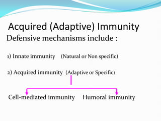
Acquired (Adaptive) Immunity
Defensivemechanisms include :
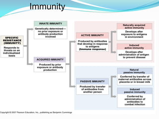
1) Innate immunity (Natural or Non specific)
2) Acquired immunity (Adaptive or Specific)
Cell-mediated immunity Humoral immunity
4.
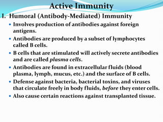
Active Immunity
I. Humoral(Antibody-Mediated) Immunity
Involves production of antibodies against foreign
antigens.
Antibodies are produced by a subset of lymphocytes
called B cells.
B cells that are stimulated will actively secrete antibodies
and are called plasma cells.
Antibodies are found in extracellular fluids (blood
plasma, lymph, mucus, etc.) and the surface of B cells.
Defense against bacteria, bacterial toxins, and viruses
that circulate freely in body fluids, before they enter cells.
Also cause certain reactions against transplanted tissue.
5.
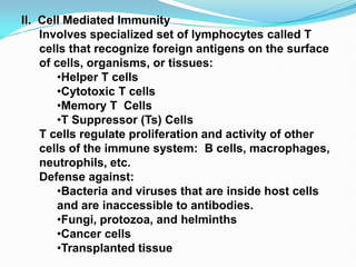
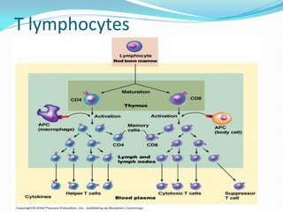
II. Cell MediatedImmunity
Involves specialized set of lymphocytes called T
cells that recognize foreign antigens on the surface
of cells, organisms, or tissues:
•Helper T cells
•Cytotoxic T cells
•Memory T Cells
•T Suppressor (Ts) Cells
T cells regulate proliferation and activity of other
cells of the immune system: B cells, macrophages,
neutrophils, etc.
Defense against:
•Bacteria and viruses that are inside host cells
and are inaccessible to antibodies.
•Fungi, protozoa, and helminths
•Cancer cells
•Transplanted tissue
6.
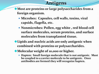
Antigens
Most are proteinsor large polysaccharides from a
foreign organism.
Microbes: Capsules, cell walls, toxins, viral
capsids, flagella, etc.
Nonmicrobes: Pollen, egg white , red blood cell
surface molecules, serum proteins, and surface
molecules from transplanted tissue.
Lipids and nucleic acids are only antigenic when
combined with proteins or polysaccharides.
Molecular weight of 10,000 or higher.
Hapten: Small foreign molecule that is not antigenic. Must
be coupled to a carrier molecule to be antigenic. Once
antibodies are formed they will recognize hapten.
7.
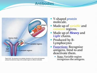
Antibodies
Y-shaped protein
molecule.
Made up of variable and
constant regions.
Made up of Heavy and
Light chains.
Produced by B-
Lymphocytes
Function: Recognize
antigens, bind to and
deactivate them.
Note: Variable region
recognizes the anitgens.
8.
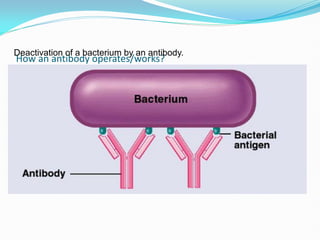
How an antibodyoperates/works?
Deactivation of a bacterium by an antibody.
9.
Immunoglobulin Classes
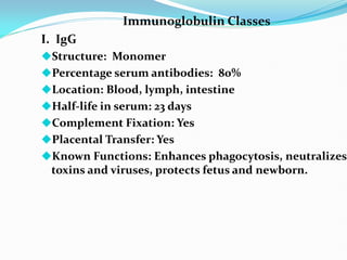
I. IgG
Structure:Monomer
Percentage serum antibodies: 80%
Location: Blood, lymph, intestine
Half-life in serum: 23 days
Complement Fixation: Yes
Placental Transfer: Yes
Known Functions: Enhances phagocytosis, neutralizes
toxins and viruses, protects fetus and newborn.
10.
Immunoglobulin Classes
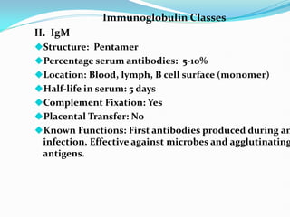
II. IgM
Structure:Pentamer
Percentage serum antibodies: 5-10%
Location: Blood, lymph, B cell surface (monomer)
Half-life in serum: 5 days
Complement Fixation: Yes
Placental Transfer: No
Known Functions: First antibodies produced during an
infection. Effective against microbes and agglutinating
antigens.
11.
Immunoglobulin Classes
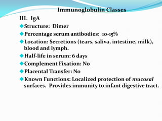
III. IgA
Structure:Dimer
Percentage serum antibodies: 10-15%
Location: Secretions (tears, saliva, intestine, milk),
blood and lymph.
Half-life in serum: 6 days
Complement Fixation: No
Placental Transfer: No
Known Functions: Localized protection of mucosal
surfaces. Provides immunity to infant digestive tract.
12.
Immunoglobulin Classes
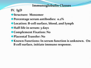
IV. IgD
Structure:Monomer
Percentage serum antibodies: 0.2%
Location: B-cell surface, blood, and lymph
Half-life in serum: 3 days
Complement Fixation: No
Placental Transfer: No
Known Functions: In serum function is unknown. On
B cell surface, initiate immune response.
13.
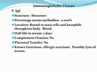
Immunoglobulin Classes
V. IgE
Structure:Monomer
Percentage serum antibodies: 0.002%
Location: Bound to mast cells and basophils
throughout body. Blood.
Half-life in serum: 2 days
Complement Fixation: No
Placental Transfer: No
Known Functions: Allergic reactions. Possibly lysis of
worms.
14.
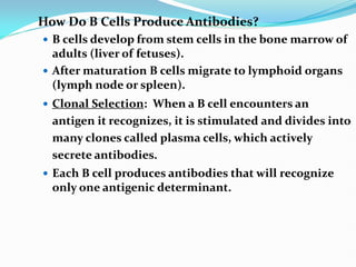
How Do BCells Produce Antibodies?
B cells develop from stem cells in the bone marrow of
adults (liver of fetuses).
After maturation B cells migrate to lymphoid organs
(lymph node or spleen).
Clonal Selection: When a B cell encounters an
antigen it recognizes, it is stimulated and divides into
many clones called plasma cells, which actively
secrete antibodies.
Each B cell produces antibodies that will recognize
only one antigenic determinant.
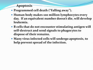
Apoptosis
Programmed celldeath (“Falling away”).
Human body makes 100 million lymphocytes every
day. If an equivalent number doesn’t die, will develop
leukemia.
B cells that do not encounter stimulating antigen will
self-destruct and send signals to phagocytes to
dispose of their remains.
Many virus infected cells will undergo apoptosis, to
help prevent spread of the infection.
17.
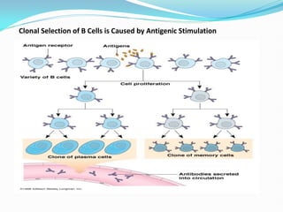
Clonal Selection
ClonalSelection: B cells (and T cells) that encounter
stimulating antigen will proliferate into a large group
of cells.
Why don’t we produce antibodies against our own
antigens? We have developed tolerance to them.
Clonal Deletion: B and T cells that react against self
antigens appear to be destroyed during fetal
development. Process is poorly understood.
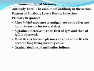
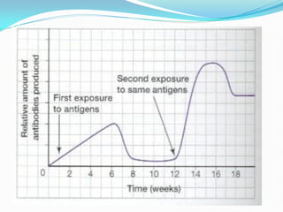
Immunological Memory
Antibody Titer:The amount of antibody in the serum.
Pattern of Antibody Levels During Infection
Primary Response:
After initial exposure to antigen, no antibodies are
found in serum for several days.
A gradual increase in titer, first of IgM and then of
IgG is observed.
Most B cells become plasma cells, but some B cells
become long living memory cells.
Gradual decline of antibodies follows.
20.
Immunological Memory (Continued)
SecondaryResponse:
Subsequent exposure to the same antigen displays a
faster and more intense antibody response.
Increased antibody response is due to the existence of
memory cells, which rapidly produce plasma cells
upon antigen stimulation.
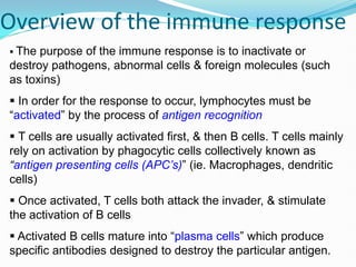
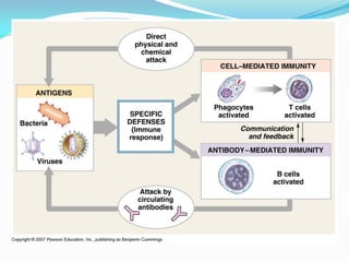
Overview of theimmune response
The purpose of the immune response is to inactivate or
destroy pathogens, abnormal cells & foreign molecules (such
as toxins)
In order for the response to occur, lymphocytes must be
“activated” by the process of antigen recognition
T cells are usually activated first, & then B cells. T cells mainly
rely on activation by phagocytic cells collectively known as
“antigen presenting cells (APC’s)” (ie. Macrophages, dendritic
cells)
Once activated, T cells both attack the invader, & stimulate
the activation of B cells
Activated B cells mature into “plasma cells” which produce
specific antibodies designed to destroy the particular antigen.
25.
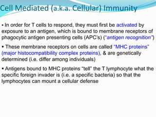
Cell Mediated (a.k.a.Cellular) Immunity
In order for T cells to respond, they must first be activated by
exposure to an antigen, which is bound to membrane receptors of
phagocytic antigen presenting cells (APC’s) (“antigen recognition”)
These membrane receptors on cells are called “MHC proteins”
(major histocompatibility complex proteins), & are genetically
determined (i.e. differ among individuals)
Antigens bound to MHC proteins “tell” the T lymphocyte what the
specific foreign invader is (i.e. a specific bacteria) so that the
lymphocytes can mount a cellular defense
26.
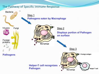
The Pathway ofSpecific Immune Response
Pathogens
Pathogens eaten by Macrophage
Displays portion of Pathogen
on surface
Helper-T cell recognizes
Pathogen
Step 1
Step 2
Step 3
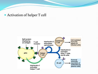
Activation ofhelper T cell
Self protein
displaying
an antigen T cell
receptor
Interleukin-2
stimulates
cell division
Cytotoxic
T cell
Interleukin-2
activates
other T cells
and B cells
Cell-mediated
immunity
(attack on
infected cells)
Humoral
immunity
(secretion of
antibodies by
plasma cells)
B cell
Helper
T cell
APC
Interleukin-1
activates
helper T cell
29.
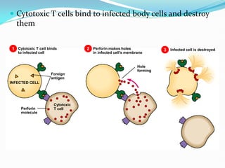
Cytotoxic Tcells bind to infected body cells and destroy
them
Cytotoxic T cell binds
to infected cell
1 2 3
Perforin makes holes
in infected cell’s membrane
Infected cell is destroyed
INFECTED CELL
Perforin
molecule
Cytotoxic
T cell
Foreign
antigen
Hole
forming
30.
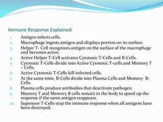
Immune Response Explained
1.Antigen infects cells.
2. Macrophage ingests antigen and displays portion on its surface.
3. Helper T- Cell recognizes antigen on the surface of the macrophage
and becomes active.
4. Active Helper T-Cell activates Cytotoxic T-Cells and B-Cells.
5. Cytotoxic T-Cells divide into Active Cytotoxic T-cells and Memory T
– Cells.
6. Active Cytotoxic T-Cells kill infected cells.
7. At the same time, B-Cells divide into Plasma Cells and Memory B-
Cells.
8. Plasma cells produce antibodies that deactivate pathogen.
9. Memory T and Memory B cells remain in the body to speed up the
response if the same antigen reappears.
10. Supressor T-Cells stop the immune response when all antigens have
been destroyed.
31.
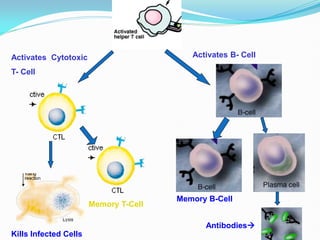
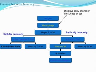
Immune Response Summary
Antigen
Macrophage
HelperT - Cell
Active Cytotoxic T-Cell Active B - Cell
Kills Infected Cells Memory T- Cell Plasma Cell Memory B-Cell
Antibodies
Deactivates Antigens
Displays copy of antigen
on surface of cell
Cellular Immunity
Antibody Immunity
32.
Autoimmune Disease
Autoimmunediseases are diseases where the immune
system begins to attack itself.
Ex:
Rheumatoid Arthritis – crippling disease of the joints.
Lupus – disease of blood and organs.
Multiple Sclerosis – disease of nervous system
Myasthenia Gravis
Cause(s): unknown
Cures/Treatments: No known cures. Usually treated with
drugs.
33.
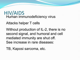
HIV/AIDS
Human immunodeficiency virus
Attackshelper T cells
Without production of IL-2, there is no
second signal, and humoral and cell
mediated immunity are shut off.
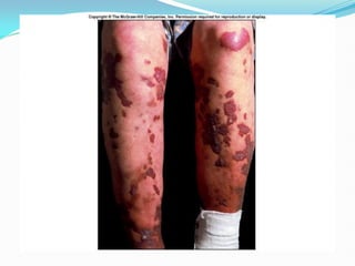
See increase in rare diseases:
TB, Kaposi sarcoma, etc.
35.
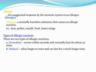
Allergies
Allergy
- An exaggeratedresponse by the immune system to an allergen.
Allergen: a normally harmless substance that causes an allergic
reaction.
ex: dust, pollen, mould, food, insect stings
Types of Allergic reactions
There are two types of allergic reactions.
a. Immediate – occurs within seconds and normally lasts for about 30
mins.
b. Delayed – takes longer to react and can last for a much longer time.
36.
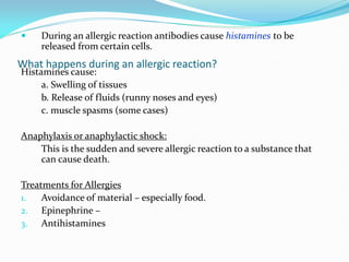
What happens duringan allergic reaction?
During an allergic reaction antibodies cause histamines to be
released from certain cells.
Histamines cause:
a. Swelling of tissues
b. Release of fluids (runny noses and eyes)
c. muscle spasms (some cases)
Anaphylaxis or anaphylactic shock:
This is the sudden and severe allergic reaction to a substance that
can cause death.
Treatments for Allergies
1. Avoidance of material – especially food.
2. Epinephrine –
3. Antihistamines
37.
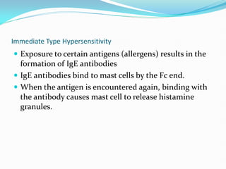
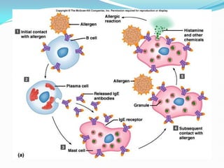
Immediate Type Hypersensitivity
Exposure to certain antigens (allergens) results in the
formation of IgE antibodies
IgE antibodies bind to mast cells by the Fc end.
When the antigen is encountered again, binding with
the antibody causes mast cell to release histamine
granules.
39.
Delayed Hypersensitivity
A typeof cell mediated immunity.
T cell – requires usual two signals
Second time antigen is encountered, T cell
produces several cytokines that attract and
activate macrophages, resulting in an
inflammatory reaction.
40.
Further reading:
Reviewof Medical Physiology. By W.F. Ganong, Lange
Medical Book. Prentice-Hall International.
Textbook of Medical Physiology, Indu Khurana,
Elsevier


![CASE_PRESENTATION_ON_subdural_hematoma(SDH)[1 FINAL PPT]-1.pptx](https://cdn.slidesharecdn.com/ss_thumbnails/casepresentationonsubduralhematomasdh1finalppt-1-260129172522-d405d375-thumbnail.jpg?width=640&height=640&fit=bounds)


