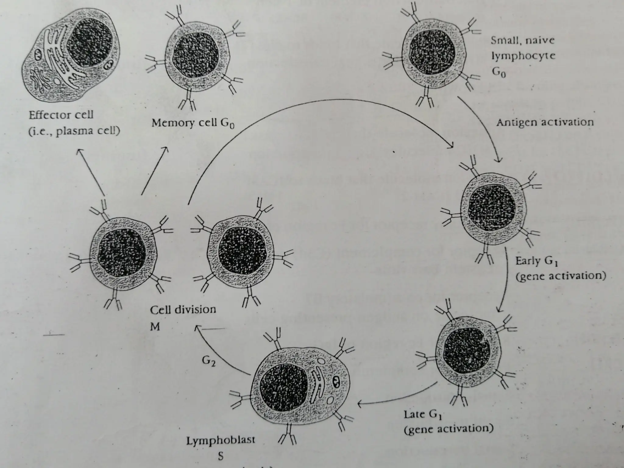
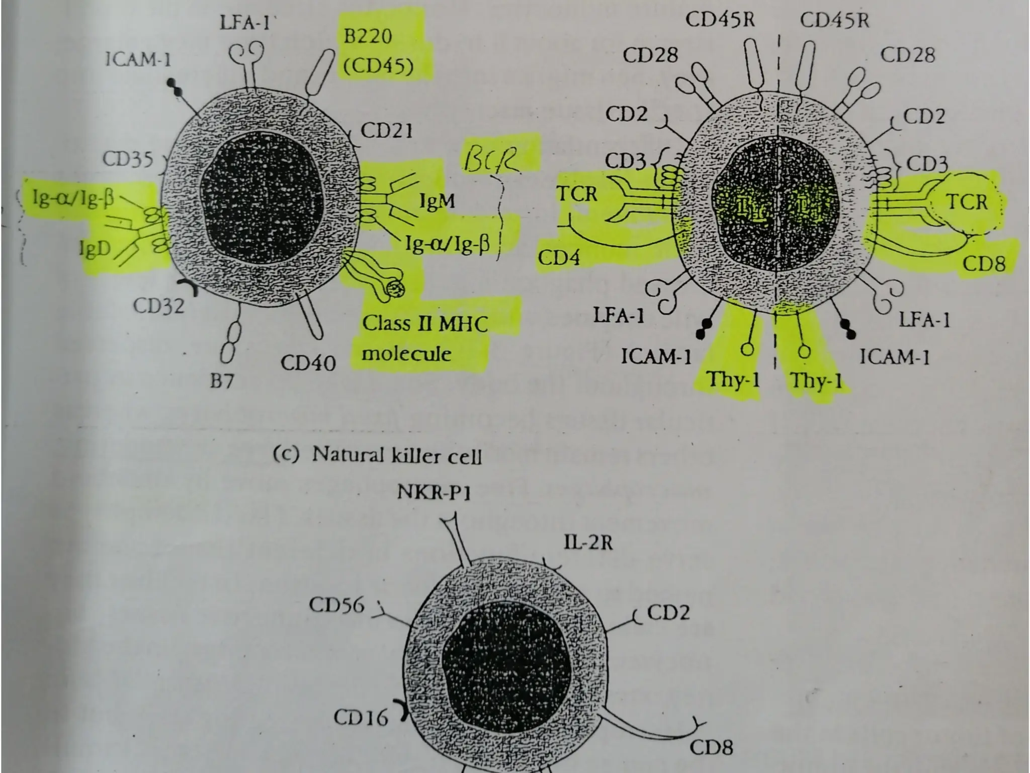
 At one time or another in life – suffered and recovered from infectious
diseases- cold, flue, measles, mumps
 Recovery- bodies are capable of protecting from harmful effects of
infectious agents
 System responsible for such protection – immune system
 The state or phenomenon of protection – immunity
 Study of immune system, immunity and immune mechanisms –
immunology
 Immunity is mainly of two types
 Innate/ Non-specific immunity
 Acquired/ Specific immunity
 Innate/ Non-specific immunity- basic/general resistance/ defense to
any disease that a species possesses by birth, four barriers are
 Anatomical/ Physical barriers: Includes skin and mucus membrane
 Skin: two distinct layers, thin-epidermis, thick- dermis
 Epidermis consists of several layers of epithelial cells, outer layers are
dead, keratinized, epidermis completely renewed in 15-30 days
 Dermis- CT, blood vessels, hair follicles, sebaceous & sweat glands
Introduction: Types of Immunity
 Skin is the first line of defense, prevents the entry of pathogens in to deeper
tissues, low pH inhibit their growth as well
 Sebaceous glands- sebum, lactic acids & fatty acids- maintain low pH of skin
between 3 to 5 – prevents the growth of most of the pathogenic bacteria
 Any wound, abrasions in intact skin leads to the entry of pathogens
 Mucus Membrane: Conjunctiva, GIT, RT, urogenital tract protected by mm,
consist of outer epithelial layer and CT
 Saliva, tears, mucus secreted by epithelial cells contains antibacterial &
antiviral substances- protects from pathogens
 Cilia in RT traps and propels microorganisms
 Microorganisms have evolved their own defense mechanisms
 Fimbriae or pilli on Neisseria gonorrhoeae interact with glycoprotein &
glycolipid receptors on epithelial cells of mm
 Physiological Barriers: includes Temperature pH and soluble factors
 Temperature: Many species are not susceptible to diseases- because of their
body temperature e.g. chicken displays natural immunity to Anthrax – 1070F
 pH: Gastric acidity – innate barrier as pathogenic bacteria grow at neutral pH
 New born more susceptible- stomach contents are less acidic, gastric flora has
not been fully established
 Soluble factors: also contribute to non-specific immunity
 Lysozyme: a hydrolytic enzyme in mucus secretions – cleave peptidoglycan
of bacterial cell wall
 Interferons: group of antiviral proteins produced by virus infected cells
 Complement: group of heat labile serum proteins- inactive pro-enzyme form
 Upon activation- destroy pathogens, help them clear from the body
 Endocytic/ Phagocytic barriers: ingestion of extracellular macromolecules
and particles through endocytosis, phagocytosis
 Endocytosis- receptor mediated endocytosis or pinocytosis, Phagocytosis-
ingestion of particulate material including whole microorganisms
 Break down into simpler products and eliminated from the cell
 Barriers created by inflammatory response: Tissue damage caused by
wounds or invasion by pathogenic microorganisms or by variety of agents/
substances like drugs, pollens, inert physical materials such as wood, pieces of
metals etc. can induce a complex sequence of events- inflammation
 In 1st century AD- Roman Physician Celsus- four cardinal signs of
inflammation- rubor (redness), tumor (swelling), calor (heat), dolor (pain)- In
2nd century AD Galen added 5th sign- functio laesa (loss of function)
Inflammation- useful process, results in clearance of pathogen followed by
tissue repair & regeneration
 Acquired or specific immunity: Reflects the presence of functional immune
system capable of specifically recognizing and selectively eliminating the
pathogens
 Four features- specificity, diversity, memory, self/non-self recognition
 Developed after birth during life time, may be acquired actively or passively
 Active immunity: When an individual is exposed to microorganisms/ foreign
substances- immune system responds- usually long lived
 Passive immunity: Through the transfer of antibodies- usually short lived
 Both types may be acquired either through natural or artificial means
 Naturally acquired immunity: Its is of further two types
 Naturally acquired active immunity: Person is exposed to an Ag in daily life/
disease
 It may be life long in some disease, for few years, sub-clinical infections-
immunity
 Naturally acquired passive immunity: Transfer of maternal antibodies
 Trans placental transfer- from placenta to fetus before birth- IgG e.g. if mother
is immune to rubella, polio, diphtheria- new born will also be temporarily
immune, Clostrum after birth- rich in IgA, in poultry through egg yolk- IgG,
amniotic fluid- IgA
 In mammals e.g. calf, now trans placental transfer, clostrum is the only source,
FCS is free of maternal Abs- used in research
 Artificially acquired immunity: Its is of further two types
 Artificially acquired active immunity: Through vaccination/ immunization,
same mechanism as pathogens but no disease
 Vaccine- live attenuated or inactivated bacterial/ viral- polio vaccine, tetanus
toxoids etc.
 Artificially acquired passive immunity: Through introduction of already
prepared purified antibodies
 Immediate immunity- short lived, Abs neutralizes Ag- catabolized
 Half life is usually up to 3 w, ATS 14-21 days
 Abs- serum, antiserum is used, study of serum, Ag-Ab interactions- serology
 Electrophoresis- two types of serum proteins, albumin & globulins
 Globulins- further three types- Alpha, Beta & gamma
 Gamma-globulins- Immunoglobulins (Igs)- Abs
 Five classes- IgG, IgM, IgA, IgD and IgE
 Immune system – structurally & functionally diverse cells, tissues & organs-
throughout the body
 Immune/ lymphoid organs – 1. Primary 2. Secondary
 Primary- appropriate micro-environment- maturation of lymphocytes
 Secondary- trap Ag from tissue & vascular spaces- site for interaction of mature
lymphocytes with Ag
 Central cells of immune system- lymphocytes- 25% of WBCs in blood and 99% of
lymph
 Approximately 1012 in human = brain + liver
 Cells of Immune System
 Lymphocytes: Only possesses the four attributes, central cells, all other cells play
accessory role- activation of lymphocytes, phagocytosis, secretion of immune
effector molecules like cytokines
 20-40% of WBCs, circulate in blood, lymph, migrate to tissue spaces and lymphoid
organs- lymphocytes re-circulation
 Three types- function and cell membrane components- B-cells/ B-lymphocytes, T-
cells/ T-lymphocytes, Null cells
 All three types- small (6µm diameter), motile, phagocytic cells- Indistinguishable
morphologically
 B/T-cells- not interacted with Ag- naïve, virgin, un-primed cells- in Go phase of
cell cycle, if don’t interact with Ag- die apoptosis (short life span-few days to few
weeks)
Cells of Immune System
 Interaction of B/T cell with Ag- enter in to cell cycle from Go to G1 (early &
late gene activation stage)- S-stage (lymphoblast stage- 15µm), S-synthesis of
DNA, finally M-division stage- proliferate & differentiate into effector cells &
memory cells
 Effector cells- short life span, B-cell lineage- plasma cells (Ab secreting cells)
 T-cell lineage- TH cells and CTL-cytotoxic T lymphocytes
 Memory cells- long lived cells- Go phase- activated with second encounter of
same Ag
 B-lymphocytes: name- site of maturation, bone marrow in mammals & bursa
of Fabricus in birds
 Distinguished from the BCR- membrane bounded Immunoglobulins (Abs)-
serve as receptor for Ag, B-220- first marker of B-cell lineage
 Express class-II MHC molecule – as antigen presenting cells APC
 Interaction Ag and BCR- a naïve B-cell together with T-cell & macrophages-
activated and divides in to plasma & memory cells, plasma cells lack
membrane bounded Igs – secrete one of the five classes of Igs (Abs)-
neutralizes Ag- Humoral immune response
 T-lymphocytes: name- site of maturation, thymus, have TCR- not membrane
bound Immunoglobulins, protein receptors which recognize an Ag only when
presented along with MHC molecule- MHC- restriction
 Fundamental difference- humoral & cell mediated branches of immunity
 The earliest marker of T-cell lineage – Thy-1, appear during maturation in
thymus & then remains throughout life span
 Two sub-populations of T-cells- T-cells that express CD4 receptors (cluster of
differentiation) – recognize Ag associated with MHC-II called CD4+ or TH
cells- MHC-II restricted
 T-cells that express CD8 receptors – recognize Ag associated with MHC-I
called CD8+ or TC cells- MHC-I restricted
 TH cells proliferate extensively- recognition of Ag-Class-II MHC complex on
APC- secrete many cytokines- activate B-cells, converted to plasma &
memory cells, plasma cells secrete Abs- humoral immune response
 TC cells activated- interaction with Ag-Class-I MHC complex on the surface
of self cells (virus infected cell)- generate CTLs which mediate killing of self/
target cells- CMI
 Ration of CD4+ : CD8+ cells is 2:1in normal peripheral blood, may be altered in
immunodeficiency or auto-immune diseases
 Null cells: Neither have TCR or BCR- lack four attributes
 One functional population – NK cells, large, granulated, 5-10% of peripheral
blood lymphocytes, play important role in defense against tumor cells
 Interact with tumor cells in two ways: Direct contact in non-specific Ab
independent process, Specific Ab-dependent cell mediated cytotoxicity
 Mononuclear Cells: include circulating monocytes- blood & macrophages- tissues
 Monocyte- macrophage- number of changes
 5-10 fold increase in size, intracellular organelles- increase in number & complexity,
cell acquires more phagocytic ability, produces more hydrolytic enzymes and
secrete more soluble factors
 Macrophages: two types 1. Fixed macrophages: Takes residence in a particular
tissues and named accordingly Liver- Kupffer cells, CT- histocytes, Lungs-
alveoler macrophages, Kidney- mesangeal cells, Brain- microglial cells
 2. Free/ wandering macrophages- move through amoeboid movement throughout
the tissues
 Phagocytosis- ingesting exogenous Ag, insoluble particles, injured & dead cells of
host, cellular debris, activated clotting factors etc.
 Granulocytes: three types on the basis of cellular morphology & cytoplasmic
staining characteristics
 Neutrophils: produced in bone marrow- hematopoiesis
 Granulated cytoplasm- stained with both acidic & basic dyes
 Polymorphonulear leukocytes- multilobed nuclei
 Released in peripheral blood circulation- 7-10 hours, migrate in to tissues- 3 days
life span
 First line of defense against infections, first cells to reach at inflammation site
 Neutrophilia – acute infection, Phagocytic activity is more than macrophages
Cells of Immune System
 Eosinophils: Biolobed nucleus, granulated cytoplasm- stained with acidic dye-
eosin Y
 Motile, phagocytic, can move from blood to tissues, Phagocytic role- less than
macrophages
 Play major role in defense against parasites- eosinophilic cytoplasmic granules
- secrete certain substances- damage parasite membrane
 Basophils: Single lobed nucleus, granulated cytoplasm- stained with basic dye-
methylene blue. Non-phagocytic
 Cytoplasmic granules- secrete- pharmacologically active substances, play
major role in allergic responses
 Migrate in to tissues- mast cells, secrete histamine – development of allergies
 Dendritic cells: named- covered with long membranous processes- resembling
dendrites of nerve cells
 Express high level of MHC-II- act as APCs help in activation of TH cells, two
types
 Non-lymphoid dendritic cells: found in tissues other than lymphoid, named
accordingly, Langerhans cells- epidermis, interstitial cells- heart, liver, kidney,
GIT etc. capture Ag from tissues spaces- regional lymph nodes
 Lymphoid dendritic cells: interdigitating dendritic cells- found in T-cell rich
areas of lymphoid organs- spleen, lymph nodes & thymus- act as APCs
 Follicular dendritic cells- found in lymphoid follicles of lymph nodes- B-cells
activation
 Two types- Primary (central) Secondary (Peripheral)
 Immature lymphocytes- hematopoiesis become mature and Ag committed within primary lymphoid
organs
 In mammals Bone marrow- B-cell maturation and Thymus- T-cell maturation, in birds Bursa of
Fabricus- B-cell maturation as no bone marrow
 Secondary organs trap Ag from tissue & vascular spaces- provide a site where mature
immunocompetent lymphocytes can interact with Ag to generate an immune response
 Main secondary lymphoid organs are Lymph Nodes and Spleen
 GIT, RT possesses MALT including Peyer’s patches, tonsils, adenoids, appendix- mucosal immunity
 Primary Lymphoid Organs:
 Thymus: flat, bilobed organ-situated above the heart
 Each lobe surrounded by fibrous tissue capsule send projections of CT- trabaculae, divide each lobe
into smaller lobules
 Outer portion of each lobe- cortex- densely packed with thymocytes (immature T-cells)
 Inner portion medulla- sparsely packed with thymocytes
 Both cortex and medulla cris-crossed with three dimensional network of thymic stromal cells
composed of epithelial cells, interdigitating dendritic cells, macrophages- make up the framework of
thymus and help in maturation of T-cells
 Cortical epithelial cells (nurse cells) have long membranous processes- hold as many as 50
thymocytes
Organs of Immune System
 Function: Maturation & selection of T-cells- Epithelial cells secrete many
hormones and cytokines (alpha 1 thymosin, beta 4 thymosin, thymopoeitin,
thymulin)
 Maturation, proliferation and differentiation of T-cells
 Express Ag binding receptors, differentiate in to sub-populations
 Selection process: two types Positive selection and negative selection
 T-cells bearing receptors that can recognize self MHC molecule are selected
and other are eliminated through apoptosis
 Self reactive T-cells- recognize self MHC with self Ag are eliminated
 Both as a result of positive and negative selection only those mature T-cells
whose TCR can recognize self MHC molecule along with foreign Ag are
selected and allowed to move into secondary lymphoid organs
 About 95-99% thymocytes die through apoptosis
 Bone marrow: Site of maturation of B-cells in mammals, Bursa of Fabricus in
birds
 Process of B-cells maturation remains unknown
 Secondary Lymphoid Organs
 Lymph Nodes: encapsulated bean-shaped structures containing reticular network- packed with lymphocytes,
macrophages and dendritic cells
 Clustered at the junction of lymphatic vessels, first organized lymphoid structure to encounter Ag that enters
in to tissue spaces
 Divided into three regions: Cortex, Para-cortex and Medulla
 Cortex: Outermost region contains Lymphocytes (B-cells) and macrophages arranged in Primary follicles, B-
cell rich area/ thymus independent area
 Following Antigenic challenge- enlarges in to secondary follicles with germinal center
 Intense B-cell maturation and differentiation into plasma and memory cells occurs in germinal center
 In childeren with B-cells deficiency cortex lacks primary follicles and germinal centers
 Para-cortex: situated beneath cortex, contains T-cells and dendritic cells
 Dendritic cells express MHC-II molecule and act as APCs for TH, Para-cortex- T-cell rich area/ thymus
dependent area
 Medulla: Inner most portion, sparsely packed with lymphocytes, mostly plasma cells actively secreting Abs
 Function: Ag carried to LN by lymph to Para-cortex, trapped, processed and presented along with MHC-II by
dendritic cells- resulting in TH activation which activates B-cells
 Ag activated TH cells, B-cells moves to primary follicles of cortex, interaction between follicular dendritic
cells, B-cells, TH cells- development of secondary follicle and germinal center
 B-cells- plasma and memory cells, plasma cells move to medulla where secrete Abs
 Multiple Afferent Lymphatic vessels pierce capsule of LN and empty lymph in sub-capsular sinuses, single
efferent vessel drains lymph away from LN- contains high conc. of Abs & 50 fold more lymphocytes
 Blood supply to LN- lymphatic artery, drainage lymphatic vein, extravasation of lymphocytes at the level of
Post-capillary venuoles
Organs of Immune System
 Spleen: Large, ovoid organ, left side of abdominal cavity, Unlike LN which trap localized Ag
from regional tissue spaces, spleen- adopted to filter blood and trap blood born Ags and thus
respond to systemic infections
 Surrounded by capsule- send trabaculae which divides in to compartments, two compartments
red pulp and white pulp separated by marginal zone
 Red Pulp: consists of network of sinusoids filled with RBCs and macrophages
 Dead and defective RBCs are destroyed in it- graveyard of RBCs
 White Pulp: contains peri-arteoler lymphatic sheath (PALS) around the splenic artery mainly
composed of T-lymphocytes, Around the PALS, marginal zone rich in B-cells organized in
primary follicles
 Upon Ag challenge primary follicle develop in to secondary follicle containing germinal
center
 Unlike LN, no lymphatic supply, blood born Ags are carried to spleen through splenic artery
which empties in to marginal zone
 Ag from marginal zone is trapped by dendritic cells- carries it to PALS, where TH are activated,
which then activates B-cells converted in memory and plasma cells
 Abs are carried by splenic vein in to the circulation where it interact with Ag- humoral
immunity
 Mucosal-associated lymphoid tissues (MALT):
 Mucus membranes lining GIT, RT, urogenital tracts- 400m2 area, protected by MALT
 Tonsils: lingual- at the base of tongue, palatine- at the side of the tongue, naso-pharangeal
(adenoids)- nasopharynx
 Payer’s patches- 30 to 40 nodules along the outer wall of intestine, BALT, RALT, NALT
 S U B S T A N C E S C A P A B L E O F I N D U C I N G A S P E C I F I C I M M U N E
R E S P O N S E
 F O U R I M M U N O L O G I C A L P R O P E R T I E S
 Immunogenicity
 Antigenicity
 Allerogenicity
 Tolerogenicity
 F O R P R O T E C T I O N A G A I N S T D I S E A S E S – I M M U N O G E N
 P R O P E R T I E S O F A N A N T I G E N
 Foreignness
 Molecular weight – 100,000 Da good, < 5000-10,000 Da poor
 Chemical composition – Protein best followed by
polysaccharides, lipids & NA are not alone
 Complexity
 Recipient genotype, dose and route of administration
ANTIGENS
 Adjuvants – Latin word adjuvare – to help
 Definition – Not immunogen by themselves, tends to increase the
immunogenicity
 Examples: Alum, Freund’s incomplete & complete, Liposome & ISCOMs
 Used when, low immunogenicity, costly
 Epitopes
 Haptens
 Mitogens: Induce cell division in B and T lymphocytes irrespective of their
antigenic specificity
 Polyclonal activators
 B-cell mitogens
 T-cell mitogens
 Both
 Lectins, super-antigens
 A N T I B O D I E S : A N T I G E N B I N D I N G P R O T E I N S
 Membrane bounded – B-cells as receptors
 Secreted by plasma cells
 Effector molecules of humoral immunity - serum
 Structure: Monomer, Y-shape, 2 heavy & 2 light chains
 Disulfide bonds, Flexible – T-shape, hinge region
 Variable region of heavy & light chain on upper ends of Y- arms
 Constant region of heavy & light chain on stem and lower ends of
Y-arms
 Fab and crystalizable fragment, Amino and carboxylic terminals
 Epitopes: Antigenic determinants on Abs – 3 types
 Isotypic, idiotypic and allotypic
Immunoglobulins: Structure & Function
Classes of Immunoglobulins: Function
Five classes/ isotypes of Igs: IgG, IgM, IgA, IgD and IgE
IgG, IgD and IgE – Monomer, IgA – Dimer, IgM – Pentamer
IgG: Most abundant, 80% of serum, 4 subclasses
IgG1 (9mg/ml), IgG2 (3mg/ml), IgG3 (1mg/ml), IgG4 (0.5mg/ml)
Monomer, can cross the blood vessels and placenta, protects fetus
Protect against circulating bacteria & viruses, neutralize toxins
Trigger complement system, enhances the effectiveness of phagocytic
cells
 IgM: Pentamer, 5-10% of serum
Monomer as receptor on B-cells, Pentamer when secreted by plasma
cells
Ist Ig produced in response to Ag
 Ist Ig synthesized by neonate
Move less freely, remain in blood
Strong activator of complement than IgG
Enhances phagocytic activity
IgA: 10-15% of serum, major secretary Igs (milk, saliva, tears,
mucus)
Blood – mostly monomer, may be dimer, trimer or tetramer
External secretions- always dimer (J-chain & secretary
component)
Mucosal immunity, clostrum – GIT pathogens
IgD: 0.2%, similar to IgG
No known function in serum, receptor on B-cells
IgE: 0.002%, slightly larger than IgG
Binds by Fc portion to basophiles & mast cells
Pollens react – histamine & other chemical mediators release
Allergic responses and defense against parasites
 TCR are MHC restricted, CD4 cells are MHC-II, CD8 cells are MHC-I
restricted
 Antigen presenting cells (APCs), synthesize & express MHC-II and
present the processed Antigenic peptide to CD4 cells
 Self cells: synthesize & express MHC-I and present Antigenic peptide to
CD8 cells
 APCs: Monocytes, macrophages, B-cells, dendritic cells, Langerhans
cells, thymic dendritic cells, epithelial cells, venular endothelial cells
 Antigen Processing:
 Extracellular pathogens – exogenous Ag
 Intracellular pathogens – endogenous Ag, posses different challenges
 Exogenous Ag – processed through endocytic pathway – Ab production
 Endogenous Ag – processed through cytosolic pathway – CMI
Antigen Processing and Presentation
Endocytic Processing Pathway
 APCs – internalize Ag through phagocytosis, endocytosis or both
 Macrophages – phagocytosis, other cells - endocytosis
 B-cells – receptor mediated endocytosis using BCR
 Ag processed in to peptides of 13-18 residues
 Three compartments: early endosome (pH 6-6.5), late endosome/
endolysosome (pH 5-6) and late lysosome (pH 4.5-5)
 Acid dependent hydrolytic enzymes in each compartments:
proteases, nucleases, lipases, glycosidases, phospholipases,
phosphatases
 Two school of thoughts
 1-3 hours – from entry till the appearance of processed peptide in
cleft of MHC-II on the surface of same APC
Cytosolic Processing Pathway
 Endogenous Ag/ Virus – self cells, processed through cytosolic
pathway
 Degraded in to peptide of 8-9 residues
 Presented by MHC-I to TC cells – activate CTLs - CMI
 Cytosolic proteolytic system: Ag binds with carrier proteins
“ubiquitin” carried to proteosome
 Large cylindrical particles containing four subunits with a central
hollow of 10-20Ao
 Degradation of Ag inside prevents the proteolysis of other
proteins in side cytoplasm
 Low Molecular mass proteins (LMP)- alternate to Cytosolic
proteolytic system
Antigen Presentation
 Assembly & Stabilization: Processed peptides fit in to the cleft of
MHC-I & MHC-II
 Both are synthesized within RER
 MHC-II: Two chains - alpha & beta – make cleft together
 MHC-I: Two chains – large alpha make cleft & small beta2
microglobulin stabilizes it
 Assembly of antigenic peptide processed through cytosolic pathway in
to the cleft of MHC-I takes place within RER – Ag moves to RER
 It may misfit in to MHC-II – Occupied by third invariant chain
 MHC-II moves from RER to Lysosome – invariant chain degraded by
enzymes and Antigen fit in to the cleft of MHC-II
 MHC-II & MHC-I along with processed Ag is displayed on the surface
of APCs & Self cell, respectively – presented to CD4 & CD8 cells for
humoral & CMI, respectively
 Clinical Applications: For the design of new vaccines, to understand the
mechanism & treatment of certain autoimmune diseases
 Used in two ways: either for detection of Ag or Ab if one of them
is known
 Highly specific, measurement of Ag or Ab – Serology
 Three categories: Primary, Secondary & Tertiary binding tests
 Primary: ELISA, RIA, IFA/FAT
 Secondary: Precipitation & Agglutination tests, CFT
 Tertiary: Neutralization & Protection tests
 Reagents required: Serum, -200C, heat inactivation at 560C in
water bath
 Complement: group of heat labile serum proteins, source guinaea
pig serum
 Antiglobulins: Anti-Ab, Anti-species Ab, Anti-isotypic Ab
 Monoclonal Ab: highly specific & pure
Antigen-Antibody Interactions/
Immunodiagnostics
Primary Binding Tests
 Direct: for detection of Ag with known Ab
 Indirect: for detection of Ab with known Ag
 Named on the basis of Indicator system used
 Radio-immunoassays (RIA): Radioisotopes are used as indicator like 3H, C14,
I125
 RIA for Ab/ Indirect: Known Ag is impregnated to nitrocellulose filter paper
 Unknown serum sample added and incubated
 Radiolabeled Anti-Ab are added and radioactivity is observed under X-ray film
 RIA for Ag/ Direct or Competitive RIA: Unlabeled Ag will displace
radiolabeled Ag from immune-complexes
 Known Ab and Radiolabeled Ag are added in test tube, incubated, radioactivity
of supernatant is observed
 Unknown Ag/ sample is added, it will replace the radiolabeled Ag which will
come in supernatant showing radioactivity
 Amount of radioactivity is directly proportional to Ag present in sample
 Highly sensitive, qualitative as well as quantitative but costly and not safe
 Immunofluorescence assays (IFA): Fluorochromes/ fluorescent dyes
are used as indicator like FITC, RITC, Oramine etc.
 FITC yellow color compound – UV invisible – fluoresce with bright
green color
 Direct FAT for Ag: Unknown Ag/ sample is attached on to
nitrocellulose paper
 Known Ab conjugated with FITC added, incubated and fluorescence is
observed under UV invisible or fluorescent microscope
 Can detect low level of Ag directly from infected body tissues like
Negri bodies in rabbied brain
 Indirect FAT for Ab: Known Ag is attached on to nitrocellulose paper
 Unknown serum sample is added, incubated
 Known Anti-Ab conjugated with FITC added, incubated and
fluorescence is observed under UV invisible or fluorescent microscope
 Highly sensitive, qualitative as well as quantitative but costly
 Enzyme immunoassays (EIA): Enzymes along with respective
substrates are used as indicator like HRP, Alkaline phosphatase etc.
 On addition of substrate, color development/ OD value is measured
 Direct ELISA for Ag: Known Ab is attached on to the surface of ELISA
plate wells, washed
 Unknown Ag/ sample is added, incubated, washed
 Known Ab conjugated with enzyme is added, incubated
 Substrate added, OD value is observed through ELISA reader
 Sandwich ELISA as Ag is sandwiched between two Ab molecules
 Indirect ELISA for Ab: Known Ag is attached on to the surface of
ELISA plate wells, washed
 Unknown serum sample is added, incubated
 Known Anti-Ab conjugated with enzyme added, incubated and OD
value is observed through ELISA reader
 Highly sensitive, qualitative as well as quantitative, comparatively
economical, safe and most commonly used
 Ag or Ab is detected on the basis of the results of primary
reaction
 Precipitation Tests: Soluble Ag + Ab, mixture cloudy after few
minutes, precipitate in 1 hour if +ve
 Slide and tube precipitation tests
 Optimum level of both is required for precipitation to occur
 AGID/ AGPT, Immuno-electrophoresis, CCIE, Western blotting
 Simple, economical, specific but less sensitive, difficult to read
 Agglutination Tests: Particulate Ag + Ab – Agglutination/
clumping
 Simple, economical, more sensitive, easy to read
 Slide & tube agglutination tests/ macro-agglutination tests
 Micro-agglutination tests – Haemagglutination tests: HA, HI,
IHA etc.
Antigen-Antibody Interactions/ Secondary
Binding Tests
Complement Fixation Test
 Two systems: Test system & Indicator system
 Major/ Test system: Ag + test serum + complement
 Minor/ Indicator system Sheep RBCs + Amboceptors
 If no hemolysis - +ve
 If hemolysis - -ve
 Tertiary Binding Tests:
 Neutralization Test:
 To test the neutralization ability of Ab against an Ag in-vitro
 Protection Test:
 To test the protective ability of Ab against an Ag in-vivo
 Development of effective immune response – lymphoid cells,
inflammatory & hematopoietic cells
 Complex interaction between these cells is mediated by a group
of low molecular weight (30KDa) regulatory proteins - cytokines
 Secreted by WBCs, particularly lymphocytes in response to
inducing stimuli
 Binds to receptors on target cells, signal transduction, biological
effect
 Hormones – messengers of endocrine system, Cytokines –
messengers of immune system
 Cytokines may act in three ways
 Autocrine: same cell act as cytokine producing & target cell
 Paracrine: target cell is near by to producing cell
 Endocrine: binding to target cells in distant part of the body
CYTOKINES
 Cytokine may exhibit four properties
 Pleotropy: Cytokine has biological effect on different target cells –
pleotropic e.g. IL-4
 Redundancy: Two or more cytokines – same biological effect on single
target cell – redundant e.g. IL-2, IL-4, IL-5
 Synergy: When combined effect of two or more cytokines is greater
than their individual effect – synergism e.g. IL4 + IL-5
 Antagonism: Effect of one cytokines is inhibited by the other –
antagonistic e.g. IL-4 by IFN-γ
 Nomenclature: Previously on source of release e.g. lymphocytes –
lymphokines, monocytes – monokines
 Technically incorrect- as monokines and lymphokines also secreted
from other cells
 Now named on the basis of function
 Interleukins: Cellular communication among leukocytes, IL1 to IL-17...
 Interferons: Glycoproteins – in response to virus infection – antiviral
 IFN-α, IFN-β, IFN-γ
 Tumor Necrosis factor: anti-tumor TNF-α, TNF-β
 Growth factors: Stimulate the growth of many cells, CSF, G-CSF, M-
CSF, GM-CSF
 Transforming Growth factors: TGF-β
 Chemokines: Inflammation, IL-8
 Structure: Proteins, four structural groups
 I: Four α helix – IL2,3,4,5,6,7,10,11,13, G-CSF, GM-CSF, Interferons
 II: Long β sheets – TNF-α, β, IL-1, TGF-β
 III: Both α helix & β sheets: Chemokines, IL-8
 IV: Mosaic, mixed structure- IL-12
 Discovery & Purification: up to 1960s chromatography
 Disadvantages: low yield- sub-nanomolar level, less purity
 Recombinant DNA technology: in 1960 1st compound – cDNA cloning
techniques
 To date genes IL-1 to 13, IFNs, TNF, TGF, LIF, Oncostatin M cloned
 Functions of Cytokines: Main source TH , macrophages, B-cells
 Development of humoral and cellular immune responses
 Induction of inflammatory response
 Regulation of hematopoiesis
 Control of cellular proliferation & differentiation
 Induction of wound healing
 In-vitro diagnostic and in-vivo therapeutic uses
 Cytokines antagonists: Inhibit the action of cytokines
 Binds directly to cytokines
 Binds to their receptors on target cells
 Antibodies produced – purified Ag - Polyclonal
 Advantages in localization, phagocytosis & complement
mediated lysis of Ag
 Due to undesirable, non-specific, cross-reacting Ab - In-vitro
diagnostic and in-vivo therapeutic uses
 Purified through chromatographic techniques – very difficult
 Alternate – purified clones of mono-specific plasma cells in-vitro
– but short life span
 Solution by George Kohler & Cesar Milstein in 1975 by fusing
B-cells, myeloma cells – hybridoma, Nobel Prize - 1984
 Formation & selection of hybrid cells: 1970s two somatic cells
fused – heterokaryon, fusion by Sendai virus, PEG
 Initially 2-5 nuclei, loss of chromosomes until stabilizes
Hybridoma and Monoclonal Antibodies
 Selection of hybridoma: HAT medium
 Denovo pathway: Phosphoribosyl pyrophosphate + uradylate –
nucleotide and DNA - blocked by aminopterin
 Salvage pathway: Hypoxanthine + Thymidine – catalyzed by HGPRT +
TK enzymes, only hybrid cell will survive
 Production of Monoclonal Antibodies: Three steps
 Generating B-cells hybridoma
 Screening for Monoclonal antibody specificity
 Propagating hybridoma secreting specific monoclonal antibodies
 Ag – mice – Primed B-cells from spleen (HGPRT+, Ig+) + Myeloma
cells ((HGPRT-, Ig-, immortal) – PEG – Heterokaryon along with un-
fused B-cell and un-fused myeloma cells
 HAT selection – only B-cells – myeloma cell hybridoma will grow
 Assay for desired Ab in culture supernatant – ELISA, RIA, FAT
 Reclone Ab+ hybridoma: Expand in Tissue culture (10-100µg/ml),
peritoneal cavity of histocompatable mice (1-25mg/ml), Alginate gel
(100 fold more than tissue culture, Fermenter (1000L – 100g in 2w)
 Purification of Proteins: Can purify any protein out of complex
mixture, present at extremely low level with 100% purity
 Previously IFN – chromatographic techniques
 Disadvantages – 1% purity, 99% impurities
 DS Secher & DC Burke prepared anti-IFN monoclonal antibodies
 Fixed on sephadex in an Immuno-adsorbant column
 Identification and isolation of lymphocytes sub-populations:
 Monoclonal antibodies can be produced to different membrane
bound receptors or proteins – surface of lymphocytes sub-
populations
 CD4+ on TH and CD8+ on TC, anti-CD8 Ab conjugated with
FITC and anti-CD4 Ab with PE
 Identified and isolated in FAC
Uses of Monoclonal Antibodies
 Tumor detection and imaging:
 Tumor specific membrane proteins/ receptors – only on tumor cells
 Monoclonal Ab are prepared against them – detect tumor
 Ab are radiolabeled and injected in the body
 Used to image metastatic tumors
 Tumor Killing: in two ways
 Complement mediated lysis: a large no of tumors are resistant
 Immunotoxins: ricin, shigella & diphtheria toxins – potent toxins
 Two components: Binding polypeptide & inhibitor chain
 Binding component is removed & replaced with specific Ab
 Monoclonal Ab will guide toxic component to tumor cell
 Safe: will not damage the normal cells
 Diagnostic Reagents: More than 100 diagnostic agents available
 Home pregnancy diagnosis using anti-HCG monoclonal Ab coated
strips
 Cancerous T-cells – Thymoma cells
 Immunity – Active and Passive
 Passive – Natural passive – Maternal Ab (tetanus, mumps, polio,
diphtheria), Artificial passive – ATS, anti-snake venom
 Active – Natural active – disease, Artificial active –
immunization/ vaccination
 Vaccine – Live modified or killed whole or a part of
microorganism which when administered in to the body do not
cause disease rather elicit a high humoral or CMI
 As prophylaxis – infectious diseases
 Whole organism vaccines – bacterial or viral
 Live attenuated vaccines – viral or bacterial
 Attenuation – losses pathogenicity, retains immunogenicity
 Bacillus Calmattee Guerin (BCG)–M. bovis, increased conc. of
bile – 13 years – live attenuated vaccine against TB, Polio
vaccine – Sabin and Salk strains
VACCINES
 Advantages: increased, prolonged immunity, no boosters required
 Both humoral and CMI
 Disadvantages: Reversion back to virulence, stability less
 Contaminants may cause problems, post-vaccination reactions
 Irreversible attenuation through gene deletion technology, losses
immunogenicity along with pathogenicity
 Inactivated bacterial / viral vaccines:
 Inactivation - mechanical, chemical
 Heat – degradation of epitopes, chemical – formaldehyde, BEI, β-
propiolactone
 Advantages: More stable, safe, no reversion back to virulent form
 Disadvantages: Require booster, short immunity, less CMI more
humoral immunity
 Post-vaccination complications
 Purified Macro-molecules as vaccines:
 Keeping in view the disadvantages of both
 Specific surface macromolecules used as vaccine
 e.g. Capsular LPS as vaccine against meningitis
 Advantages: more specific, less un toward reactions
 Disadvantages: activates B-cells directly, no activation of TH cells
 Poor immunogenic - LPS + Protein carriers
 e.g. Capsular LPS + Tetanus toxoids – more immunogenic,
vaccine against two or more diseases in single shot
 But difficulty in obtaining purified macromolecules in large
quantity
 Recombinant Antigen Vaccine – Most immunogenic Ag is
cloned in to an appropriate cloning vector, expressed in an
expression host and used as vaccine after purification
 First ever – FMDV, VPI cDNA – plasmid – E. coli
 First in human – HBV – gene for Major surface protein – YAC –
Yeast cells, used after harvesting and purification
 Advantages: Specific immune response, no un to ward reaction,
minimum post-vaccination complications, safe , stable
 Disadvantages: Exogenous Ag – only humoral immune response
 Recombinant Vector Vaccine – Biological vectors are used –
attenuated poliovirus, adenovirus, Salmonella, BCG etc.
 Recombinant vaccinia vector vaccine of small pox
 Advantages: specific, safe, stable, CMI
 Disadvantages: Less immunogenic
RECOMBINANT VACCINES
 Synthetic peptide vaccine: Most immunogenic peptide is identified
 Amino acid sequence of highly conserved region is identified which
must have
 Receptors on cell surface
 Capable of producing neutralizing Ab
 Influenza – HA sialic acid residues
 HBV, malaria, diphtheria, HIV in process
 Multivalent Subunit Vaccine:
 SMAA - Immunogenic peptide of multiple Ag activating B-cells and T-
cells are attached to their Monoclonal Abs fixed on sephadex
 Liposomes – Ag is incorporated in to lipid vesicles using detergents
 ISCOMs – Ag is incorporated in to protein miscles
 Advantages: Better immunogenicity, both humoral and CMI,
 Anti-idiotypic vaccine:
 Unique a.a. sequence of VH and VL chain of Ab not only act as Ag
binding site but also antigenic determinant
 Ab against VH and VL chain of a specific Ab can be used as vaccine
 Adjuvants:
 Freund’s incomplete & Freund’s complete adjuvants
 Alum adsorbed vaccines, Liposome and ISCOMs
 Oil adjuvants: surfactants to prepare stable emulsion, Tween 80, Span
80, HLB = 7
 Methodology:
 Isolation, identification, characterization, purification
 Seed cultivation, attenuation, inactivation
 Quantification (CFU/ml, PFU/ml)
 Infectivity testing (LD50/ EID50/ TCID50)
 Adjuvant, HLB
 Tests (Safety, stability, sterility, potency/ efficacy)
 Lyophilization, packing, labeling
 Marketing

immunology.pdf immunity immune system mechanisms

  • 1.
     At onetime or another in life – suffered and recovered from infectious diseases- cold, flue, measles, mumps  Recovery- bodies are capable of protecting from harmful effects of infectious agents  System responsible for such protection – immune system  The state or phenomenon of protection – immunity  Study of immune system, immunity and immune mechanisms – immunology  Immunity is mainly of two types  Innate/ Non-specific immunity  Acquired/ Specific immunity  Innate/ Non-specific immunity- basic/general resistance/ defense to any disease that a species possesses by birth, four barriers are  Anatomical/ Physical barriers: Includes skin and mucus membrane  Skin: two distinct layers, thin-epidermis, thick- dermis  Epidermis consists of several layers of epithelial cells, outer layers are dead, keratinized, epidermis completely renewed in 15-30 days  Dermis- CT, blood vessels, hair follicles, sebaceous & sweat glands Introduction: Types of Immunity
  • 2.
     Skin isthe first line of defense, prevents the entry of pathogens in to deeper tissues, low pH inhibit their growth as well  Sebaceous glands- sebum, lactic acids & fatty acids- maintain low pH of skin between 3 to 5 – prevents the growth of most of the pathogenic bacteria  Any wound, abrasions in intact skin leads to the entry of pathogens  Mucus Membrane: Conjunctiva, GIT, RT, urogenital tract protected by mm, consist of outer epithelial layer and CT  Saliva, tears, mucus secreted by epithelial cells contains antibacterial & antiviral substances- protects from pathogens  Cilia in RT traps and propels microorganisms  Microorganisms have evolved their own defense mechanisms  Fimbriae or pilli on Neisseria gonorrhoeae interact with glycoprotein & glycolipid receptors on epithelial cells of mm  Physiological Barriers: includes Temperature pH and soluble factors  Temperature: Many species are not susceptible to diseases- because of their body temperature e.g. chicken displays natural immunity to Anthrax – 1070F  pH: Gastric acidity – innate barrier as pathogenic bacteria grow at neutral pH  New born more susceptible- stomach contents are less acidic, gastric flora has not been fully established
  • 3.
     Soluble factors:also contribute to non-specific immunity  Lysozyme: a hydrolytic enzyme in mucus secretions – cleave peptidoglycan of bacterial cell wall  Interferons: group of antiviral proteins produced by virus infected cells  Complement: group of heat labile serum proteins- inactive pro-enzyme form  Upon activation- destroy pathogens, help them clear from the body  Endocytic/ Phagocytic barriers: ingestion of extracellular macromolecules and particles through endocytosis, phagocytosis  Endocytosis- receptor mediated endocytosis or pinocytosis, Phagocytosis- ingestion of particulate material including whole microorganisms  Break down into simpler products and eliminated from the cell  Barriers created by inflammatory response: Tissue damage caused by wounds or invasion by pathogenic microorganisms or by variety of agents/ substances like drugs, pollens, inert physical materials such as wood, pieces of metals etc. can induce a complex sequence of events- inflammation  In 1st century AD- Roman Physician Celsus- four cardinal signs of inflammation- rubor (redness), tumor (swelling), calor (heat), dolor (pain)- In 2nd century AD Galen added 5th sign- functio laesa (loss of function) Inflammation- useful process, results in clearance of pathogen followed by tissue repair & regeneration
  • 4.
     Acquired orspecific immunity: Reflects the presence of functional immune system capable of specifically recognizing and selectively eliminating the pathogens  Four features- specificity, diversity, memory, self/non-self recognition  Developed after birth during life time, may be acquired actively or passively  Active immunity: When an individual is exposed to microorganisms/ foreign substances- immune system responds- usually long lived  Passive immunity: Through the transfer of antibodies- usually short lived  Both types may be acquired either through natural or artificial means  Naturally acquired immunity: Its is of further two types  Naturally acquired active immunity: Person is exposed to an Ag in daily life/ disease  It may be life long in some disease, for few years, sub-clinical infections- immunity  Naturally acquired passive immunity: Transfer of maternal antibodies  Trans placental transfer- from placenta to fetus before birth- IgG e.g. if mother is immune to rubella, polio, diphtheria- new born will also be temporarily immune, Clostrum after birth- rich in IgA, in poultry through egg yolk- IgG, amniotic fluid- IgA  In mammals e.g. calf, now trans placental transfer, clostrum is the only source, FCS is free of maternal Abs- used in research
  • 5.
     Artificially acquiredimmunity: Its is of further two types  Artificially acquired active immunity: Through vaccination/ immunization, same mechanism as pathogens but no disease  Vaccine- live attenuated or inactivated bacterial/ viral- polio vaccine, tetanus toxoids etc.  Artificially acquired passive immunity: Through introduction of already prepared purified antibodies  Immediate immunity- short lived, Abs neutralizes Ag- catabolized  Half life is usually up to 3 w, ATS 14-21 days  Abs- serum, antiserum is used, study of serum, Ag-Ab interactions- serology  Electrophoresis- two types of serum proteins, albumin & globulins  Globulins- further three types- Alpha, Beta & gamma  Gamma-globulins- Immunoglobulins (Igs)- Abs  Five classes- IgG, IgM, IgA, IgD and IgE
  • 6.
     Immune system– structurally & functionally diverse cells, tissues & organs- throughout the body  Immune/ lymphoid organs – 1. Primary 2. Secondary  Primary- appropriate micro-environment- maturation of lymphocytes  Secondary- trap Ag from tissue & vascular spaces- site for interaction of mature lymphocytes with Ag  Central cells of immune system- lymphocytes- 25% of WBCs in blood and 99% of lymph  Approximately 1012 in human = brain + liver  Cells of Immune System  Lymphocytes: Only possesses the four attributes, central cells, all other cells play accessory role- activation of lymphocytes, phagocytosis, secretion of immune effector molecules like cytokines  20-40% of WBCs, circulate in blood, lymph, migrate to tissue spaces and lymphoid organs- lymphocytes re-circulation  Three types- function and cell membrane components- B-cells/ B-lymphocytes, T- cells/ T-lymphocytes, Null cells  All three types- small (6µm diameter), motile, phagocytic cells- Indistinguishable morphologically  B/T-cells- not interacted with Ag- naïve, virgin, un-primed cells- in Go phase of cell cycle, if don’t interact with Ag- die apoptosis (short life span-few days to few weeks) Cells of Immune System
  • 7.
     Interaction ofB/T cell with Ag- enter in to cell cycle from Go to G1 (early & late gene activation stage)- S-stage (lymphoblast stage- 15µm), S-synthesis of DNA, finally M-division stage- proliferate & differentiate into effector cells & memory cells  Effector cells- short life span, B-cell lineage- plasma cells (Ab secreting cells)  T-cell lineage- TH cells and CTL-cytotoxic T lymphocytes  Memory cells- long lived cells- Go phase- activated with second encounter of same Ag  B-lymphocytes: name- site of maturation, bone marrow in mammals & bursa of Fabricus in birds  Distinguished from the BCR- membrane bounded Immunoglobulins (Abs)- serve as receptor for Ag, B-220- first marker of B-cell lineage  Express class-II MHC molecule – as antigen presenting cells APC  Interaction Ag and BCR- a naïve B-cell together with T-cell & macrophages- activated and divides in to plasma & memory cells, plasma cells lack membrane bounded Igs – secrete one of the five classes of Igs (Abs)- neutralizes Ag- Humoral immune response  T-lymphocytes: name- site of maturation, thymus, have TCR- not membrane bound Immunoglobulins, protein receptors which recognize an Ag only when presented along with MHC molecule- MHC- restriction  Fundamental difference- humoral & cell mediated branches of immunity
  • 8.
     The earliestmarker of T-cell lineage – Thy-1, appear during maturation in thymus & then remains throughout life span  Two sub-populations of T-cells- T-cells that express CD4 receptors (cluster of differentiation) – recognize Ag associated with MHC-II called CD4+ or TH cells- MHC-II restricted  T-cells that express CD8 receptors – recognize Ag associated with MHC-I called CD8+ or TC cells- MHC-I restricted  TH cells proliferate extensively- recognition of Ag-Class-II MHC complex on APC- secrete many cytokines- activate B-cells, converted to plasma & memory cells, plasma cells secrete Abs- humoral immune response  TC cells activated- interaction with Ag-Class-I MHC complex on the surface of self cells (virus infected cell)- generate CTLs which mediate killing of self/ target cells- CMI  Ration of CD4+ : CD8+ cells is 2:1in normal peripheral blood, may be altered in immunodeficiency or auto-immune diseases  Null cells: Neither have TCR or BCR- lack four attributes  One functional population – NK cells, large, granulated, 5-10% of peripheral blood lymphocytes, play important role in defense against tumor cells  Interact with tumor cells in two ways: Direct contact in non-specific Ab independent process, Specific Ab-dependent cell mediated cytotoxicity
  • 11.
     Mononuclear Cells:include circulating monocytes- blood & macrophages- tissues  Monocyte- macrophage- number of changes  5-10 fold increase in size, intracellular organelles- increase in number & complexity, cell acquires more phagocytic ability, produces more hydrolytic enzymes and secrete more soluble factors  Macrophages: two types 1. Fixed macrophages: Takes residence in a particular tissues and named accordingly Liver- Kupffer cells, CT- histocytes, Lungs- alveoler macrophages, Kidney- mesangeal cells, Brain- microglial cells  2. Free/ wandering macrophages- move through amoeboid movement throughout the tissues  Phagocytosis- ingesting exogenous Ag, insoluble particles, injured & dead cells of host, cellular debris, activated clotting factors etc.  Granulocytes: three types on the basis of cellular morphology & cytoplasmic staining characteristics  Neutrophils: produced in bone marrow- hematopoiesis  Granulated cytoplasm- stained with both acidic & basic dyes  Polymorphonulear leukocytes- multilobed nuclei  Released in peripheral blood circulation- 7-10 hours, migrate in to tissues- 3 days life span  First line of defense against infections, first cells to reach at inflammation site  Neutrophilia – acute infection, Phagocytic activity is more than macrophages Cells of Immune System
  • 12.
     Eosinophils: Biolobednucleus, granulated cytoplasm- stained with acidic dye- eosin Y  Motile, phagocytic, can move from blood to tissues, Phagocytic role- less than macrophages  Play major role in defense against parasites- eosinophilic cytoplasmic granules - secrete certain substances- damage parasite membrane  Basophils: Single lobed nucleus, granulated cytoplasm- stained with basic dye- methylene blue. Non-phagocytic  Cytoplasmic granules- secrete- pharmacologically active substances, play major role in allergic responses  Migrate in to tissues- mast cells, secrete histamine – development of allergies  Dendritic cells: named- covered with long membranous processes- resembling dendrites of nerve cells  Express high level of MHC-II- act as APCs help in activation of TH cells, two types  Non-lymphoid dendritic cells: found in tissues other than lymphoid, named accordingly, Langerhans cells- epidermis, interstitial cells- heart, liver, kidney, GIT etc. capture Ag from tissues spaces- regional lymph nodes  Lymphoid dendritic cells: interdigitating dendritic cells- found in T-cell rich areas of lymphoid organs- spleen, lymph nodes & thymus- act as APCs  Follicular dendritic cells- found in lymphoid follicles of lymph nodes- B-cells activation
  • 15.
     Two types-Primary (central) Secondary (Peripheral)  Immature lymphocytes- hematopoiesis become mature and Ag committed within primary lymphoid organs  In mammals Bone marrow- B-cell maturation and Thymus- T-cell maturation, in birds Bursa of Fabricus- B-cell maturation as no bone marrow  Secondary organs trap Ag from tissue & vascular spaces- provide a site where mature immunocompetent lymphocytes can interact with Ag to generate an immune response  Main secondary lymphoid organs are Lymph Nodes and Spleen  GIT, RT possesses MALT including Peyer’s patches, tonsils, adenoids, appendix- mucosal immunity  Primary Lymphoid Organs:  Thymus: flat, bilobed organ-situated above the heart  Each lobe surrounded by fibrous tissue capsule send projections of CT- trabaculae, divide each lobe into smaller lobules  Outer portion of each lobe- cortex- densely packed with thymocytes (immature T-cells)  Inner portion medulla- sparsely packed with thymocytes  Both cortex and medulla cris-crossed with three dimensional network of thymic stromal cells composed of epithelial cells, interdigitating dendritic cells, macrophages- make up the framework of thymus and help in maturation of T-cells  Cortical epithelial cells (nurse cells) have long membranous processes- hold as many as 50 thymocytes Organs of Immune System
  • 16.
     Function: Maturation& selection of T-cells- Epithelial cells secrete many hormones and cytokines (alpha 1 thymosin, beta 4 thymosin, thymopoeitin, thymulin)  Maturation, proliferation and differentiation of T-cells  Express Ag binding receptors, differentiate in to sub-populations  Selection process: two types Positive selection and negative selection  T-cells bearing receptors that can recognize self MHC molecule are selected and other are eliminated through apoptosis  Self reactive T-cells- recognize self MHC with self Ag are eliminated  Both as a result of positive and negative selection only those mature T-cells whose TCR can recognize self MHC molecule along with foreign Ag are selected and allowed to move into secondary lymphoid organs  About 95-99% thymocytes die through apoptosis  Bone marrow: Site of maturation of B-cells in mammals, Bursa of Fabricus in birds  Process of B-cells maturation remains unknown
  • 19.
     Secondary LymphoidOrgans  Lymph Nodes: encapsulated bean-shaped structures containing reticular network- packed with lymphocytes, macrophages and dendritic cells  Clustered at the junction of lymphatic vessels, first organized lymphoid structure to encounter Ag that enters in to tissue spaces  Divided into three regions: Cortex, Para-cortex and Medulla  Cortex: Outermost region contains Lymphocytes (B-cells) and macrophages arranged in Primary follicles, B- cell rich area/ thymus independent area  Following Antigenic challenge- enlarges in to secondary follicles with germinal center  Intense B-cell maturation and differentiation into plasma and memory cells occurs in germinal center  In childeren with B-cells deficiency cortex lacks primary follicles and germinal centers  Para-cortex: situated beneath cortex, contains T-cells and dendritic cells  Dendritic cells express MHC-II molecule and act as APCs for TH, Para-cortex- T-cell rich area/ thymus dependent area  Medulla: Inner most portion, sparsely packed with lymphocytes, mostly plasma cells actively secreting Abs  Function: Ag carried to LN by lymph to Para-cortex, trapped, processed and presented along with MHC-II by dendritic cells- resulting in TH activation which activates B-cells  Ag activated TH cells, B-cells moves to primary follicles of cortex, interaction between follicular dendritic cells, B-cells, TH cells- development of secondary follicle and germinal center  B-cells- plasma and memory cells, plasma cells move to medulla where secrete Abs  Multiple Afferent Lymphatic vessels pierce capsule of LN and empty lymph in sub-capsular sinuses, single efferent vessel drains lymph away from LN- contains high conc. of Abs & 50 fold more lymphocytes  Blood supply to LN- lymphatic artery, drainage lymphatic vein, extravasation of lymphocytes at the level of Post-capillary venuoles Organs of Immune System
  • 23.
     Spleen: Large,ovoid organ, left side of abdominal cavity, Unlike LN which trap localized Ag from regional tissue spaces, spleen- adopted to filter blood and trap blood born Ags and thus respond to systemic infections  Surrounded by capsule- send trabaculae which divides in to compartments, two compartments red pulp and white pulp separated by marginal zone  Red Pulp: consists of network of sinusoids filled with RBCs and macrophages  Dead and defective RBCs are destroyed in it- graveyard of RBCs  White Pulp: contains peri-arteoler lymphatic sheath (PALS) around the splenic artery mainly composed of T-lymphocytes, Around the PALS, marginal zone rich in B-cells organized in primary follicles  Upon Ag challenge primary follicle develop in to secondary follicle containing germinal center  Unlike LN, no lymphatic supply, blood born Ags are carried to spleen through splenic artery which empties in to marginal zone  Ag from marginal zone is trapped by dendritic cells- carries it to PALS, where TH are activated, which then activates B-cells converted in memory and plasma cells  Abs are carried by splenic vein in to the circulation where it interact with Ag- humoral immunity  Mucosal-associated lymphoid tissues (MALT):  Mucus membranes lining GIT, RT, urogenital tracts- 400m2 area, protected by MALT  Tonsils: lingual- at the base of tongue, palatine- at the side of the tongue, naso-pharangeal (adenoids)- nasopharynx  Payer’s patches- 30 to 40 nodules along the outer wall of intestine, BALT, RALT, NALT
  • 25.
     S UB S T A N C E S C A P A B L E O F I N D U C I N G A S P E C I F I C I M M U N E R E S P O N S E  F O U R I M M U N O L O G I C A L P R O P E R T I E S  Immunogenicity  Antigenicity  Allerogenicity  Tolerogenicity  F O R P R O T E C T I O N A G A I N S T D I S E A S E S – I M M U N O G E N  P R O P E R T I E S O F A N A N T I G E N  Foreignness  Molecular weight – 100,000 Da good, < 5000-10,000 Da poor  Chemical composition – Protein best followed by polysaccharides, lipids & NA are not alone  Complexity  Recipient genotype, dose and route of administration ANTIGENS
  • 26.
     Adjuvants –Latin word adjuvare – to help  Definition – Not immunogen by themselves, tends to increase the immunogenicity  Examples: Alum, Freund’s incomplete & complete, Liposome & ISCOMs  Used when, low immunogenicity, costly  Epitopes  Haptens  Mitogens: Induce cell division in B and T lymphocytes irrespective of their antigenic specificity  Polyclonal activators  B-cell mitogens  T-cell mitogens  Both  Lectins, super-antigens
  • 27.
     A NT I B O D I E S : A N T I G E N B I N D I N G P R O T E I N S  Membrane bounded – B-cells as receptors  Secreted by plasma cells  Effector molecules of humoral immunity - serum  Structure: Monomer, Y-shape, 2 heavy & 2 light chains  Disulfide bonds, Flexible – T-shape, hinge region  Variable region of heavy & light chain on upper ends of Y- arms  Constant region of heavy & light chain on stem and lower ends of Y-arms  Fab and crystalizable fragment, Amino and carboxylic terminals  Epitopes: Antigenic determinants on Abs – 3 types  Isotypic, idiotypic and allotypic Immunoglobulins: Structure & Function
  • 28.
    Classes of Immunoglobulins:Function Five classes/ isotypes of Igs: IgG, IgM, IgA, IgD and IgE IgG, IgD and IgE – Monomer, IgA – Dimer, IgM – Pentamer IgG: Most abundant, 80% of serum, 4 subclasses IgG1 (9mg/ml), IgG2 (3mg/ml), IgG3 (1mg/ml), IgG4 (0.5mg/ml) Monomer, can cross the blood vessels and placenta, protects fetus Protect against circulating bacteria & viruses, neutralize toxins Trigger complement system, enhances the effectiveness of phagocytic cells  IgM: Pentamer, 5-10% of serum Monomer as receptor on B-cells, Pentamer when secreted by plasma cells Ist Ig produced in response to Ag  Ist Ig synthesized by neonate
  • 29.
    Move less freely,remain in blood Strong activator of complement than IgG Enhances phagocytic activity IgA: 10-15% of serum, major secretary Igs (milk, saliva, tears, mucus) Blood – mostly monomer, may be dimer, trimer or tetramer External secretions- always dimer (J-chain & secretary component) Mucosal immunity, clostrum – GIT pathogens IgD: 0.2%, similar to IgG No known function in serum, receptor on B-cells IgE: 0.002%, slightly larger than IgG Binds by Fc portion to basophiles & mast cells Pollens react – histamine & other chemical mediators release Allergic responses and defense against parasites
  • 30.
     TCR areMHC restricted, CD4 cells are MHC-II, CD8 cells are MHC-I restricted  Antigen presenting cells (APCs), synthesize & express MHC-II and present the processed Antigenic peptide to CD4 cells  Self cells: synthesize & express MHC-I and present Antigenic peptide to CD8 cells  APCs: Monocytes, macrophages, B-cells, dendritic cells, Langerhans cells, thymic dendritic cells, epithelial cells, venular endothelial cells  Antigen Processing:  Extracellular pathogens – exogenous Ag  Intracellular pathogens – endogenous Ag, posses different challenges  Exogenous Ag – processed through endocytic pathway – Ab production  Endogenous Ag – processed through cytosolic pathway – CMI Antigen Processing and Presentation
  • 31.
    Endocytic Processing Pathway APCs – internalize Ag through phagocytosis, endocytosis or both  Macrophages – phagocytosis, other cells - endocytosis  B-cells – receptor mediated endocytosis using BCR  Ag processed in to peptides of 13-18 residues  Three compartments: early endosome (pH 6-6.5), late endosome/ endolysosome (pH 5-6) and late lysosome (pH 4.5-5)  Acid dependent hydrolytic enzymes in each compartments: proteases, nucleases, lipases, glycosidases, phospholipases, phosphatases  Two school of thoughts  1-3 hours – from entry till the appearance of processed peptide in cleft of MHC-II on the surface of same APC
  • 32.
    Cytosolic Processing Pathway Endogenous Ag/ Virus – self cells, processed through cytosolic pathway  Degraded in to peptide of 8-9 residues  Presented by MHC-I to TC cells – activate CTLs - CMI  Cytosolic proteolytic system: Ag binds with carrier proteins “ubiquitin” carried to proteosome  Large cylindrical particles containing four subunits with a central hollow of 10-20Ao  Degradation of Ag inside prevents the proteolysis of other proteins in side cytoplasm  Low Molecular mass proteins (LMP)- alternate to Cytosolic proteolytic system
  • 33.
    Antigen Presentation  Assembly& Stabilization: Processed peptides fit in to the cleft of MHC-I & MHC-II  Both are synthesized within RER  MHC-II: Two chains - alpha & beta – make cleft together  MHC-I: Two chains – large alpha make cleft & small beta2 microglobulin stabilizes it  Assembly of antigenic peptide processed through cytosolic pathway in to the cleft of MHC-I takes place within RER – Ag moves to RER  It may misfit in to MHC-II – Occupied by third invariant chain  MHC-II moves from RER to Lysosome – invariant chain degraded by enzymes and Antigen fit in to the cleft of MHC-II  MHC-II & MHC-I along with processed Ag is displayed on the surface of APCs & Self cell, respectively – presented to CD4 & CD8 cells for humoral & CMI, respectively  Clinical Applications: For the design of new vaccines, to understand the mechanism & treatment of certain autoimmune diseases
  • 34.
     Used intwo ways: either for detection of Ag or Ab if one of them is known  Highly specific, measurement of Ag or Ab – Serology  Three categories: Primary, Secondary & Tertiary binding tests  Primary: ELISA, RIA, IFA/FAT  Secondary: Precipitation & Agglutination tests, CFT  Tertiary: Neutralization & Protection tests  Reagents required: Serum, -200C, heat inactivation at 560C in water bath  Complement: group of heat labile serum proteins, source guinaea pig serum  Antiglobulins: Anti-Ab, Anti-species Ab, Anti-isotypic Ab  Monoclonal Ab: highly specific & pure Antigen-Antibody Interactions/ Immunodiagnostics
  • 35.
    Primary Binding Tests Direct: for detection of Ag with known Ab  Indirect: for detection of Ab with known Ag  Named on the basis of Indicator system used  Radio-immunoassays (RIA): Radioisotopes are used as indicator like 3H, C14, I125  RIA for Ab/ Indirect: Known Ag is impregnated to nitrocellulose filter paper  Unknown serum sample added and incubated  Radiolabeled Anti-Ab are added and radioactivity is observed under X-ray film  RIA for Ag/ Direct or Competitive RIA: Unlabeled Ag will displace radiolabeled Ag from immune-complexes  Known Ab and Radiolabeled Ag are added in test tube, incubated, radioactivity of supernatant is observed  Unknown Ag/ sample is added, it will replace the radiolabeled Ag which will come in supernatant showing radioactivity  Amount of radioactivity is directly proportional to Ag present in sample  Highly sensitive, qualitative as well as quantitative but costly and not safe
  • 36.
     Immunofluorescence assays(IFA): Fluorochromes/ fluorescent dyes are used as indicator like FITC, RITC, Oramine etc.  FITC yellow color compound – UV invisible – fluoresce with bright green color  Direct FAT for Ag: Unknown Ag/ sample is attached on to nitrocellulose paper  Known Ab conjugated with FITC added, incubated and fluorescence is observed under UV invisible or fluorescent microscope  Can detect low level of Ag directly from infected body tissues like Negri bodies in rabbied brain  Indirect FAT for Ab: Known Ag is attached on to nitrocellulose paper  Unknown serum sample is added, incubated  Known Anti-Ab conjugated with FITC added, incubated and fluorescence is observed under UV invisible or fluorescent microscope  Highly sensitive, qualitative as well as quantitative but costly
  • 37.
     Enzyme immunoassays(EIA): Enzymes along with respective substrates are used as indicator like HRP, Alkaline phosphatase etc.  On addition of substrate, color development/ OD value is measured  Direct ELISA for Ag: Known Ab is attached on to the surface of ELISA plate wells, washed  Unknown Ag/ sample is added, incubated, washed  Known Ab conjugated with enzyme is added, incubated  Substrate added, OD value is observed through ELISA reader  Sandwich ELISA as Ag is sandwiched between two Ab molecules  Indirect ELISA for Ab: Known Ag is attached on to the surface of ELISA plate wells, washed  Unknown serum sample is added, incubated  Known Anti-Ab conjugated with enzyme added, incubated and OD value is observed through ELISA reader  Highly sensitive, qualitative as well as quantitative, comparatively economical, safe and most commonly used
  • 38.
     Ag orAb is detected on the basis of the results of primary reaction  Precipitation Tests: Soluble Ag + Ab, mixture cloudy after few minutes, precipitate in 1 hour if +ve  Slide and tube precipitation tests  Optimum level of both is required for precipitation to occur  AGID/ AGPT, Immuno-electrophoresis, CCIE, Western blotting  Simple, economical, specific but less sensitive, difficult to read  Agglutination Tests: Particulate Ag + Ab – Agglutination/ clumping  Simple, economical, more sensitive, easy to read  Slide & tube agglutination tests/ macro-agglutination tests  Micro-agglutination tests – Haemagglutination tests: HA, HI, IHA etc. Antigen-Antibody Interactions/ Secondary Binding Tests
  • 39.
    Complement Fixation Test Two systems: Test system & Indicator system  Major/ Test system: Ag + test serum + complement  Minor/ Indicator system Sheep RBCs + Amboceptors  If no hemolysis - +ve  If hemolysis - -ve  Tertiary Binding Tests:  Neutralization Test:  To test the neutralization ability of Ab against an Ag in-vitro  Protection Test:  To test the protective ability of Ab against an Ag in-vivo
  • 40.
     Development ofeffective immune response – lymphoid cells, inflammatory & hematopoietic cells  Complex interaction between these cells is mediated by a group of low molecular weight (30KDa) regulatory proteins - cytokines  Secreted by WBCs, particularly lymphocytes in response to inducing stimuli  Binds to receptors on target cells, signal transduction, biological effect  Hormones – messengers of endocrine system, Cytokines – messengers of immune system  Cytokines may act in three ways  Autocrine: same cell act as cytokine producing & target cell  Paracrine: target cell is near by to producing cell  Endocrine: binding to target cells in distant part of the body CYTOKINES
  • 41.
     Cytokine mayexhibit four properties  Pleotropy: Cytokine has biological effect on different target cells – pleotropic e.g. IL-4  Redundancy: Two or more cytokines – same biological effect on single target cell – redundant e.g. IL-2, IL-4, IL-5  Synergy: When combined effect of two or more cytokines is greater than their individual effect – synergism e.g. IL4 + IL-5  Antagonism: Effect of one cytokines is inhibited by the other – antagonistic e.g. IL-4 by IFN-γ  Nomenclature: Previously on source of release e.g. lymphocytes – lymphokines, monocytes – monokines  Technically incorrect- as monokines and lymphokines also secreted from other cells  Now named on the basis of function  Interleukins: Cellular communication among leukocytes, IL1 to IL-17...  Interferons: Glycoproteins – in response to virus infection – antiviral  IFN-α, IFN-β, IFN-γ
  • 42.
     Tumor Necrosisfactor: anti-tumor TNF-α, TNF-β  Growth factors: Stimulate the growth of many cells, CSF, G-CSF, M- CSF, GM-CSF  Transforming Growth factors: TGF-β  Chemokines: Inflammation, IL-8  Structure: Proteins, four structural groups  I: Four α helix – IL2,3,4,5,6,7,10,11,13, G-CSF, GM-CSF, Interferons  II: Long β sheets – TNF-α, β, IL-1, TGF-β  III: Both α helix & β sheets: Chemokines, IL-8  IV: Mosaic, mixed structure- IL-12  Discovery & Purification: up to 1960s chromatography  Disadvantages: low yield- sub-nanomolar level, less purity  Recombinant DNA technology: in 1960 1st compound – cDNA cloning techniques  To date genes IL-1 to 13, IFNs, TNF, TGF, LIF, Oncostatin M cloned
  • 43.
     Functions ofCytokines: Main source TH , macrophages, B-cells  Development of humoral and cellular immune responses  Induction of inflammatory response  Regulation of hematopoiesis  Control of cellular proliferation & differentiation  Induction of wound healing  In-vitro diagnostic and in-vivo therapeutic uses  Cytokines antagonists: Inhibit the action of cytokines  Binds directly to cytokines  Binds to their receptors on target cells
  • 44.
     Antibodies produced– purified Ag - Polyclonal  Advantages in localization, phagocytosis & complement mediated lysis of Ag  Due to undesirable, non-specific, cross-reacting Ab - In-vitro diagnostic and in-vivo therapeutic uses  Purified through chromatographic techniques – very difficult  Alternate – purified clones of mono-specific plasma cells in-vitro – but short life span  Solution by George Kohler & Cesar Milstein in 1975 by fusing B-cells, myeloma cells – hybridoma, Nobel Prize - 1984  Formation & selection of hybrid cells: 1970s two somatic cells fused – heterokaryon, fusion by Sendai virus, PEG  Initially 2-5 nuclei, loss of chromosomes until stabilizes Hybridoma and Monoclonal Antibodies
  • 45.
     Selection ofhybridoma: HAT medium  Denovo pathway: Phosphoribosyl pyrophosphate + uradylate – nucleotide and DNA - blocked by aminopterin  Salvage pathway: Hypoxanthine + Thymidine – catalyzed by HGPRT + TK enzymes, only hybrid cell will survive  Production of Monoclonal Antibodies: Three steps  Generating B-cells hybridoma  Screening for Monoclonal antibody specificity  Propagating hybridoma secreting specific monoclonal antibodies  Ag – mice – Primed B-cells from spleen (HGPRT+, Ig+) + Myeloma cells ((HGPRT-, Ig-, immortal) – PEG – Heterokaryon along with un- fused B-cell and un-fused myeloma cells  HAT selection – only B-cells – myeloma cell hybridoma will grow  Assay for desired Ab in culture supernatant – ELISA, RIA, FAT  Reclone Ab+ hybridoma: Expand in Tissue culture (10-100µg/ml), peritoneal cavity of histocompatable mice (1-25mg/ml), Alginate gel (100 fold more than tissue culture, Fermenter (1000L – 100g in 2w)
  • 46.
     Purification ofProteins: Can purify any protein out of complex mixture, present at extremely low level with 100% purity  Previously IFN – chromatographic techniques  Disadvantages – 1% purity, 99% impurities  DS Secher & DC Burke prepared anti-IFN monoclonal antibodies  Fixed on sephadex in an Immuno-adsorbant column  Identification and isolation of lymphocytes sub-populations:  Monoclonal antibodies can be produced to different membrane bound receptors or proteins – surface of lymphocytes sub- populations  CD4+ on TH and CD8+ on TC, anti-CD8 Ab conjugated with FITC and anti-CD4 Ab with PE  Identified and isolated in FAC Uses of Monoclonal Antibodies
  • 47.
     Tumor detectionand imaging:  Tumor specific membrane proteins/ receptors – only on tumor cells  Monoclonal Ab are prepared against them – detect tumor  Ab are radiolabeled and injected in the body  Used to image metastatic tumors  Tumor Killing: in two ways  Complement mediated lysis: a large no of tumors are resistant  Immunotoxins: ricin, shigella & diphtheria toxins – potent toxins  Two components: Binding polypeptide & inhibitor chain  Binding component is removed & replaced with specific Ab  Monoclonal Ab will guide toxic component to tumor cell  Safe: will not damage the normal cells  Diagnostic Reagents: More than 100 diagnostic agents available  Home pregnancy diagnosis using anti-HCG monoclonal Ab coated strips  Cancerous T-cells – Thymoma cells
  • 48.
     Immunity –Active and Passive  Passive – Natural passive – Maternal Ab (tetanus, mumps, polio, diphtheria), Artificial passive – ATS, anti-snake venom  Active – Natural active – disease, Artificial active – immunization/ vaccination  Vaccine – Live modified or killed whole or a part of microorganism which when administered in to the body do not cause disease rather elicit a high humoral or CMI  As prophylaxis – infectious diseases  Whole organism vaccines – bacterial or viral  Live attenuated vaccines – viral or bacterial  Attenuation – losses pathogenicity, retains immunogenicity  Bacillus Calmattee Guerin (BCG)–M. bovis, increased conc. of bile – 13 years – live attenuated vaccine against TB, Polio vaccine – Sabin and Salk strains VACCINES
  • 49.
     Advantages: increased,prolonged immunity, no boosters required  Both humoral and CMI  Disadvantages: Reversion back to virulence, stability less  Contaminants may cause problems, post-vaccination reactions  Irreversible attenuation through gene deletion technology, losses immunogenicity along with pathogenicity  Inactivated bacterial / viral vaccines:  Inactivation - mechanical, chemical  Heat – degradation of epitopes, chemical – formaldehyde, BEI, β- propiolactone  Advantages: More stable, safe, no reversion back to virulent form  Disadvantages: Require booster, short immunity, less CMI more humoral immunity  Post-vaccination complications
  • 50.
     Purified Macro-moleculesas vaccines:  Keeping in view the disadvantages of both  Specific surface macromolecules used as vaccine  e.g. Capsular LPS as vaccine against meningitis  Advantages: more specific, less un toward reactions  Disadvantages: activates B-cells directly, no activation of TH cells  Poor immunogenic - LPS + Protein carriers  e.g. Capsular LPS + Tetanus toxoids – more immunogenic, vaccine against two or more diseases in single shot  But difficulty in obtaining purified macromolecules in large quantity
  • 51.
     Recombinant AntigenVaccine – Most immunogenic Ag is cloned in to an appropriate cloning vector, expressed in an expression host and used as vaccine after purification  First ever – FMDV, VPI cDNA – plasmid – E. coli  First in human – HBV – gene for Major surface protein – YAC – Yeast cells, used after harvesting and purification  Advantages: Specific immune response, no un to ward reaction, minimum post-vaccination complications, safe , stable  Disadvantages: Exogenous Ag – only humoral immune response  Recombinant Vector Vaccine – Biological vectors are used – attenuated poliovirus, adenovirus, Salmonella, BCG etc.  Recombinant vaccinia vector vaccine of small pox  Advantages: specific, safe, stable, CMI  Disadvantages: Less immunogenic RECOMBINANT VACCINES
  • 52.
     Synthetic peptidevaccine: Most immunogenic peptide is identified  Amino acid sequence of highly conserved region is identified which must have  Receptors on cell surface  Capable of producing neutralizing Ab  Influenza – HA sialic acid residues  HBV, malaria, diphtheria, HIV in process  Multivalent Subunit Vaccine:  SMAA - Immunogenic peptide of multiple Ag activating B-cells and T- cells are attached to their Monoclonal Abs fixed on sephadex  Liposomes – Ag is incorporated in to lipid vesicles using detergents  ISCOMs – Ag is incorporated in to protein miscles  Advantages: Better immunogenicity, both humoral and CMI,
  • 53.
     Anti-idiotypic vaccine: Unique a.a. sequence of VH and VL chain of Ab not only act as Ag binding site but also antigenic determinant  Ab against VH and VL chain of a specific Ab can be used as vaccine  Adjuvants:  Freund’s incomplete & Freund’s complete adjuvants  Alum adsorbed vaccines, Liposome and ISCOMs  Oil adjuvants: surfactants to prepare stable emulsion, Tween 80, Span 80, HLB = 7  Methodology:  Isolation, identification, characterization, purification  Seed cultivation, attenuation, inactivation  Quantification (CFU/ml, PFU/ml)  Infectivity testing (LD50/ EID50/ TCID50)  Adjuvant, HLB  Tests (Safety, stability, sterility, potency/ efficacy)  Lyophilization, packing, labeling  Marketing