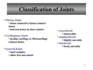
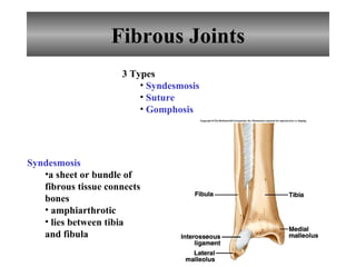
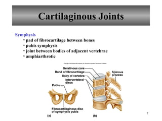
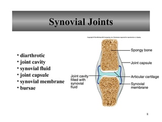
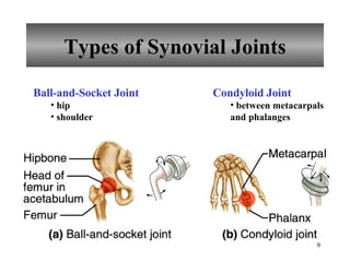
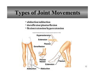
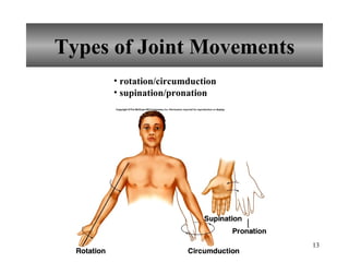
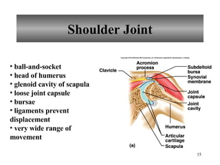
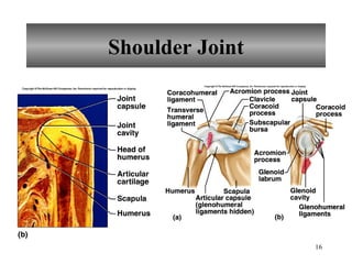
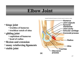
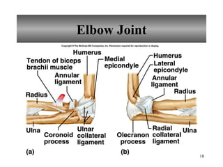
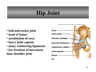
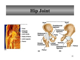
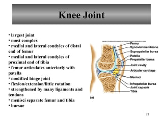
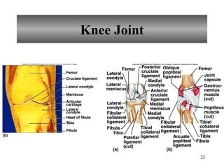
PowerPoint Lecture Outlines  to accompany Hole’s Human Anatomy and Physiology Eleventh Edition Shier    Butler    Lewis Chapter  8 Copyright © The McGraw-Hill Companies, Inc. Permission required for reproduction or display.
Chapter 8 Joints of the Skeletal System Articulations Functional junctions between bones Bind parts of skeletal system together Make bone growth possible Permit parts of the skeleton to change shape during childbirth Enable body to move in response to skeletal muscle contraction
Classification of Joints Fibrous Joints dense connective tissues connect bones between bones in close contact Cartilaginous Joints hyaline cartilage or fibrocartilage connect bones Synovial Joints most complex allow free movement synarthrotic immovable amphiarthrotic slightly movable diarthrotic freely movable
Fibrous Joints 3 Types Syndesmosis Suture Gomphosis Syndesmosis   a sheet or bundle of fibrous tissue connects bones amphiarthrotic lies between tibia and fibula
Fibrous Joints Suture between flat bones synarthrotic thin layer of connective tissue connects bones Gomphosis cone-shaped bony process in a socket tooth in jawbone synarthrotic
Cartilaginous Joints 2 Types Synchondrosis Symphysis Synchondrosis bands of hyaline cartilage unite bones epiphyseal plate (temporary) between manubrium and first rib synarthrotic
Cartilaginous Joints Symphysis pad of fibrocartilage between bones pubis symphysis joint between bodies of adjacent vertebrae amphiarthrotic
Synovial Joints diarthrotic joint cavity synovial fluid joint capsule synovial membrane bursae
Types of Synovial Joints Ball-and-Socket Joint hip shoulder Condyloid Joint between metacarpals and phalanges
Types of Synovial Joints Gliding Joint between carpals between tarsals Hinge Joint elbow between phalanges
Types of Synovial Joints Pivot Joint between proximal ends of radius and ulna Saddle Joint between carpal and metacarpal of thumb
Types of Joint Movements abduction/adduction dorsiflexion/plantarflexion flexion/extension/hyperextension
Types of Joint Movements rotation/circumduction supination/pronation
Types of Joint Movements eversion/inversion protraction/retraction elevation/depression
Shoulder Joint ball-and-socket head of humerus glenoid cavity of scapula loose joint capsule bursae ligaments prevent displacement very wide range of movement
Shoulder Joint
Elbow Joint hinge joint trochlea of humerus trochlear notch of ulna gliding joint capitulum of humerus head of radius flexion and extension many reinforcing ligaments stable joint
Elbow Joint
Hip Joint ball-and-socket joint head of femur acetabulum of coxa heavy joint capsule many reinforcing ligaments less freedom of movement than shoulder joint
Hip Joint
Knee Joint largest joint most complex medial and lateral condyles of distal end of femur medial and lateral condyles of proximal end of tibia femur articulates anteriorly with patella modified hinge joint flexion/extension/little rotation strengthened by many ligaments and tendons menisci separate femur and tibia bursae
Knee Joint
Life-Span Changes Joint stiffness is an early sign of aging Fibrous joints first to change; can strengthen over a lifetime Changes in symphysis joints of vertebral column diminish flexibility and decrease height  Synovial joints lose elasticity Disuse hampers the blood supply Activity and exercise can keep joints functional longer
Clinical Application Joint Disorders Sprains damage to cartilage, ligaments, or tendons associated with joints forceful twisting of joint Bursitis inflammation of a bursa overuse of a joint Arthritis inflamed, swollen, painful joints Rheumatoid Arthritis Osteoarthritis Gout

Skeletal System Lecture

  • 1.
    PowerPoint Lecture Outlines to accompany Hole’s Human Anatomy and Physiology Eleventh Edition Shier  Butler  Lewis Chapter 8 Copyright © The McGraw-Hill Companies, Inc. Permission required for reproduction or display.
  • 2.
    Chapter 8 Jointsof the Skeletal System Articulations Functional junctions between bones Bind parts of skeletal system together Make bone growth possible Permit parts of the skeleton to change shape during childbirth Enable body to move in response to skeletal muscle contraction
  • 3.
    Classification of JointsFibrous Joints dense connective tissues connect bones between bones in close contact Cartilaginous Joints hyaline cartilage or fibrocartilage connect bones Synovial Joints most complex allow free movement synarthrotic immovable amphiarthrotic slightly movable diarthrotic freely movable
  • 4.
    Fibrous Joints 3Types Syndesmosis Suture Gomphosis Syndesmosis a sheet or bundle of fibrous tissue connects bones amphiarthrotic lies between tibia and fibula
  • 5.
    Fibrous Joints Suturebetween flat bones synarthrotic thin layer of connective tissue connects bones Gomphosis cone-shaped bony process in a socket tooth in jawbone synarthrotic
  • 6.
    Cartilaginous Joints 2Types Synchondrosis Symphysis Synchondrosis bands of hyaline cartilage unite bones epiphyseal plate (temporary) between manubrium and first rib synarthrotic
  • 7.
    Cartilaginous Joints Symphysispad of fibrocartilage between bones pubis symphysis joint between bodies of adjacent vertebrae amphiarthrotic
  • 8.
    Synovial Joints diarthroticjoint cavity synovial fluid joint capsule synovial membrane bursae
  • 9.
    Types of SynovialJoints Ball-and-Socket Joint hip shoulder Condyloid Joint between metacarpals and phalanges
  • 10.
    Types of SynovialJoints Gliding Joint between carpals between tarsals Hinge Joint elbow between phalanges
  • 11.
    Types of SynovialJoints Pivot Joint between proximal ends of radius and ulna Saddle Joint between carpal and metacarpal of thumb
  • 12.
    Types of JointMovements abduction/adduction dorsiflexion/plantarflexion flexion/extension/hyperextension
  • 13.
    Types of JointMovements rotation/circumduction supination/pronation
  • 14.
    Types of JointMovements eversion/inversion protraction/retraction elevation/depression
  • 15.
    Shoulder Joint ball-and-sockethead of humerus glenoid cavity of scapula loose joint capsule bursae ligaments prevent displacement very wide range of movement
  • 16.
  • 17.
    Elbow Joint hingejoint trochlea of humerus trochlear notch of ulna gliding joint capitulum of humerus head of radius flexion and extension many reinforcing ligaments stable joint
  • 18.
  • 19.
    Hip Joint ball-and-socketjoint head of femur acetabulum of coxa heavy joint capsule many reinforcing ligaments less freedom of movement than shoulder joint
  • 20.
  • 21.
    Knee Joint largestjoint most complex medial and lateral condyles of distal end of femur medial and lateral condyles of proximal end of tibia femur articulates anteriorly with patella modified hinge joint flexion/extension/little rotation strengthened by many ligaments and tendons menisci separate femur and tibia bursae
  • 22.
  • 23.
    Life-Span Changes Jointstiffness is an early sign of aging Fibrous joints first to change; can strengthen over a lifetime Changes in symphysis joints of vertebral column diminish flexibility and decrease height Synovial joints lose elasticity Disuse hampers the blood supply Activity and exercise can keep joints functional longer
  • 24.
    Clinical Application JointDisorders Sprains damage to cartilage, ligaments, or tendons associated with joints forceful twisting of joint Bursitis inflammation of a bursa overuse of a joint Arthritis inflamed, swollen, painful joints Rheumatoid Arthritis Osteoarthritis Gout