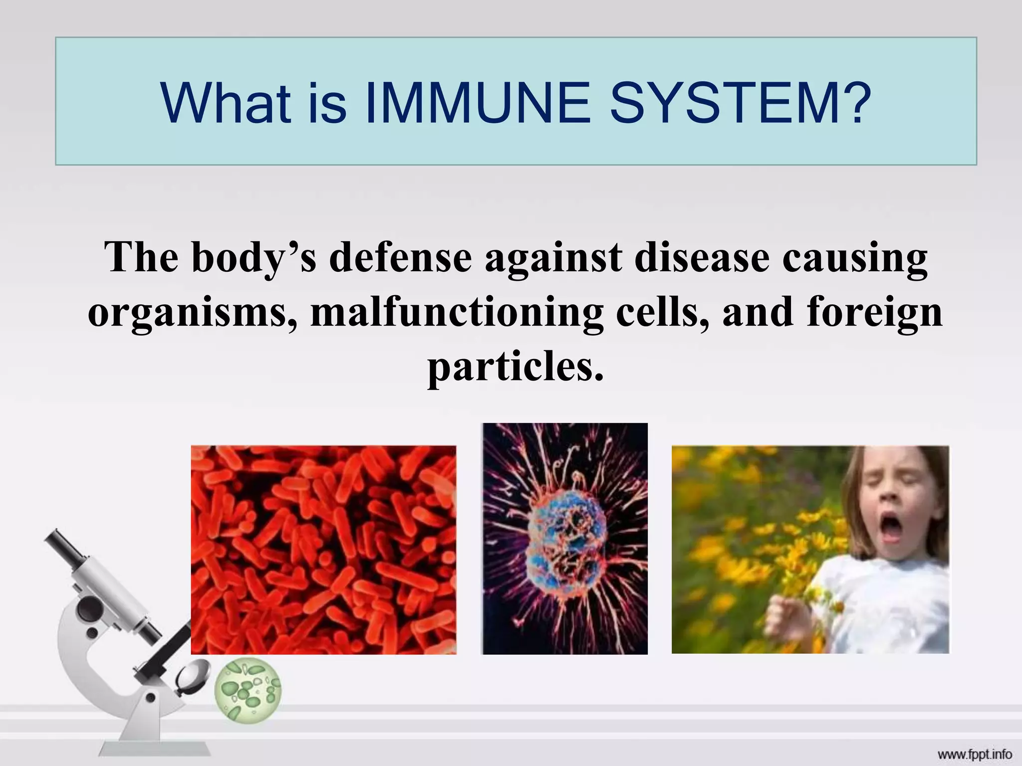
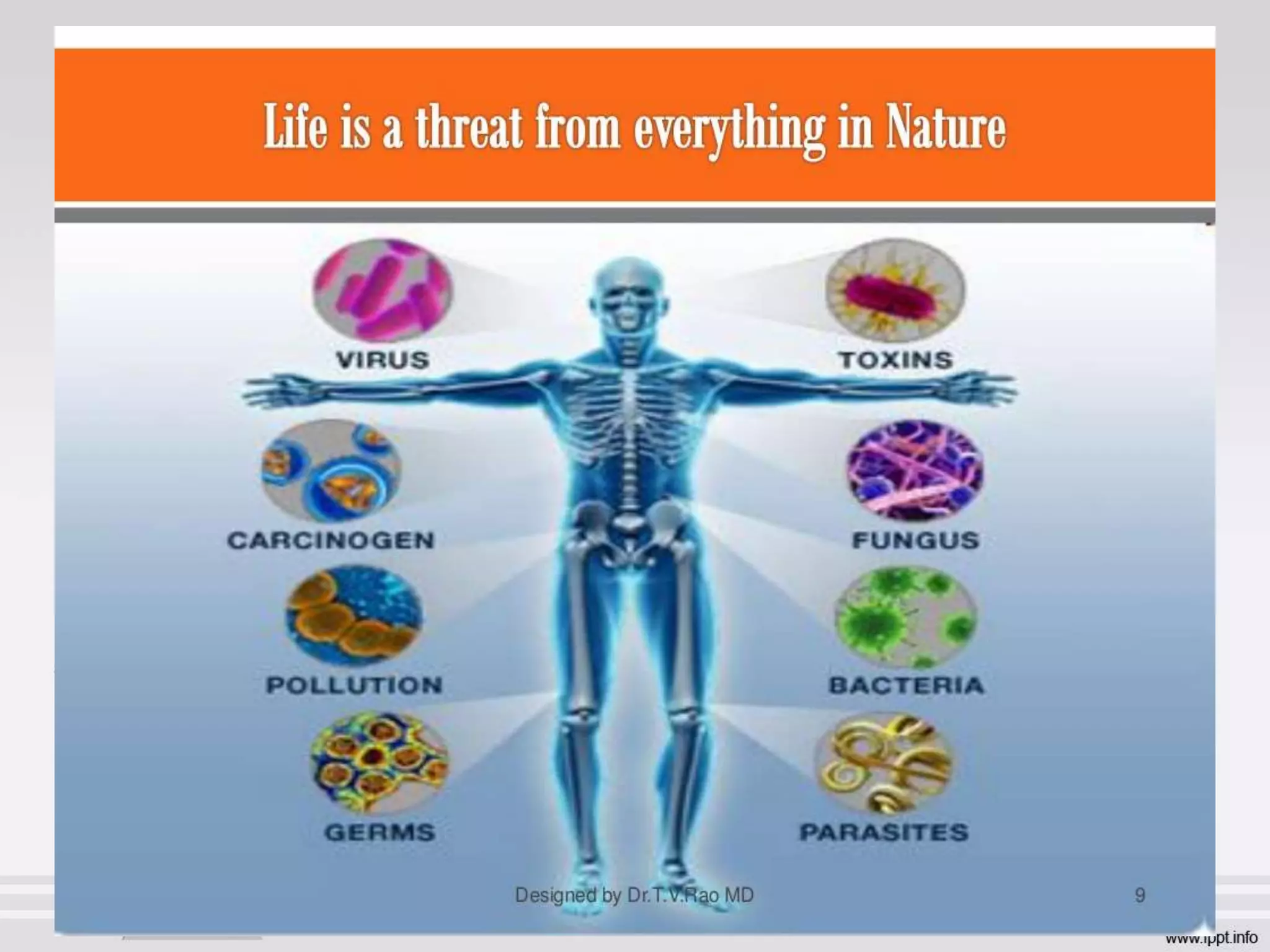
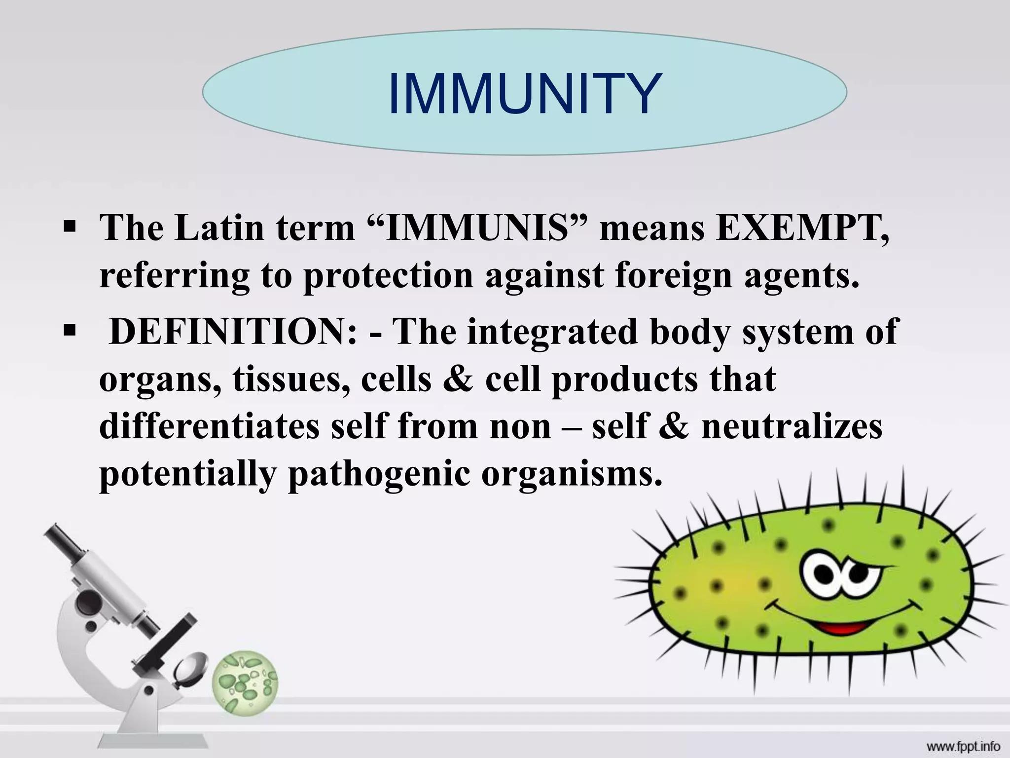
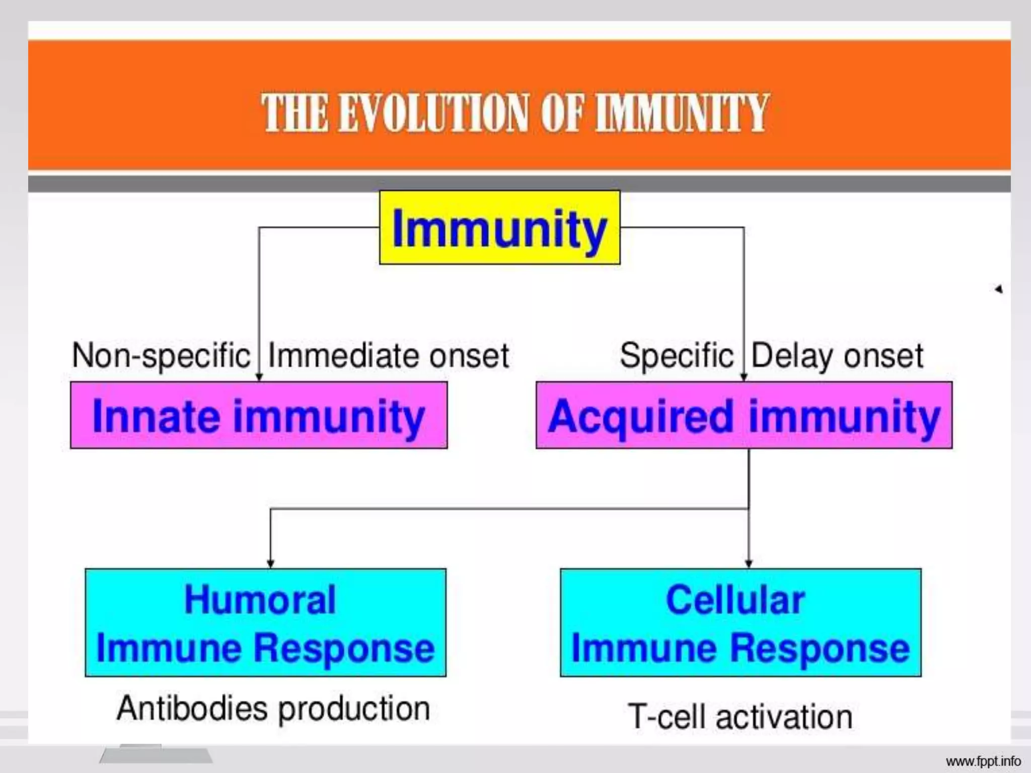
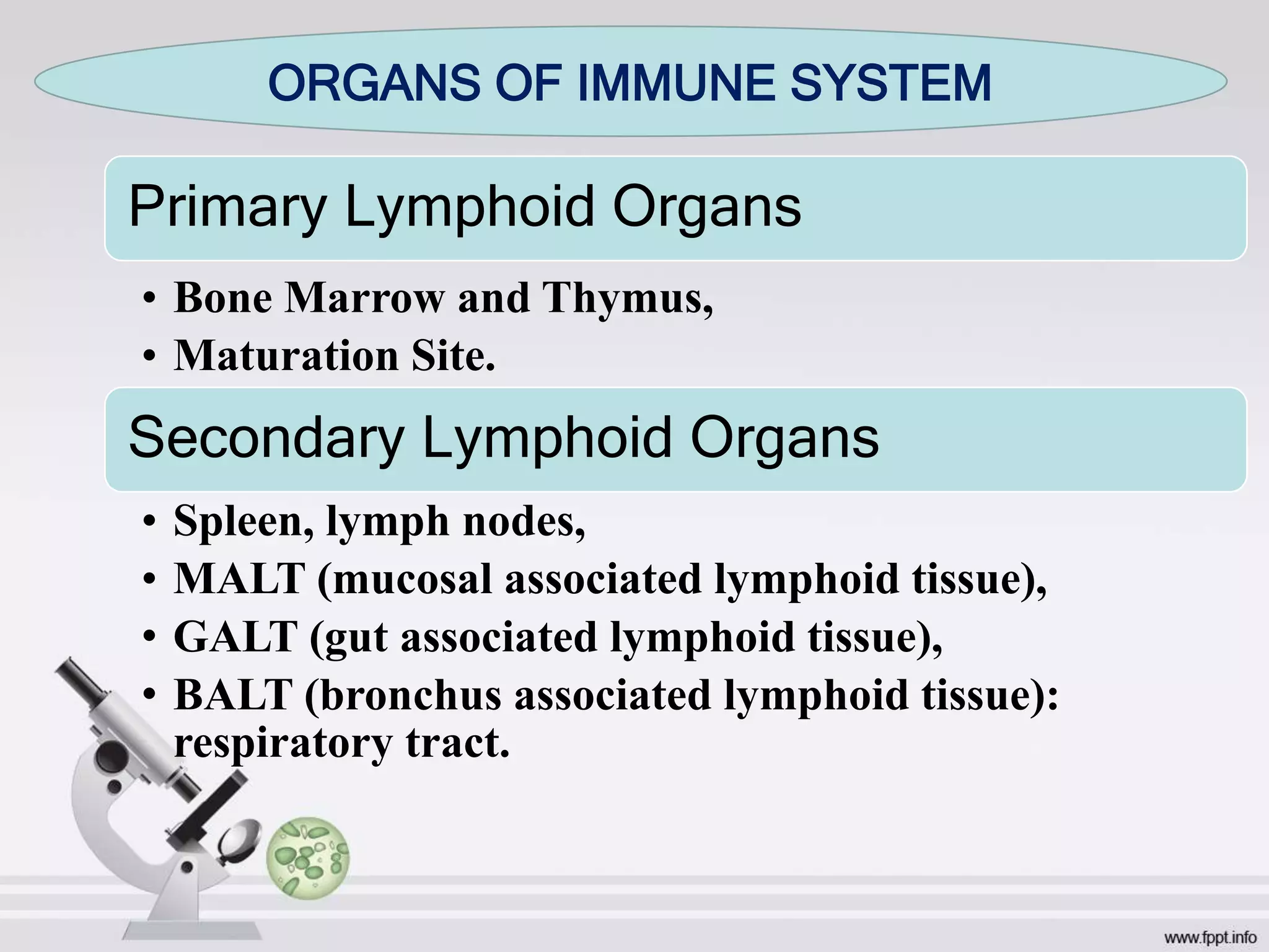
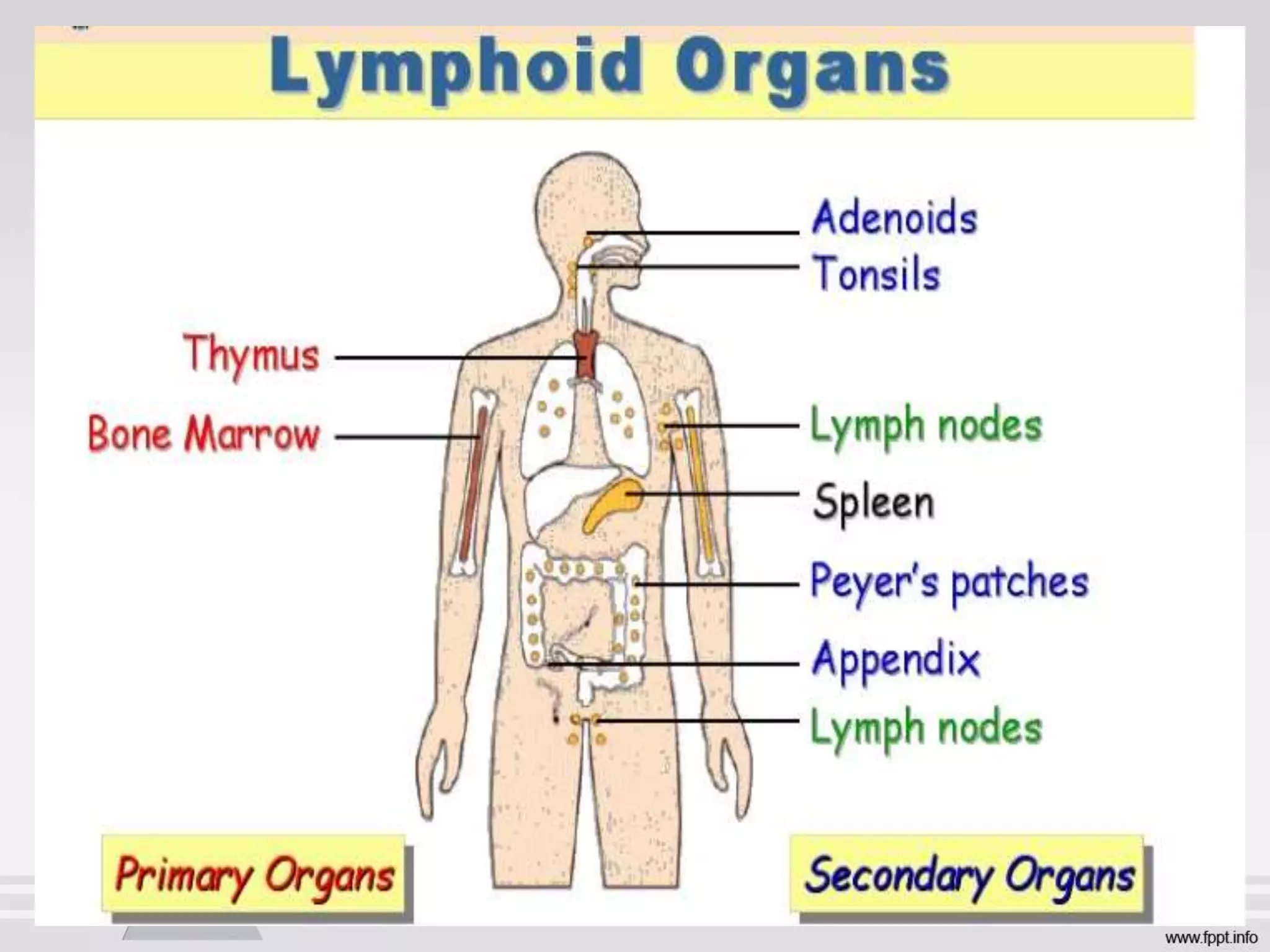
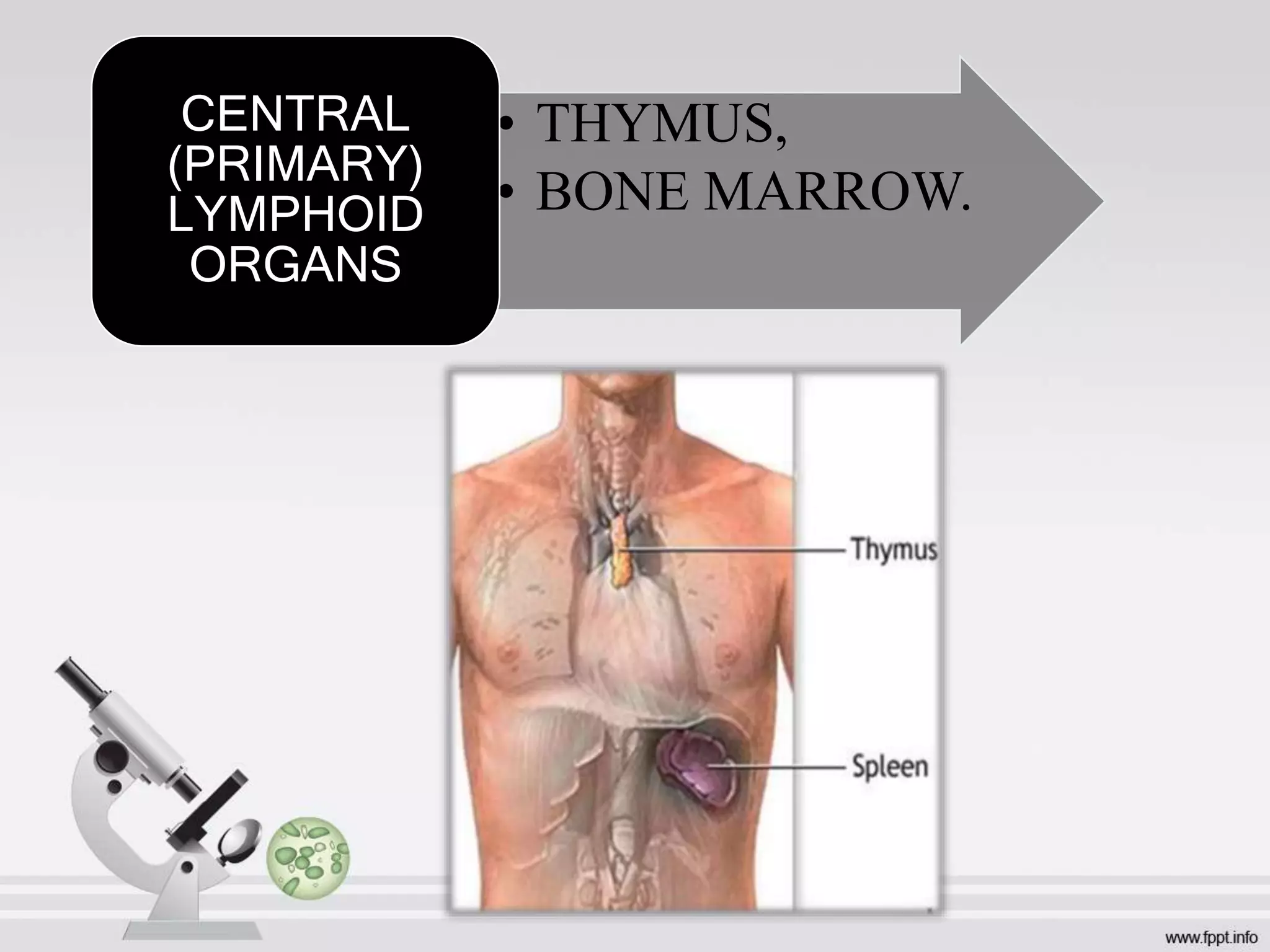
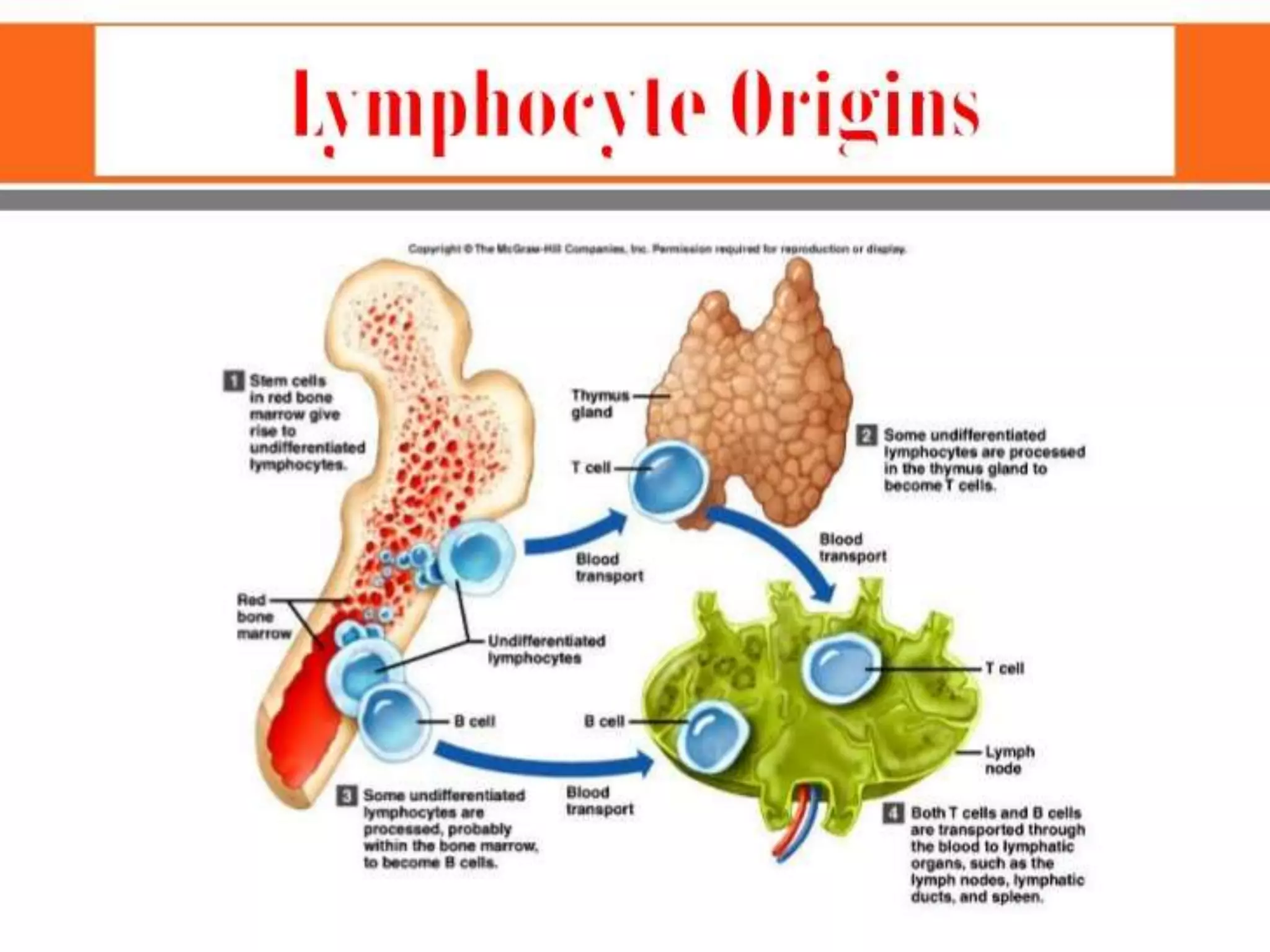
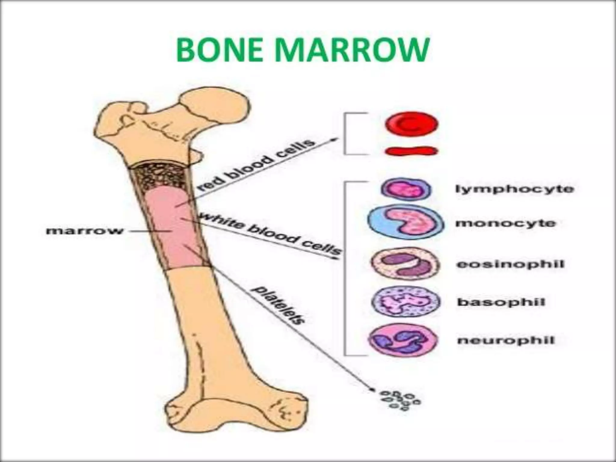
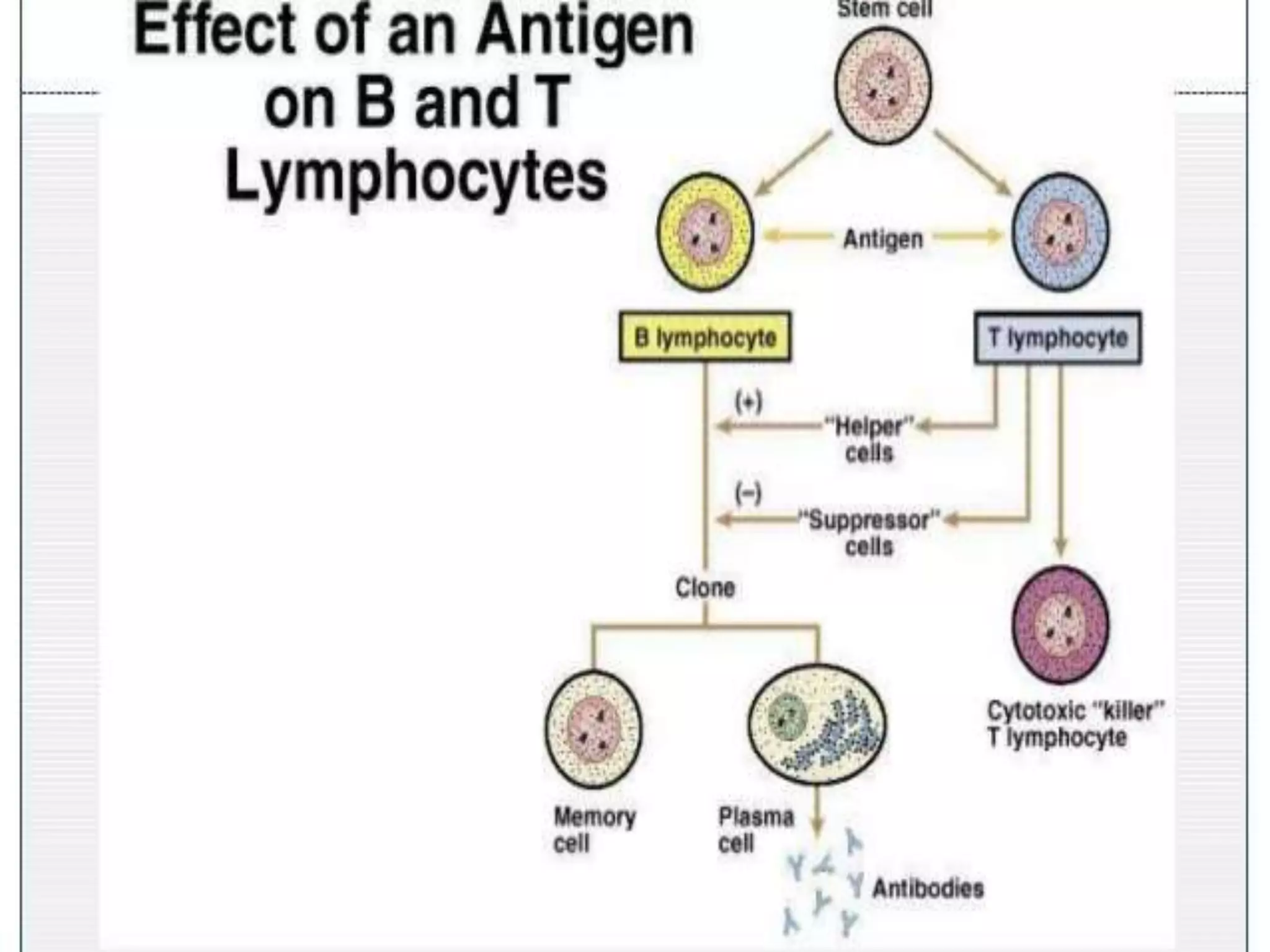
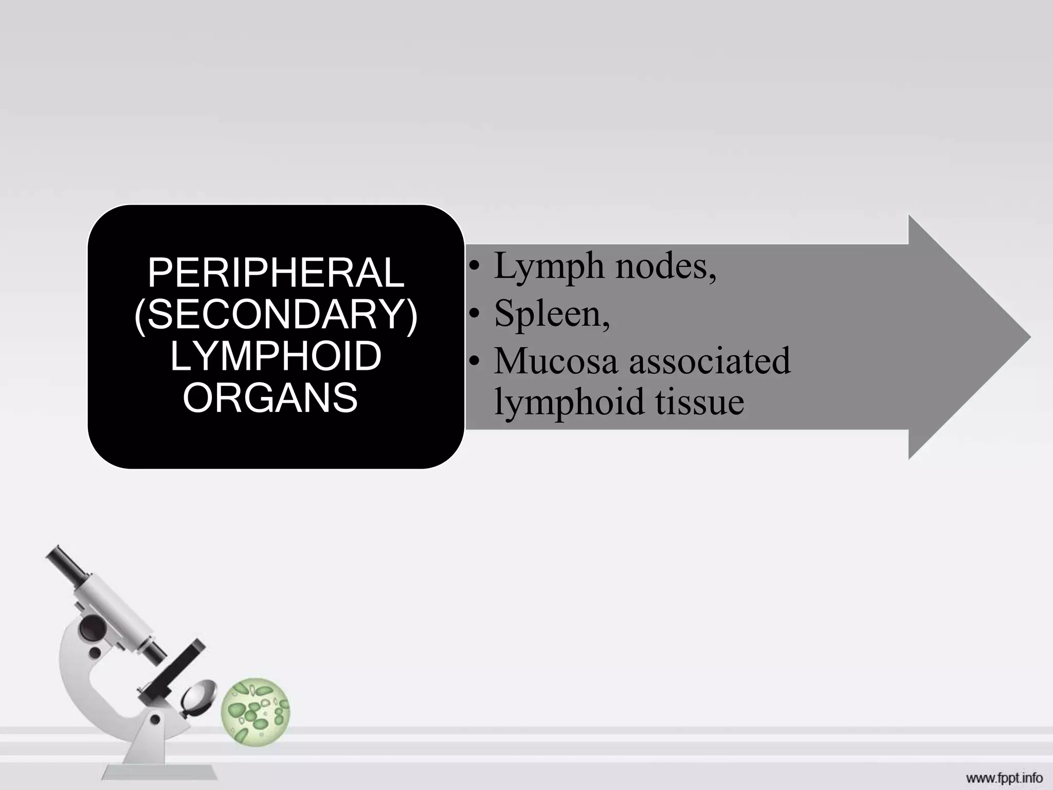
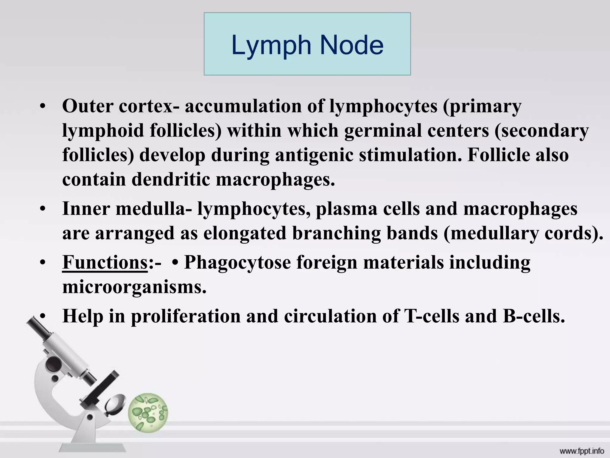
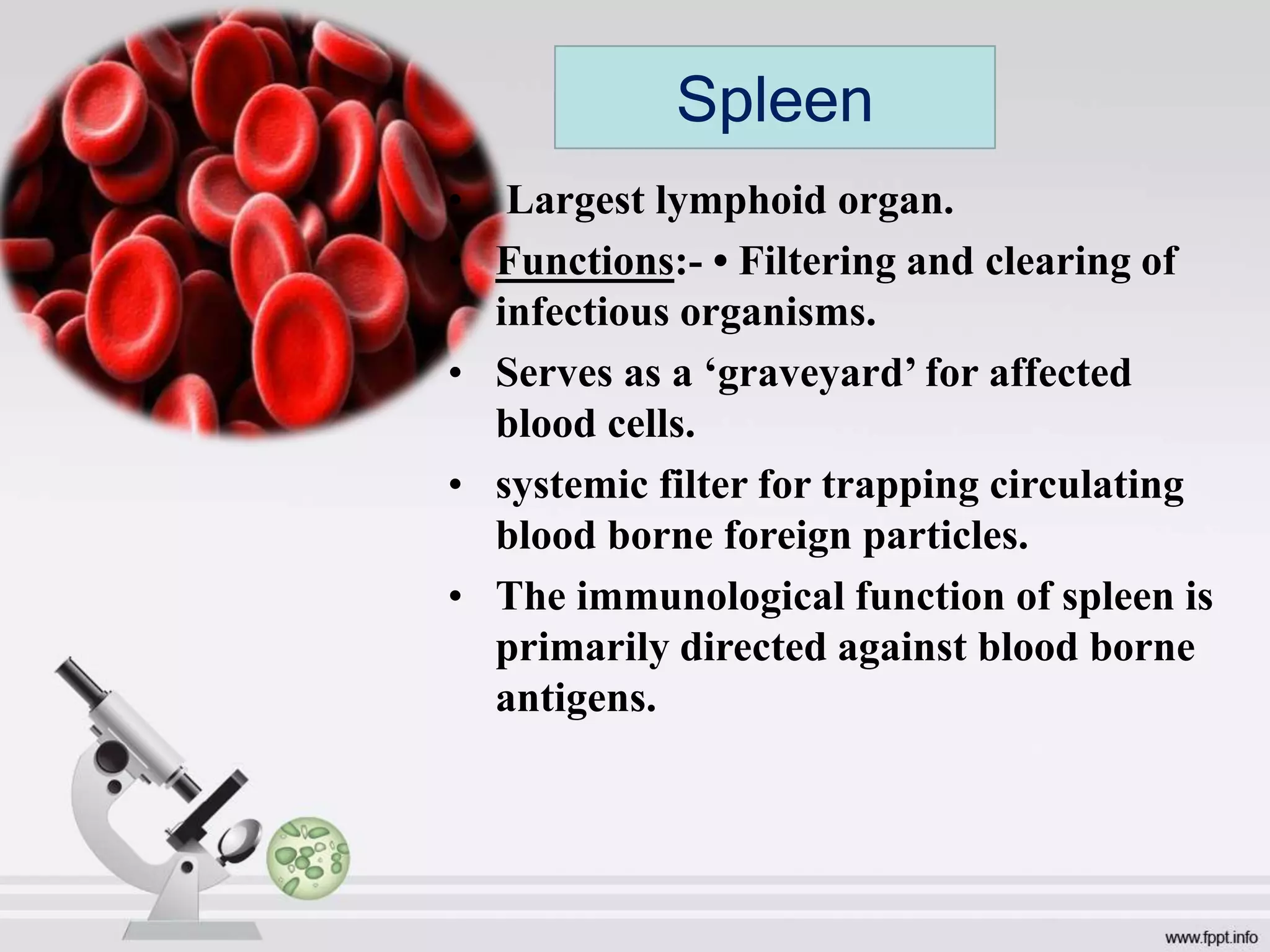
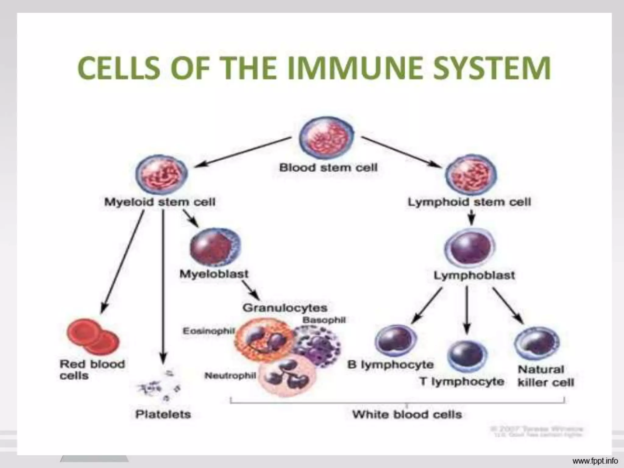
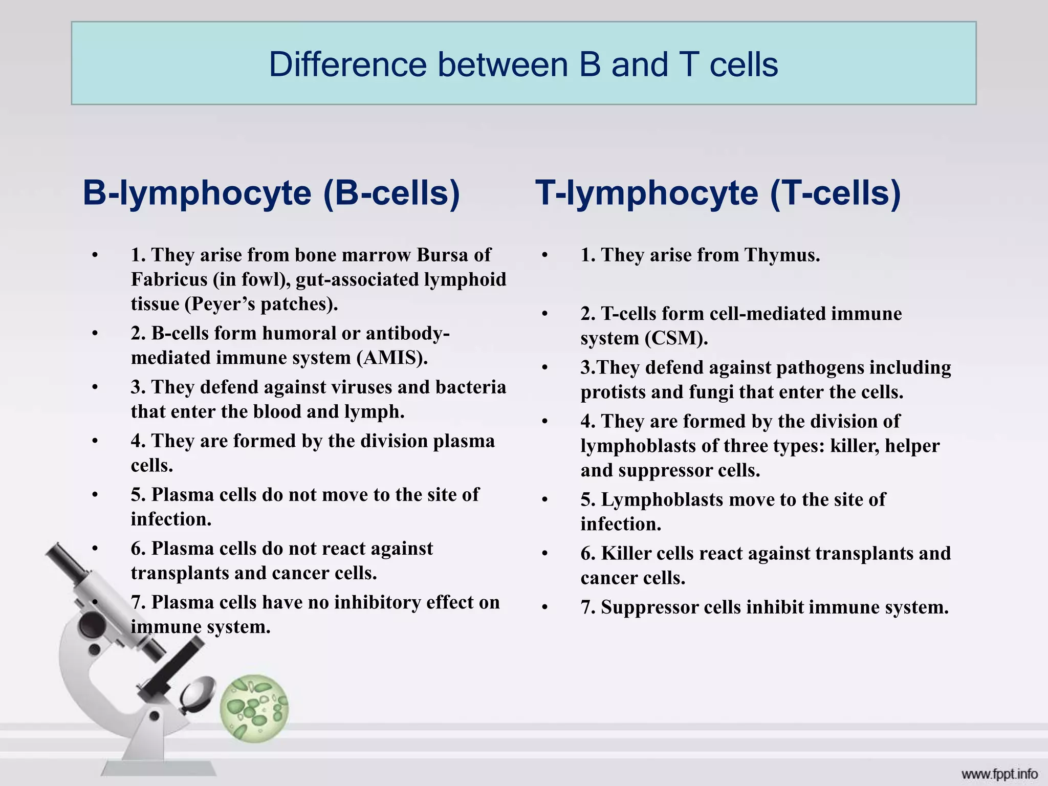
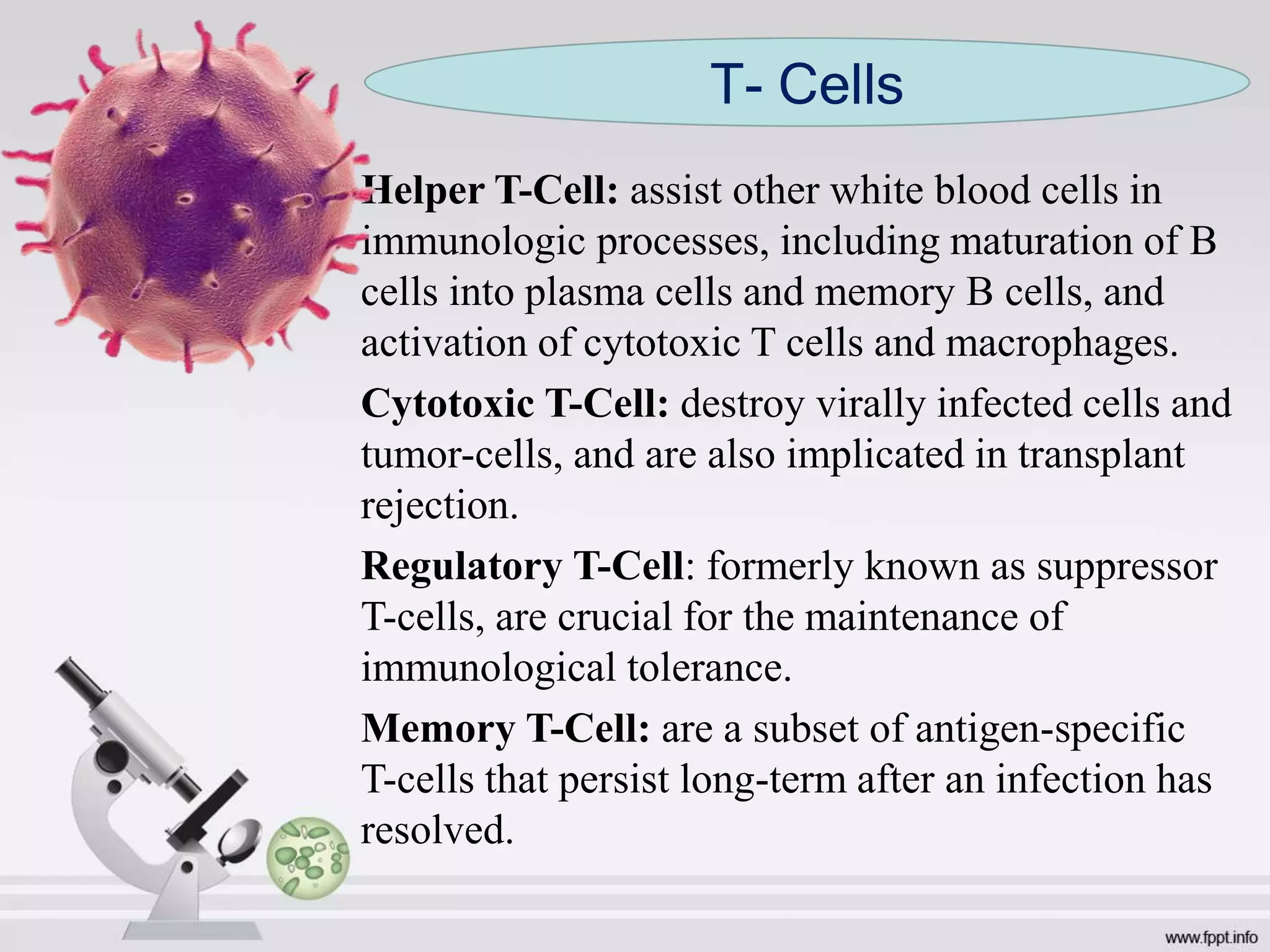
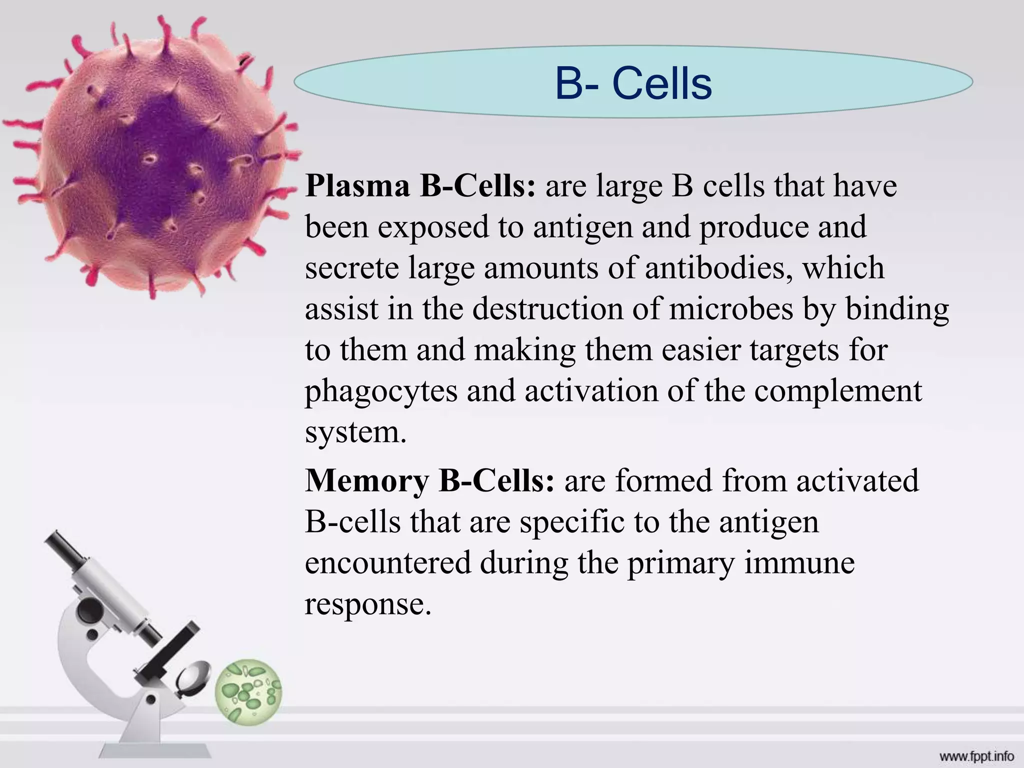
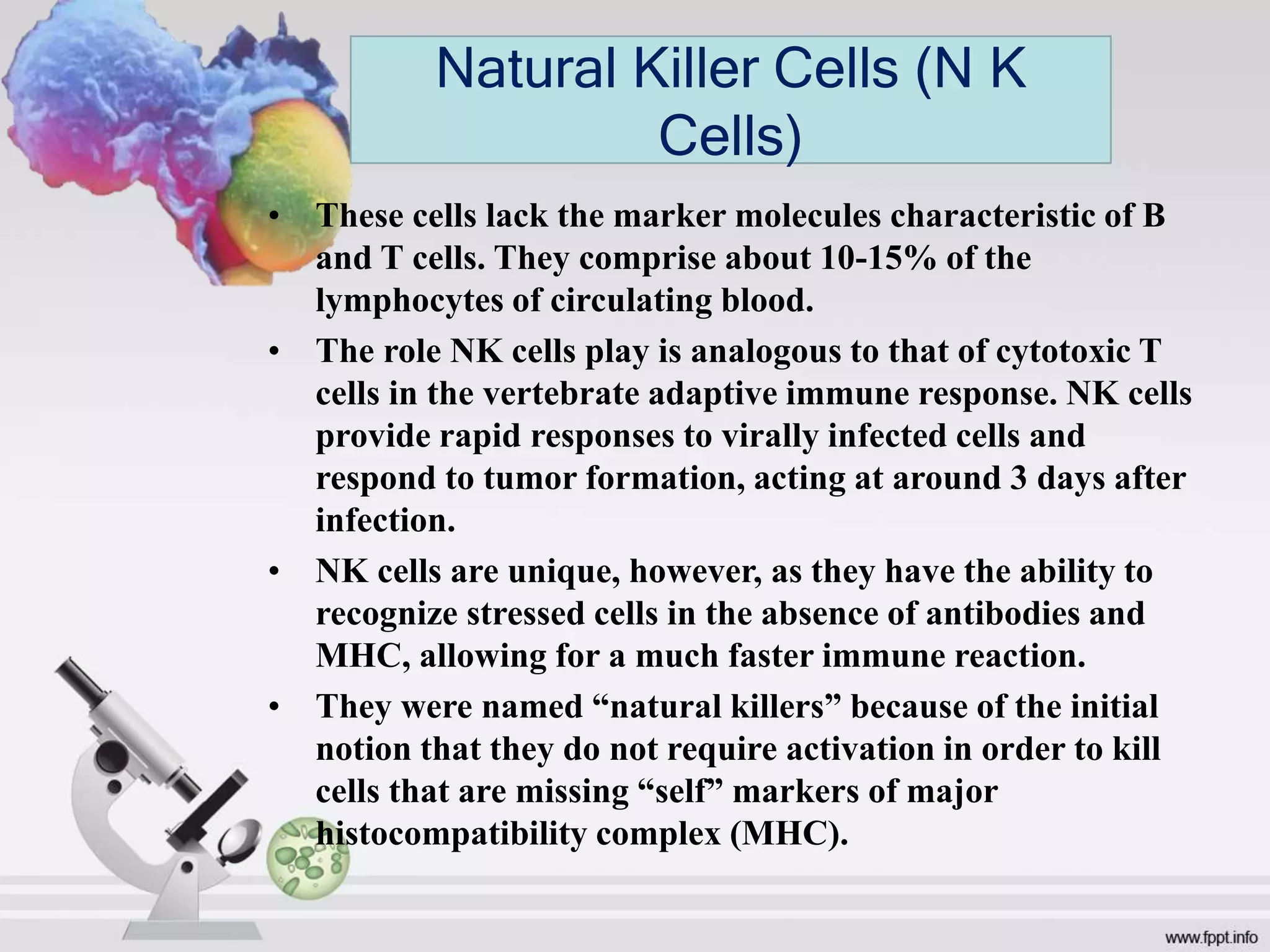
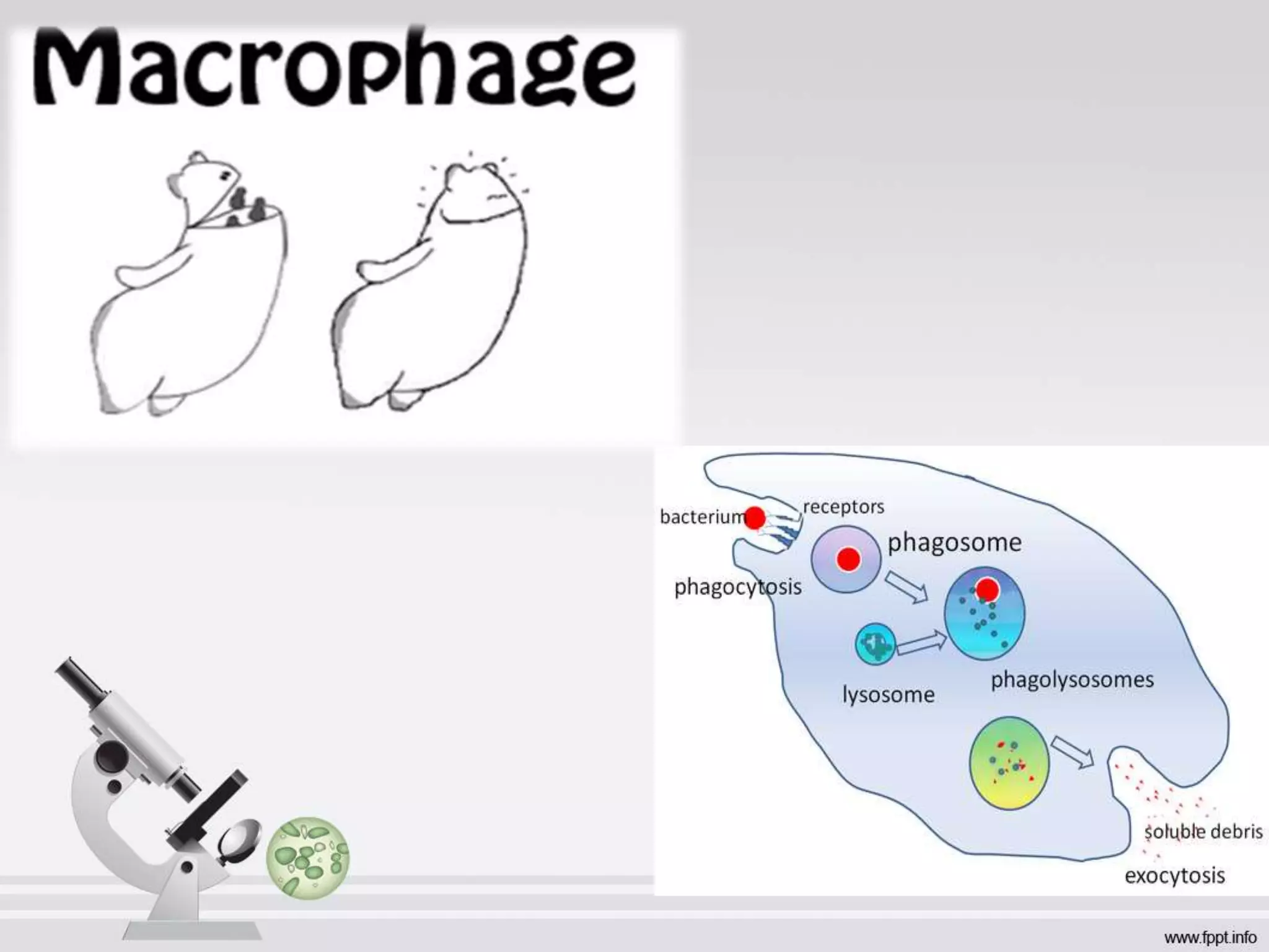
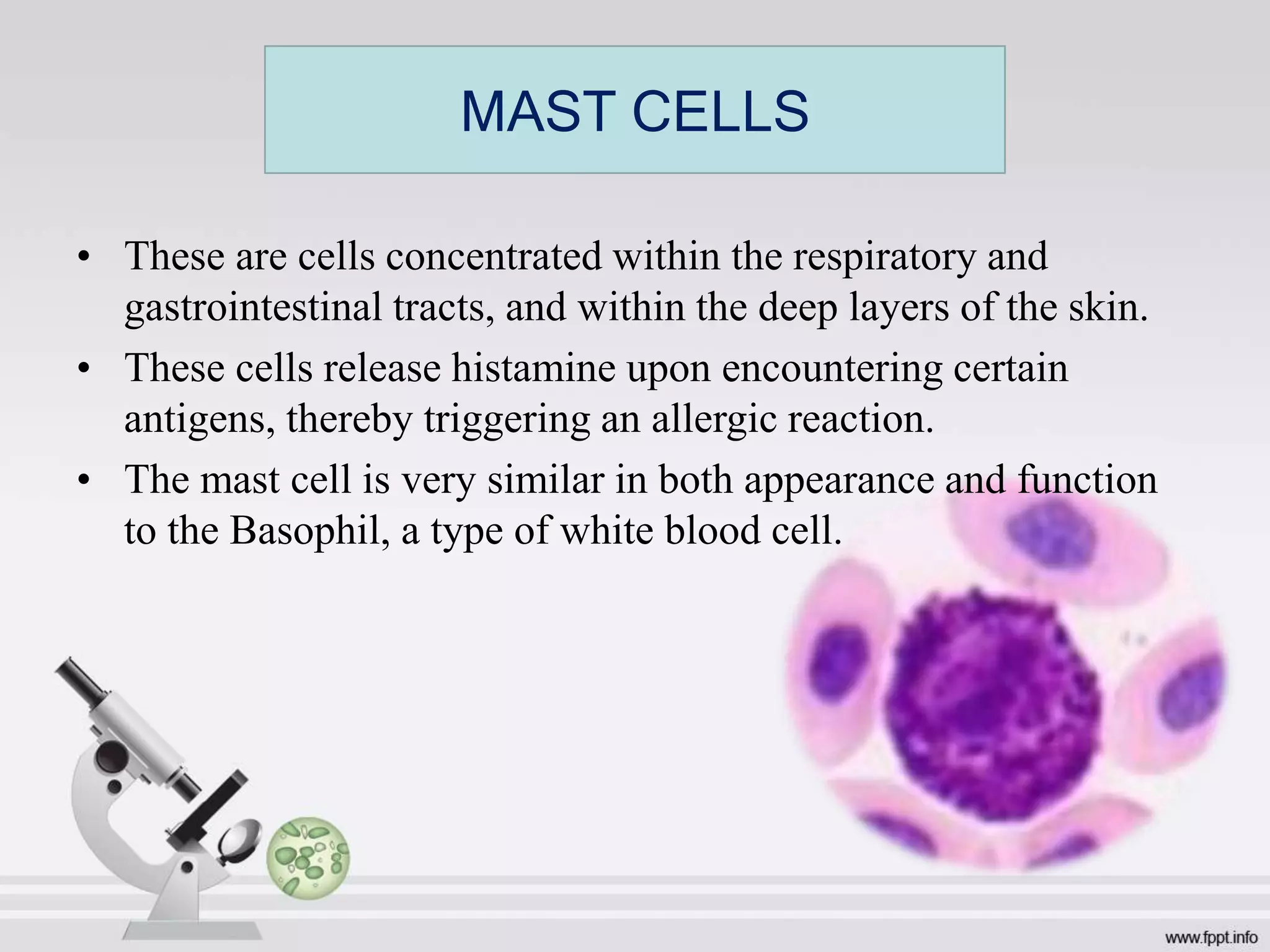
💎VVIP Kolkata Call Girls Parganas🩱7001035870🩱Independent Girl ( Ac Rooms Avai...
Structure and Functions of immune system- MICROBIOLOGY
1. Structure and functions of
immune system
Submitted by:
ANKUSHA ARORA
(Saraswati Dental College,
Lucknow)
2. The body’s defense against disease causing
organisms, malfunctioning cells, and foreign
particles.
What is IMMUNE SYSTEM?
3.
4. The Latin term “IMMUNIS” means EXEMPT,
referring to protection against foreign agents.
DEFINITION: - The integrated body system of
organs, tissues, cells & cell products that
differentiates self from non – self & neutralizes
potentially pathogenic organisms.
IMMUNITY
9. • Thymus is the site where a large diversity of T cells
are produced – (so they can recognize and act
against a large number of antigen)
• Thymus induces the death of those T cells that
cannot recognize antigen.
• Also induces death of those T cells that react with
self- Ag.
• More than 90% of all thymocytes die by apoptosis in
the thymus without ever reaching maturity.
THYMUS
10. • Primary function:- production of thymic lymphocytes.
• A major organ for proliferation of lymphocytes in body.
• Plays key role in determining the differentiation of T cell.
• Lymphocytes during maturation acquire new surface
antigens (Thy Ag)& are called as T lymphocytes or T cells
(thymus dependent).
• Lymphocyte proliferation in thymus is not dependent on
antigenic stimulus (unlike in peripheral lymphoid organs).
• T lymphocytes –primarily responsible for Cell-Mediated
Immunity.
Functions of thymus
11.
12. • Some lymphocytes originate, develop, & mature within the
bone marrow – B cells. (B for Bursa of Fabricus or bone
marrow).
• Site for proliferation of stem cells.
• Site for the origin of pre-B cells & their maturation to
become immunoglobulin producing lymphocytes
(PLASMA CELL).
• B lymphocytes are transformed into plasma cells & secrete
antibodies, & are primarily responsible for antibody-
mediated immunity.
BONE MARROW
16. • Outer cortex- accumulation of lymphocytes (primary
lymphoid follicles) within which germinal centers (secondary
follicles) develop during antigenic stimulation. Follicle also
contain dendritic macrophages.
• Inner medulla- lymphocytes, plasma cells and macrophages
are arranged as elongated branching bands (medullary cords).
• Functions:- • Phagocytose foreign materials including
microorganisms.
• Help in proliferation and circulation of T-cells and B-cells.
Lymph Node
17. • Largest lymphoid organ.
• Functions:- • Filtering and clearing of
infectious organisms.
• Serves as a ‘graveyard’ for affected
blood cells.
• systemic filter for trapping circulating
blood borne foreign particles.
• The immunological function of spleen is
primarily directed against blood borne
antigens.
Spleen
18. • Depends on the age at which the spleen is removed.
• In children, splenectomy often leads to an increased
incidence of bacterial sepsis caused primarily by
Streptococcus pneumoniae, Neisseria meningitidis, and
Haemophilus influenzae.
• Splenectomy in adults has less adverse effects, although in
some, it makes the host more susceptible to blood-borne
bacterial infection.
EFFECTS OF SPLENECTOMY
19. • The mucosa lining the alimentary, respiratory and other
lumina are endowed with a rich collection of lymphoid cell
aggregates like the Peyer’s patches or scattered isolated
follicles- collectively called MALT.
• MALT contains lymphoid as well as phagocytic cells.
• Both B and T cells are present.
• While the predominant immunoglobulin produced in the
mucosa is secretory IgA, other immunoglobulin classes,
IgG, IgM and IgE are also formed locally.
MUCOSA ASSOCIATED LYMPHOID TISSUE (MALT)
20.
21. B-lymphocyte (B-cells)
• 1. They arise from bone marrow Bursa of
Fabricus (in fowl), gut-associated lymphoid
tissue (Peyer’s patches).
• 2. В-cells form humoral or antibody-
mediated immune system (AMIS).
• 3. They defend against viruses and bacteria
that enter the blood and lymph.
• 4. They are formed by the division plasma
cells.
• 5. Plasma cells do not move to the site of
infection.
• 6. Plasma cells do not react against
transplants and cancer cells.
• 7. Plasma cells have no inhibitory effect on
immune system.
T-lymphocyte (T-cells)
• 1. They arise from Thymus.
• 2. T-cells form cell-mediated immune
system (CSM).
• 3.They defend against pathogens including
protists and fungi that enter the cells.
• 4. They are formed by the division of
lymphoblasts of three types: killer, helper
and suppressor cells.
• 5. Lymphoblasts move to the site of
infection.
• 6. Killer cells react against transplants and
cancer cells.
• 7. Suppressor cells inhibit immune system.
Difference between B and T cells
22. Helper T-Cell: assist other white blood cells in
immunologic processes, including maturation of B
cells into plasma cells and memory B cells, and
activation of cytotoxic T cells and macrophages.
Cytotoxic T-Cell: destroy virally infected cells and
tumor-cells, and are also implicated in transplant
rejection.
Regulatory T-Cell: formerly known as suppressor
T-cells, are crucial for the maintenance of
immunological tolerance.
Memory T-Cell: are a subset of antigen-specific
T-cells that persist long-term after an infection has
resolved.
T- Cells
23. Plasma B-Cells: are large B cells that have
been exposed to antigen and produce and
secrete large amounts of antibodies, which
assist in the destruction of microbes by binding
to them and making them easier targets for
phagocytes and activation of the complement
system.
Memory B-Cells: are formed from activated
B-cells that are specific to the antigen
encountered during the primary immune
response.
B- Cells
24. • These cells lack the marker molecules characteristic of B
and T cells. They comprise about 10-15% of the
lymphocytes of circulating blood.
• The role NK cells play is analogous to that of cytotoxic T
cells in the vertebrate adaptive immune response. NK cells
provide rapid responses to virally infected cells and
respond to tumor formation, acting at around 3 days after
infection.
• NK cells are unique, however, as they have the ability to
recognize stressed cells in the absence of antibodies and
MHC, allowing for a much faster immune reaction.
• They were named “natural killers” because of the initial
notion that they do not require activation in order to kill
cells that are missing “self” markers of major
histocompatibility complex (MHC).
Natural Killer Cells (N K
Cells)
25. • These are group of diverse cell types that assists other
cells in the Immune Response.
• Cells which do not have antigen-specific receptors.
Instead, they capture and process antigens, present
them to T cell receptors.
• These cells include Macrophages, B Cells and
Dentritic cells.
Antigen- Presenting Cells
26. • These cells are immune cells forming part of the Mammalian Immune System.
• Their main function is to process antigen material and present it on the surface to other cells
of the immune system.
• They act as messengers between the innate and adaptive immunity.
• At certain development stages they grow branched projections, the dendrites.
• While similar in appearance, these are distinct structures from the dendrites of neurons.
Immature dendritic cells are also called veiled cells, as they possess large cytoplasmic “veils”
rather than dendrites.
• Dendritic cells are present in tissues in contact with the external environment, such as the skin
and the inner lining of the nose, lungs, stomach and intestines. They can also be found in an
immature state in the blood.
• Once activated, they migrate to the lymph nodes where they interact with T cells and B cells
to initiate and shape the adaptive immune response.
Dendritic Cell
27. • Macrophages are cells produced by the differentiation of monocytes in
tissues. It function in both non-specific defense, Innate Immunity, as well as
help initiate specific defense mechanisms, Adaptive Immunity, of vertebrate
animals.
• Their role is to ”phagocytose” (engulf and then digest) cellular debris and
pathogens, either as stationary or as mobile cells.
• They also stimulate lymphocytes and other immune cells to respond to
pathogens. They are specialized phagocytic cells that attack foreign
substances, infectious microbes and cancer cells through destruction and
ingestion.
Macrophages
28.
29. • These are cells concentrated within the respiratory and
gastrointestinal tracts, and within the deep layers of the skin.
• These cells release histamine upon encountering certain
antigens, thereby triggering an allergic reaction.
• The mast cell is very similar in both appearance and function
to the Basophil, a type of white blood cell.
MAST CELLS