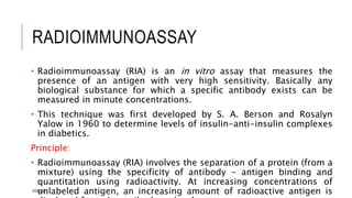
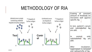
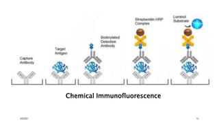
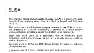
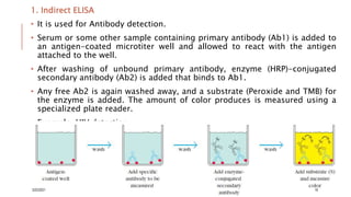
IMMUNOLOGICAL
TECHNIQUES
Nidhi S.
Dept. of Microbiology
3/22/2021 1
RADIOIMMUNOASSAY
• Radioimmunoassay (RIA) is an in vitro assay that measures the
presence of an antigen with very high sensitivity. Basically any
biological substance for which a specific antibody exists can be
measured in minute concentrations.
• This technique was first developed by S. A. Berson and Rosalyn
Yalow in 1960 to determine levels of insulin-anti-insulin complexes
in diabetics.
Principle:
• Radioimmunoassay (RIA) involves the separation of a protein (from a
mixture) using the specificity of antibody - antigen binding and
quantitation using radioactivity. At increasing concentrations of
unlabeled antigen, an increasing amount of radioactive antigen is
3/22/2021 2
Radioisotopes used in RIA:
1. Beta emitters – H-3, C-14, O-15, N-13 and F-18 (useful for in-
vitro experiments)
2. Gamma emitters – I-125, I-123 (useful for in-vivo imaging)
In all of them I-125 is mostly used because-
a) Shelf life for labelled Ag is long (half life is 60.2 days).
b) Natural constituent of thyroxine and tri-iodothyronine.
c) Easily introduced into peptide molecules, steroids.
d) Gamma radiation emission permits the use of simple radioactivity
counting chamber.
3/22/2021 3
METHODOLOGY OF RIA
Test Contr
ol
Coating of constant
amount of Antibody on
microtitre well against
specific Ag
In test, patient’s serum
and radiolabelled Ag
are add.
In control, only
radiolabelled Ag are
add.
After incubation,
supernatant is removed
and radioactivity of Ag-
3/22/2021 4
ADVANTAGES & DISADVANTAGES
OF RIA
Advantages:
1. Radio immuno assay is very
sensitive technique used to
measure concentrations of
antigen without the need to use
a bioassay. It can measure one
trillionth (10-12) of a gram of
material per milliliter of blood.
2. It is structurally specific as
antigen: antibody reaction are
highly specific.
3. It is indirect method of analysis.
4. It is a saturation analysis as
active reagent added in smaller
Disadvantages:
1. Prolonged reaction time (in days)
as a consequence highly diluted
reagent is used.
2. Radioactive Iodine used in is not a
cheap reagent.
3. Possible health hazards due to
handling of radioisotopes.
4. All the reagents must be added
precisely.
5. Limited assay range.
6. Lack of direct linear relationship
between analyte concentration
and signal response.
7. Difficulty of automation.
3/22/2021 5
APPLICATIONS
1. Narcotics (drug) detection,
2. Blood bank screening for the hepatitis (a highly contagious
condition) virus,
3. Early cancer detection,
4. Measurement of growth hormone levels,
5. Tracking of the leukemia virus,
6. Diagnosis and treatment of peptic ulcers,
7. Research with brain chemicals called neurotransmitter
3/22/2021 6
ROCKET
ELECTROIMMUNODIFFUSION
• Rocket Immunoelectrophoresis, also known as electro-
immunodiffusion, is a simple, quick and reproducible method for
determining the concentration of antigen in an unknown sample.
• This quantitative one dimensional immunoelectrophoresis
method involves a comparison of antigen sample of unknown
concentration with a series of dilutions of a known concentration
of the antigen and requires a monospecific antibody against the
antigen under investigation.
• In this method, antigen migrates from the well through agarose
gel containing antiserum, forming rocket shaped precipitin peaks.
The height of this peak is proportional to the concentration of the
antigen loaded in the corresponding well.
3/22/2021 7
Principle: In Rocket Immunoelectrophoresis, negatively charged antigen
samples are electrophoresed in an agarose gel containing antibody
which is specific to that antigen.
As the antigen moves out of the well and enters the agarose gel, it
combines with the antibody to form immune complex which is visible as
white precipitin arcs.
Because the antigen is migrated through the gel under the influence of
an applied electric current, it moves in one direction.
In initial antigen is in excess over antibody so that no visible
precipitation occurs, as the antigen sample migrates further through the
agarose gel, more antibody molecules are encountered that interact with
the antigen to form immune complex and when this immune complexes
become large enough to be retained within the gel, movement of the
antigen stops.
The area of precipitin has the shape of a rocket and its height is
proportional to the concentration of antigen in the corresponding well.
3/22/2021 8
3/22/2021 9
Applications:
1. Rocket electrophoresis is used mainly for quantitative estimation
of antigen in the serum.
2. The method has been used for quantitation of human serum
proteins before automated methods became available.
3. Determining the concentration of a specific protein in a protein
mixture.
4. In estimation of immunoglobulin protease activity.
5. Studies dealing with antigenic relationships between organisms.
3/22/2021 10
CHEMICAL
IMMUNOFLUORESCENCE/IMMUNOA
SSAY
• Chemiluminescence refers to the emission of light as a result of an
electrical, biochemical, or chemical reaction .
• Some organic compounds become exited when oxidized and emit
light as they revert to the ground state.
• Chemiluminescence immunoassay are most sensitive immunoassays
with detection limits as low as attomole (10-18) or zeptomole (10-21)
level .
• Chemiluminescence can be applied as direct label or
chemiluminescent compound can be used as a substrate for an
enzyme (HRP)-labeled immunoreactant (Ab).
• HRP reacts with Hydrogen peroxide and produces nescent oxygen
which reacts with luminol and produces light.
3/22/2021 11
Most common chemiluminescent compounds are:
Luminol:
• It is the first chemiluminescent label used in immunoassays; it emits
light energy under alkaline conditions in the presence of peroxide
and peroxidase.
• Because peroxidase can serve as the catalyst, assays may use this
enzyme as the label; the chemiluminogenic substrate, luminol, will
produce light that is directly proportional to the amount of
peroxidase present.
Luminol + 2H2O2 + OH- 3-aminophthalate + light
( 425 nm )
Principle:
In the presence of complimentary antigen and antibody, the paratope
of the antibody binds to the epitope of the antigen to form an
antigen-antibody or an immune complex. Estimating the levels of
such immune complex by use of labeled antibodies form the basis of
Peroxidas
e
3/22/2021 12
Chemical Immunofluorescence
3/22/2021 13
Applications:
1. Detection of Hormones: insulin, thyroxin, estradiol, testosterone,
progesterone.
2. Detection of Vitamin: vit B12
3. Detection of tumor markers: bone morphogenic protein-2, carcino
embryonic antigen (CEA), alpha fetoprotein (AFP)
4. Detection of human beta chorionic gonadotropin, C-reactive protein and
Tumor necrosis factor
3/22/2021 14
ELISA
The enzyme-linked immunosorbent assay (ELISA) is a commonly used
analytical biochemistry assay, first described by Engvall and Perlmann
in 1971.
The assay uses a solid-phase enzyme immunoassay (EIA) to detect
the presence of a ligand (commonly a protein) in a liquid sample
using antibodies directed against the protein to be measured.
ELISA has been used as a diagnostic tool in medicine, plant
pathology, and biotechnology, as well as a quality control check in
various industries.
Enzymes used in ELISA are HRP (Horse Redish Peroxidase), Alkaline
phosphatase etc.
It is mainly are of 3 types: Direct, Sandwich and competitive.
3/22/2021 15
1. Indirect ELISA
• It is used for Antibody detection.
• Serum or some other sample containing primary antibody (Ab1) is added to
an antigen-coated microtiter well and allowed to react with the antigen
attached to the well.
• After washing of unbound primary antibody, enzyme (HRP)-conjugated
secondary antibody (Ab2) is added that binds to Ab1.
• Any free Ab2 is again washed away, and a substrate (Peroxide and TMB) for
the enzyme is added. The amount of color produces is measured using a
specialized plate reader.
• Example: HIV detection.
3/22/2021 16
2. Sandwich ELISA
• Antigen can be detected or measured by a sandwich ELISA.
• In this technique, the antibody is immobilized on a microtiter well.
A sample containing unknown amounts of antigen is allowed to
react with the immobilized antibody.
• After wells washing, a second enzyme (HRP) -linked antibody
(specific to Fc region) is added and allowed to react with the bound
antigen.
• After washing, substrate (peroxide and TMB) is added, and the
colored reaction product is measured.
3/22/2021 17
3. Competitive ELISA:
• In this technique, antibody is first incubated in solution with a sample
containing antigen, then antigen-antibody mixture is added to an
antigen-coated microtiter well.
• The more antigen present in the initial solution-phase sample, the
less free antibody will be available to bind to the antigen-coated well.
• After washing off the unbound antibody, an enzyme (HRP)-conjugated
Ab2 specific for the Ab1 can be added to determine the amount of
Ab1 bound to the well.
• In the competitive assay, the higher the concentration of antigen in
the original sample, the lower the final signal.
3/22/2021 18
ALLERGEN TEST
Allergy testing can help confirm or rule out allergies and
consequently reduce adverse reactions and limit unnecessary
avoidance and medications.
A positive allergy test indicates a potential readiness to develop an
allergic reaction upon exposure to a specific allergen, but this does
not mean that a clinically significant reaction would necessarily occur.
Increasingly strong allergic tests may correlate with increased disease
activity.
Allergic (Hypersensitivity) Reactions include 4 main types:
1) Immediate (Anaphylactic) (IgE- Mediated) Reaction
2) Cytotoxic Reaction (as incompatible blood transfusion)
3) Immune Complex Reaction (as post- streptococcal GN)
3/22/2021 19
1. Provocative tests (in vivo test)
A. Skin provocation test:
i. Skin prick test:
• The device used to prick the skin may have a single or multiple heads.
• in a single time several antigens are tested. To avoid cross contamination,
the antigens should be applied at least 2 cm apart and a separate lancet
used for every antigen application.
• A drop of allergen extract is placed on the skin and the needle or lancet is
gently passed through it to penetrate the epidermis without causing any
bleeding. The lancet is held against the skin for 1 second, with equal
pressure applied for each application.
ii. Intra- Dermal Test (IDT):
• Using a hypodermic (insulin) syringe and needle, the skin is held tense and
the needle is inserted just under the dermis, almost parallel to the skin
surface, just far enough to cover the beveled portion.
• Little amount of allergen extract is injected to raise a small bleb.
3/22/2021 20
Skin prick
test
Intra- Dermal
Test
3/22/2021 21
B. Nasal provocation test:
• Each specific organ or location may have its own pattern of acquiring
IgE sensitized mast cells.
• Specific IgE may be locally produced within the nasal mucosa without
being detectable in skin or blood tests. So, NPT may occasionally be
+ve for a specific Ag while allergic skin tests and serologic IgE
assays related to that Ag are –ve.
• NPT is intended to reproduce pathologic reactions of allergic nasal
mucosa to defined aeroallergens and occupationally relevant
substances under standardized conditions.
• NPT may be a safer alternative to bronchial provocation.
3/22/2021 22
Serological tests (in vitro test)
A. Total IgE:
• Generally elevated in allergic individuals
• Concentrations fluctuate widely.
• It is helpful in diagnosis of Allergic bronchopulmonary aspergillosis
and Atopic dermatitis
B. Allergen specific IgE:
• May be markedly elevated even if total IgE is normal or mildly
elevated, so it may occasionally be used as alternative or
complementary to skin testing.
• Assays are expensive and not readily available for many antigens.
• Helpful in diagnosis of Extensive skin disease or dermographism.
3/22/2021 23
REFERENCES
1. Kuby, J., Goldsby, R. A, Kindt T. J., Osborne B. A. (2013). Immunology 7th
edition, W.H. Freeman and Company, New York.
2. Lyolyard, P. M., Whelan, A., Fanger. M. (2011) Instant Notes in Immunology.
3rd edition. Garland Science Taylor and Francis Group, Newyork
3. A. K. Abbas, A. H. H.Lichtman, S. Pillai. (2017).Molecular and Cellular
Immunity. 9th edition. Elsevier
4. C. A. Janeway, P. Travers, M. Walport, M. J. Shlomchick. (2005). Immunology –
the immune system in health and Diseases. 6th edition. Garland Science
Taylor and Francis Group, Newyork
5. K. Murphy, P. Travers, M. Walport. (2008). Janeway’s Immunology. 7th edition.
Garland Science Taylor and Francis Group, Newyork
6. J. M.Cruse, R. E. Lewis. (2009). Illustrated Dictionary of Immunology. 3rd
edition. CRC Press Taylor and Francis Group, New York.
7. Google
3/22/2021 24

Immunological techniques

  • 1.
  • 2.
    RADIOIMMUNOASSAY • Radioimmunoassay (RIA)is an in vitro assay that measures the presence of an antigen with very high sensitivity. Basically any biological substance for which a specific antibody exists can be measured in minute concentrations. • This technique was first developed by S. A. Berson and Rosalyn Yalow in 1960 to determine levels of insulin-anti-insulin complexes in diabetics. Principle: • Radioimmunoassay (RIA) involves the separation of a protein (from a mixture) using the specificity of antibody - antigen binding and quantitation using radioactivity. At increasing concentrations of unlabeled antigen, an increasing amount of radioactive antigen is 3/22/2021 2
  • 3.
    Radioisotopes used inRIA: 1. Beta emitters – H-3, C-14, O-15, N-13 and F-18 (useful for in- vitro experiments) 2. Gamma emitters – I-125, I-123 (useful for in-vivo imaging) In all of them I-125 is mostly used because- a) Shelf life for labelled Ag is long (half life is 60.2 days). b) Natural constituent of thyroxine and tri-iodothyronine. c) Easily introduced into peptide molecules, steroids. d) Gamma radiation emission permits the use of simple radioactivity counting chamber. 3/22/2021 3
  • 4.
    METHODOLOGY OF RIA TestContr ol Coating of constant amount of Antibody on microtitre well against specific Ag In test, patient’s serum and radiolabelled Ag are add. In control, only radiolabelled Ag are add. After incubation, supernatant is removed and radioactivity of Ag- 3/22/2021 4
  • 5.
    ADVANTAGES & DISADVANTAGES OFRIA Advantages: 1. Radio immuno assay is very sensitive technique used to measure concentrations of antigen without the need to use a bioassay. It can measure one trillionth (10-12) of a gram of material per milliliter of blood. 2. It is structurally specific as antigen: antibody reaction are highly specific. 3. It is indirect method of analysis. 4. It is a saturation analysis as active reagent added in smaller Disadvantages: 1. Prolonged reaction time (in days) as a consequence highly diluted reagent is used. 2. Radioactive Iodine used in is not a cheap reagent. 3. Possible health hazards due to handling of radioisotopes. 4. All the reagents must be added precisely. 5. Limited assay range. 6. Lack of direct linear relationship between analyte concentration and signal response. 7. Difficulty of automation. 3/22/2021 5
  • 6.
    APPLICATIONS 1. Narcotics (drug)detection, 2. Blood bank screening for the hepatitis (a highly contagious condition) virus, 3. Early cancer detection, 4. Measurement of growth hormone levels, 5. Tracking of the leukemia virus, 6. Diagnosis and treatment of peptic ulcers, 7. Research with brain chemicals called neurotransmitter 3/22/2021 6
  • 7.
    ROCKET ELECTROIMMUNODIFFUSION • Rocket Immunoelectrophoresis,also known as electro- immunodiffusion, is a simple, quick and reproducible method for determining the concentration of antigen in an unknown sample. • This quantitative one dimensional immunoelectrophoresis method involves a comparison of antigen sample of unknown concentration with a series of dilutions of a known concentration of the antigen and requires a monospecific antibody against the antigen under investigation. • In this method, antigen migrates from the well through agarose gel containing antiserum, forming rocket shaped precipitin peaks. The height of this peak is proportional to the concentration of the antigen loaded in the corresponding well. 3/22/2021 7
  • 8.
    Principle: In RocketImmunoelectrophoresis, negatively charged antigen samples are electrophoresed in an agarose gel containing antibody which is specific to that antigen. As the antigen moves out of the well and enters the agarose gel, it combines with the antibody to form immune complex which is visible as white precipitin arcs. Because the antigen is migrated through the gel under the influence of an applied electric current, it moves in one direction. In initial antigen is in excess over antibody so that no visible precipitation occurs, as the antigen sample migrates further through the agarose gel, more antibody molecules are encountered that interact with the antigen to form immune complex and when this immune complexes become large enough to be retained within the gel, movement of the antigen stops. The area of precipitin has the shape of a rocket and its height is proportional to the concentration of antigen in the corresponding well. 3/22/2021 8
  • 9.
  • 10.
    Applications: 1. Rocket electrophoresisis used mainly for quantitative estimation of antigen in the serum. 2. The method has been used for quantitation of human serum proteins before automated methods became available. 3. Determining the concentration of a specific protein in a protein mixture. 4. In estimation of immunoglobulin protease activity. 5. Studies dealing with antigenic relationships between organisms. 3/22/2021 10
  • 11.
    CHEMICAL IMMUNOFLUORESCENCE/IMMUNOA SSAY • Chemiluminescence refersto the emission of light as a result of an electrical, biochemical, or chemical reaction . • Some organic compounds become exited when oxidized and emit light as they revert to the ground state. • Chemiluminescence immunoassay are most sensitive immunoassays with detection limits as low as attomole (10-18) or zeptomole (10-21) level . • Chemiluminescence can be applied as direct label or chemiluminescent compound can be used as a substrate for an enzyme (HRP)-labeled immunoreactant (Ab). • HRP reacts with Hydrogen peroxide and produces nescent oxygen which reacts with luminol and produces light. 3/22/2021 11
  • 12.
    Most common chemiluminescentcompounds are: Luminol: • It is the first chemiluminescent label used in immunoassays; it emits light energy under alkaline conditions in the presence of peroxide and peroxidase. • Because peroxidase can serve as the catalyst, assays may use this enzyme as the label; the chemiluminogenic substrate, luminol, will produce light that is directly proportional to the amount of peroxidase present. Luminol + 2H2O2 + OH- 3-aminophthalate + light ( 425 nm ) Principle: In the presence of complimentary antigen and antibody, the paratope of the antibody binds to the epitope of the antigen to form an antigen-antibody or an immune complex. Estimating the levels of such immune complex by use of labeled antibodies form the basis of Peroxidas e 3/22/2021 12
  • 13.
  • 14.
    Applications: 1. Detection ofHormones: insulin, thyroxin, estradiol, testosterone, progesterone. 2. Detection of Vitamin: vit B12 3. Detection of tumor markers: bone morphogenic protein-2, carcino embryonic antigen (CEA), alpha fetoprotein (AFP) 4. Detection of human beta chorionic gonadotropin, C-reactive protein and Tumor necrosis factor 3/22/2021 14
  • 15.
    ELISA The enzyme-linked immunosorbentassay (ELISA) is a commonly used analytical biochemistry assay, first described by Engvall and Perlmann in 1971. The assay uses a solid-phase enzyme immunoassay (EIA) to detect the presence of a ligand (commonly a protein) in a liquid sample using antibodies directed against the protein to be measured. ELISA has been used as a diagnostic tool in medicine, plant pathology, and biotechnology, as well as a quality control check in various industries. Enzymes used in ELISA are HRP (Horse Redish Peroxidase), Alkaline phosphatase etc. It is mainly are of 3 types: Direct, Sandwich and competitive. 3/22/2021 15
  • 16.
    1. Indirect ELISA •It is used for Antibody detection. • Serum or some other sample containing primary antibody (Ab1) is added to an antigen-coated microtiter well and allowed to react with the antigen attached to the well. • After washing of unbound primary antibody, enzyme (HRP)-conjugated secondary antibody (Ab2) is added that binds to Ab1. • Any free Ab2 is again washed away, and a substrate (Peroxide and TMB) for the enzyme is added. The amount of color produces is measured using a specialized plate reader. • Example: HIV detection. 3/22/2021 16
  • 17.
    2. Sandwich ELISA •Antigen can be detected or measured by a sandwich ELISA. • In this technique, the antibody is immobilized on a microtiter well. A sample containing unknown amounts of antigen is allowed to react with the immobilized antibody. • After wells washing, a second enzyme (HRP) -linked antibody (specific to Fc region) is added and allowed to react with the bound antigen. • After washing, substrate (peroxide and TMB) is added, and the colored reaction product is measured. 3/22/2021 17
  • 18.
    3. Competitive ELISA: •In this technique, antibody is first incubated in solution with a sample containing antigen, then antigen-antibody mixture is added to an antigen-coated microtiter well. • The more antigen present in the initial solution-phase sample, the less free antibody will be available to bind to the antigen-coated well. • After washing off the unbound antibody, an enzyme (HRP)-conjugated Ab2 specific for the Ab1 can be added to determine the amount of Ab1 bound to the well. • In the competitive assay, the higher the concentration of antigen in the original sample, the lower the final signal. 3/22/2021 18
  • 19.
    ALLERGEN TEST Allergy testingcan help confirm or rule out allergies and consequently reduce adverse reactions and limit unnecessary avoidance and medications. A positive allergy test indicates a potential readiness to develop an allergic reaction upon exposure to a specific allergen, but this does not mean that a clinically significant reaction would necessarily occur. Increasingly strong allergic tests may correlate with increased disease activity. Allergic (Hypersensitivity) Reactions include 4 main types: 1) Immediate (Anaphylactic) (IgE- Mediated) Reaction 2) Cytotoxic Reaction (as incompatible blood transfusion) 3) Immune Complex Reaction (as post- streptococcal GN) 3/22/2021 19
  • 20.
    1. Provocative tests(in vivo test) A. Skin provocation test: i. Skin prick test: • The device used to prick the skin may have a single or multiple heads. • in a single time several antigens are tested. To avoid cross contamination, the antigens should be applied at least 2 cm apart and a separate lancet used for every antigen application. • A drop of allergen extract is placed on the skin and the needle or lancet is gently passed through it to penetrate the epidermis without causing any bleeding. The lancet is held against the skin for 1 second, with equal pressure applied for each application. ii. Intra- Dermal Test (IDT): • Using a hypodermic (insulin) syringe and needle, the skin is held tense and the needle is inserted just under the dermis, almost parallel to the skin surface, just far enough to cover the beveled portion. • Little amount of allergen extract is injected to raise a small bleb. 3/22/2021 20
  • 21.
  • 22.
    B. Nasal provocationtest: • Each specific organ or location may have its own pattern of acquiring IgE sensitized mast cells. • Specific IgE may be locally produced within the nasal mucosa without being detectable in skin or blood tests. So, NPT may occasionally be +ve for a specific Ag while allergic skin tests and serologic IgE assays related to that Ag are –ve. • NPT is intended to reproduce pathologic reactions of allergic nasal mucosa to defined aeroallergens and occupationally relevant substances under standardized conditions. • NPT may be a safer alternative to bronchial provocation. 3/22/2021 22
  • 23.
    Serological tests (invitro test) A. Total IgE: • Generally elevated in allergic individuals • Concentrations fluctuate widely. • It is helpful in diagnosis of Allergic bronchopulmonary aspergillosis and Atopic dermatitis B. Allergen specific IgE: • May be markedly elevated even if total IgE is normal or mildly elevated, so it may occasionally be used as alternative or complementary to skin testing. • Assays are expensive and not readily available for many antigens. • Helpful in diagnosis of Extensive skin disease or dermographism. 3/22/2021 23
  • 24.
    REFERENCES 1. Kuby, J.,Goldsby, R. A, Kindt T. J., Osborne B. A. (2013). Immunology 7th edition, W.H. Freeman and Company, New York. 2. Lyolyard, P. M., Whelan, A., Fanger. M. (2011) Instant Notes in Immunology. 3rd edition. Garland Science Taylor and Francis Group, Newyork 3. A. K. Abbas, A. H. H.Lichtman, S. Pillai. (2017).Molecular and Cellular Immunity. 9th edition. Elsevier 4. C. A. Janeway, P. Travers, M. Walport, M. J. Shlomchick. (2005). Immunology – the immune system in health and Diseases. 6th edition. Garland Science Taylor and Francis Group, Newyork 5. K. Murphy, P. Travers, M. Walport. (2008). Janeway’s Immunology. 7th edition. Garland Science Taylor and Francis Group, Newyork 6. J. M.Cruse, R. E. Lewis. (2009). Illustrated Dictionary of Immunology. 3rd edition. CRC Press Taylor and Francis Group, New York. 7. Google 3/22/2021 24