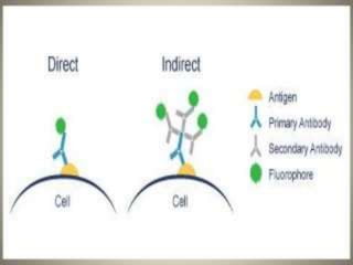
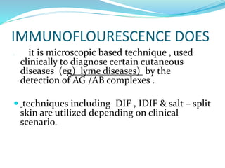
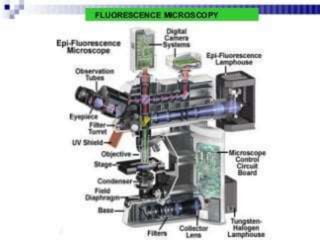
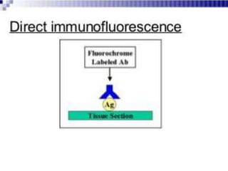
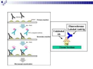
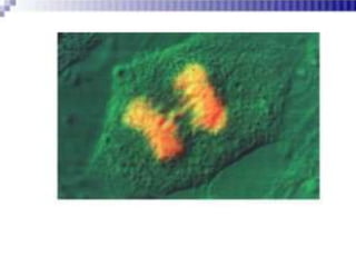
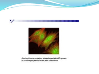
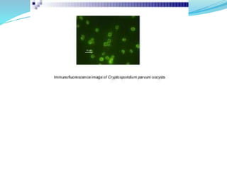
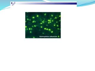
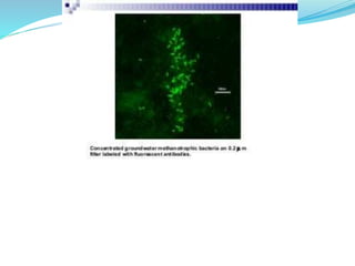
INTRODUCTION
immunoflourescence is a technique is visualization of
a specific antigen by binding a specific antibody
chemically conjugated with a flourescent dye such as
flourescein isothiocyanate ( FITC).
the specific antibodies are labeled with a compound
(FITC) that makes them glow an apple green colour
when observed microscopically under ultraviolet light.
FLOURESCENSE
flourescence is the property of certain molecules to
absorb light at one wavelength and emit light at longer
wave length when it is illuminated by light of a
different wavelength.
the flourescence can be visualized using flourescence
microscopy . The if technique allows for a visualization
of the presence as well as distribution of target
molecules in a sample.
 flourescene is a dye which emits greenish
flourescence under uv light .
 this technique is sometimes used to make viral
plaques more readily visible to the humen eye .
 flourescent antibodies is visible to flourescent
microscope .
IMMUNOFLOURESCENCE STAINING
 2 TYPES
 1) DIRECT IMMUNOFLOURESCENCE
 2) IN DIRECT IMMUNOFLOURESCENCE
 1) DIRECT IMMUNOFLOURESCENCE
 staining in the primary antibody is labeled with
flourescence dye.
 2) INDIRECT IMMUNOFLOURESCENCE
 staining in which a secondary antibody labeled with
flourochrome is used to recognize a primary antibody.
DIFFERENCE
DIRECT INDIRECT

IMMUNOFLOURESCENCE DOES
• it is microscopic based technique , used
clinically to diagnose certain cutaneous
diseases (eg) lyme diseases) by the
detection of AG /AB complexes .
 techniques including DIF , IDIF & salt – split
skin are utilized depending on clinical
scenario.
DIF is performed on patients skin
using flourophore labeled antiodies
that directly bind to pathogenic
autoantibody – antigen complexes In
the skin.
 IDIF techniques are used in
dermatology primarily to detect
circulating pathogenic auto
antibodies
/
LIMITATIONS
 Flourescence signals depend on the quality &
concentration of antibodies , proper handling of
specimen & detection with appropriate secondary
antibodies.
APPLICATIONS
 immunoflourescense can be used to tissue sections ,
cultured cell lines , or individual cells, and may be
used to analyse the distribution of proteins and small
biological and non biological molecules
 Immunoflourescence can be used in combination with
other non antibody method s of flourescence staining ,
for (ex) use of DAPI, (4,6 – diamidino -2-
phenylindole ) is a flourescent stain that binds
strongly to A-T rich regions in DNA to lable DNA .
 the technique has a numer of different biological
applications including evalution of cells in suspention
, cultured cells , tissue , beads.
 it also play a key role in the diagnosis of autoimmune
disorder.
IMMUNOFLOURESCENCE.pptx

IMMUNOFLOURESCENCE.pptx

  • 2.
    INTRODUCTION immunoflourescence is atechnique is visualization of a specific antigen by binding a specific antibody chemically conjugated with a flourescent dye such as flourescein isothiocyanate ( FITC). the specific antibodies are labeled with a compound (FITC) that makes them glow an apple green colour when observed microscopically under ultraviolet light.
  • 3.
    FLOURESCENSE flourescence is theproperty of certain molecules to absorb light at one wavelength and emit light at longer wave length when it is illuminated by light of a different wavelength. the flourescence can be visualized using flourescence microscopy . The if technique allows for a visualization of the presence as well as distribution of target molecules in a sample.
  • 4.
     flourescene isa dye which emits greenish flourescence under uv light .  this technique is sometimes used to make viral plaques more readily visible to the humen eye .  flourescent antibodies is visible to flourescent microscope .
  • 5.
    IMMUNOFLOURESCENCE STAINING  2TYPES  1) DIRECT IMMUNOFLOURESCENCE  2) IN DIRECT IMMUNOFLOURESCENCE
  • 6.
     1) DIRECTIMMUNOFLOURESCENCE  staining in the primary antibody is labeled with flourescence dye.  2) INDIRECT IMMUNOFLOURESCENCE  staining in which a secondary antibody labeled with flourochrome is used to recognize a primary antibody.
  • 8.
  • 9.
    IMMUNOFLOURESCENCE DOES • itis microscopic based technique , used clinically to diagnose certain cutaneous diseases (eg) lyme diseases) by the detection of AG /AB complexes .  techniques including DIF , IDIF & salt – split skin are utilized depending on clinical scenario.
  • 10.
    DIF is performedon patients skin using flourophore labeled antiodies that directly bind to pathogenic autoantibody – antigen complexes In the skin.  IDIF techniques are used in dermatology primarily to detect circulating pathogenic auto antibodies
  • 12.
  • 20.
    LIMITATIONS  Flourescence signalsdepend on the quality & concentration of antibodies , proper handling of specimen & detection with appropriate secondary antibodies.
  • 21.
    APPLICATIONS  immunoflourescense canbe used to tissue sections , cultured cell lines , or individual cells, and may be used to analyse the distribution of proteins and small biological and non biological molecules  Immunoflourescence can be used in combination with other non antibody method s of flourescence staining , for (ex) use of DAPI, (4,6 – diamidino -2- phenylindole ) is a flourescent stain that binds strongly to A-T rich regions in DNA to lable DNA .
  • 22.
     the techniquehas a numer of different biological applications including evalution of cells in suspention , cultured cells , tissue , beads.  it also play a key role in the diagnosis of autoimmune disorder.