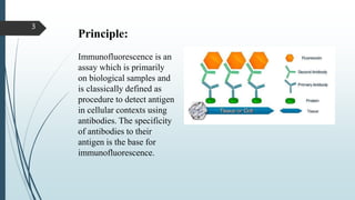
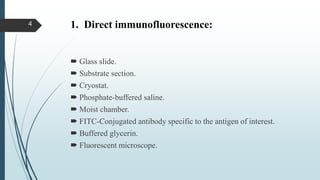
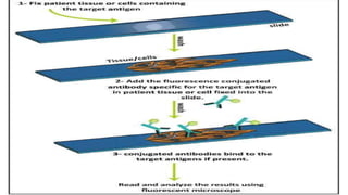
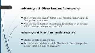
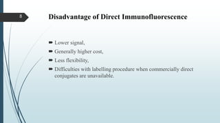
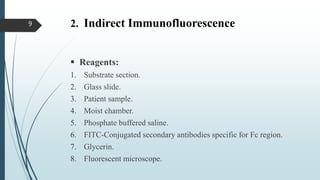
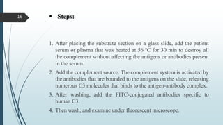
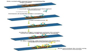
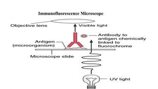
FLUORESCENCE ANTIBODY TEST
By: KINZA HAROON
SAEED JAMAL
Immunofluorescence
 Immunofluorescence is a Microscopic-based technique, used
clinically to diagnose certain cutaneous diseases by the detection of
Antigen-Antibody complexes.
 Techniques including Direct, Indirect, and Complement indirect
immunofluorescence are utilized depending on clinical scenario.
 IF studies are considering the “Gold Standard” for autoimmune
blistering diseases.
 Examples included; thyrotoxicosis.
2
Principle:
Immunofluorescence is an
assay which is primarily
on biological samples and
is classically defined as
procedure to detect antigen
in cellular contexts using
antibodies. The specificity
of antibodies to their
antigen is the base for
immunofluorescence.
3
1. Direct immunofluorescence:
 Glass slide.
 Substrate section.
 Cryostat.
 Phosphate-buffered saline.
 Moist chamber.
 FITC-Conjugated antibody specific to the antigen of interest.
 Buffered glycerin.
 Fluorescent microscope.
4
Steps:
1. Patients sample (skin or mucosal biopsy).
2. About 4-6ul of the snap-frozen sample is sectioned using a cryostat
and placed on a glass slide, and air-dried foe 15 min.
3. After washing by phosphate-buffered saline, the conjugated
antibodies that are specific to the antigens of interest in the patient
sample are added into the slide and incubated in a moist chamber.
4. Wash, mount with glycerin, place on the cover slip, and examine
under fluorescent microscope.
5
6
Advantages of Direct Immunofluorescence:
This technique is used to detect viral, parasitic, tumor antigens
from patient specimens.
Anatomic identification of anatomic distribution of an antigen
within tissue or compartments of cell.
Advantages of Direct Immunofluorescence:
Shorter sample staining times.
 In case where one has multiple Ab raised in the same species,
a direct labelling may be necessary.
7
Disadvantage of Direct Immunofluorescence
 Lower signal,
 Generally higher cost,
 Less flexibility,
 Difficulties with labelling procedure when commercially direct
conjugates are unavailable.
8
2. Indirect Immunofluorescence
 Reagents:
1. Substrate section.
2. Glass slide.
3. Patient sample.
4. Moist chamber.
5. Phosphate buffered saline.
6. FITC-Conjugated secondary antibodies specific for Fc region.
7. Glycerin.
8. Fluorescent microscope.
9
 Steps:
1. After placing the substrate section on a glass slide, add the serial
diluted patient serum, and incubate in a moist for 30 min. positive
and negative control sera must be used to test the antibody
reactivity.
2. Wash with phosphate-buffered saline, and add the conjugated
antibodies that are specific to the human antibody Fc region.
3. Wash for at least 10 times with phosphate-buffered, mount and
examine under the fluorescent microscope.
10
Indirect
immunofluorescence
Steps involve in indirect
immunofluorescence.
11
Applications of Indirect Immunofluorescence:
 It is often used to detect autoantibodies in serum or other body
fluids.
 Used in Dermatology primarily to detect circulating pathogenic
autoantibodies.
 Commonly used in the detection;
 Anti-nuclear antibodies,
 Systematic lupus erythematous,
 Antithyroid antibodies.
12
Advantages of Indirect immunofluorescence:
 Greater sensitivity than direct immunofluorescence
 For every antibody there is a characteristics fluorescence pattern.
Disadvantages of Indirect immunofluorescence:
 Potential cross reactivity.
 Finding labeled primary Ab which is more difficult to get especially
for multiple labeling experiments.
13
3. Complement Indirect
Immunofluorescence:
 Reagents:
1. Tissue substrate.
2. Glass slide.
3. Phosphate-buffered saline.
4. Patient sample.
5. Heating source.
6. Complement source such as fresh human serum.
14
Continue….
7. FITC-conjugated anti-human C3 antibodies.
8. Glycerin.
9. Cover slip.
10. Fluorescent microscope.
15
 Steps:
1. After placing the substrate section on a glass slide, add the patient
serum or plasma that was heated at 56 ºC for 30 min to destroy all
the complement without affecting the antigens or antibodies present
in the serum.
2. Add the complement source. The complement system is activated by
the antibodies that are bounded to the antigens on the slide, releasing
numerous C3 molecules that binds to the antigen-antibody complex.
3. After washing, add the FITC-conjugated antibodies specific to
human C3.
4. Then wash, and examine under fluorescent microscope.
16
17
Applications of Complement Indirect
Immunofluorescence:
 Used on tissue sections.
 Cultured cell lines or individual cells.
 To analyze the distribution of proteins, glycan's, and small
biological molecules.
18
Result Interpretation:
 If no fluorescence is detected under the fluorescent microscope, it is
a negative sample.
 According to the biding pattern of the anti-nuclear antibodies and
the intensity of the fluorescence (1+, 2+,3+ or 4+), the autoimmune
disease can be determined in correlation with the ELISA results for
double-stranded DNA, single-stranded DNA, and histone.
19
20
Immunofluorescence Microscope:
21
22

Fluorescence antibody test

  • 1.
    FLUORESCENCE ANTIBODY TEST By:KINZA HAROON SAEED JAMAL
  • 2.
    Immunofluorescence  Immunofluorescence isa Microscopic-based technique, used clinically to diagnose certain cutaneous diseases by the detection of Antigen-Antibody complexes.  Techniques including Direct, Indirect, and Complement indirect immunofluorescence are utilized depending on clinical scenario.  IF studies are considering the “Gold Standard” for autoimmune blistering diseases.  Examples included; thyrotoxicosis. 2
  • 3.
    Principle: Immunofluorescence is an assaywhich is primarily on biological samples and is classically defined as procedure to detect antigen in cellular contexts using antibodies. The specificity of antibodies to their antigen is the base for immunofluorescence. 3
  • 4.
    1. Direct immunofluorescence: Glass slide.  Substrate section.  Cryostat.  Phosphate-buffered saline.  Moist chamber.  FITC-Conjugated antibody specific to the antigen of interest.  Buffered glycerin.  Fluorescent microscope. 4
  • 5.
    Steps: 1. Patients sample(skin or mucosal biopsy). 2. About 4-6ul of the snap-frozen sample is sectioned using a cryostat and placed on a glass slide, and air-dried foe 15 min. 3. After washing by phosphate-buffered saline, the conjugated antibodies that are specific to the antigens of interest in the patient sample are added into the slide and incubated in a moist chamber. 4. Wash, mount with glycerin, place on the cover slip, and examine under fluorescent microscope. 5
  • 6.
  • 7.
    Advantages of DirectImmunofluorescence: This technique is used to detect viral, parasitic, tumor antigens from patient specimens. Anatomic identification of anatomic distribution of an antigen within tissue or compartments of cell. Advantages of Direct Immunofluorescence: Shorter sample staining times.  In case where one has multiple Ab raised in the same species, a direct labelling may be necessary. 7
  • 8.
    Disadvantage of DirectImmunofluorescence  Lower signal,  Generally higher cost,  Less flexibility,  Difficulties with labelling procedure when commercially direct conjugates are unavailable. 8
  • 9.
    2. Indirect Immunofluorescence Reagents: 1. Substrate section. 2. Glass slide. 3. Patient sample. 4. Moist chamber. 5. Phosphate buffered saline. 6. FITC-Conjugated secondary antibodies specific for Fc region. 7. Glycerin. 8. Fluorescent microscope. 9
  • 10.
     Steps: 1. Afterplacing the substrate section on a glass slide, add the serial diluted patient serum, and incubate in a moist for 30 min. positive and negative control sera must be used to test the antibody reactivity. 2. Wash with phosphate-buffered saline, and add the conjugated antibodies that are specific to the human antibody Fc region. 3. Wash for at least 10 times with phosphate-buffered, mount and examine under the fluorescent microscope. 10
  • 11.
    Indirect immunofluorescence Steps involve inindirect immunofluorescence. 11
  • 12.
    Applications of IndirectImmunofluorescence:  It is often used to detect autoantibodies in serum or other body fluids.  Used in Dermatology primarily to detect circulating pathogenic autoantibodies.  Commonly used in the detection;  Anti-nuclear antibodies,  Systematic lupus erythematous,  Antithyroid antibodies. 12
  • 13.
    Advantages of Indirectimmunofluorescence:  Greater sensitivity than direct immunofluorescence  For every antibody there is a characteristics fluorescence pattern. Disadvantages of Indirect immunofluorescence:  Potential cross reactivity.  Finding labeled primary Ab which is more difficult to get especially for multiple labeling experiments. 13
  • 14.
    3. Complement Indirect Immunofluorescence: Reagents: 1. Tissue substrate. 2. Glass slide. 3. Phosphate-buffered saline. 4. Patient sample. 5. Heating source. 6. Complement source such as fresh human serum. 14
  • 15.
    Continue…. 7. FITC-conjugated anti-humanC3 antibodies. 8. Glycerin. 9. Cover slip. 10. Fluorescent microscope. 15
  • 16.
     Steps: 1. Afterplacing the substrate section on a glass slide, add the patient serum or plasma that was heated at 56 ºC for 30 min to destroy all the complement without affecting the antigens or antibodies present in the serum. 2. Add the complement source. The complement system is activated by the antibodies that are bounded to the antigens on the slide, releasing numerous C3 molecules that binds to the antigen-antibody complex. 3. After washing, add the FITC-conjugated antibodies specific to human C3. 4. Then wash, and examine under fluorescent microscope. 16
  • 17.
  • 18.
    Applications of ComplementIndirect Immunofluorescence:  Used on tissue sections.  Cultured cell lines or individual cells.  To analyze the distribution of proteins, glycan's, and small biological molecules. 18
  • 19.
    Result Interpretation:  Ifno fluorescence is detected under the fluorescent microscope, it is a negative sample.  According to the biding pattern of the anti-nuclear antibodies and the intensity of the fluorescence (1+, 2+,3+ or 4+), the autoimmune disease can be determined in correlation with the ELISA results for double-stranded DNA, single-stranded DNA, and histone. 19
  • 20.
  • 21.
  • 22.