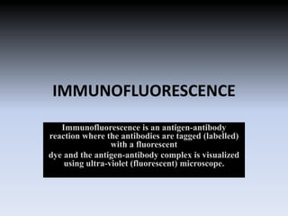
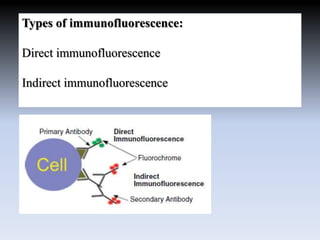
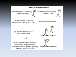
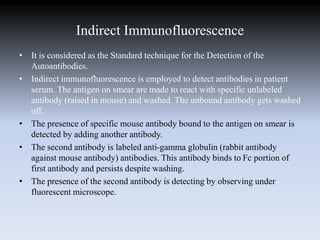
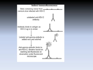
Presentation on
Immunofluorescence
Presented by:
Farhan ali
Naveen bhatt
Msc Biotechnology II sem
IMMUNOFLUORESCENCE
Immunofluorescence is an antigen-antibody
reaction where the antibodies are tagged (labelled)
with a fluorescent
dye and the antigen-antibody complex is visualized
using ultra-violet (fluorescent) microscope.
Anti-alpha smooth muscle Actin antibody.(left)
A fluorescent stain for actin in the smooth muscle of the skin.(right)
History
• Immunofluorescence studies are considered
the ‘gold standard’ for the diagnosis of
autoimmune blistering diseases.
• Coons et al. (1941) developed the
immunofluorescence techniques for the first
time, a discovery which made possible to
observe microscopically antigens, antibodies
and their related substances on tissue sections
or on cell smears
PRINCIPLE
• Fluorescence and phosphorescence are both
types of luminescence.
• In fluorescence the emission of light occurs
extremely rapidly after the absorption of
excitation light.
• phosphorescence emission continues for
milliseconds to minutes after the energy source
has been removed.
Fluorochromes are dyes that absorb ultra-
violet rays and emit visible light. This
process is called fluorescence.
The fluorochromes commonly used
in immunofluorescence are
fluorescein isothiocyanate (green)
and and tetramethyl rhodamine
isothiocyanate (red).
Types of immunofluorescence:
Direct immunofluorescence
Indirect immunofluorescence
Direct immunofluorescence:
• This is the one step histological staining process for
identifying in vivo antibodies that are bound to tissues
antigen.
• This technique is used to detect antigen in clinical
specimens using specific fluorochrome labeled
antibody. The steps involved are:
• Fixation of smear on the slide, treating with labeled
antibody, incubation, washing to remove unbound
excess labeled antibody and visualization under
fluorescent microscope. When viewed under
fluorescent microscope, the field is dark and areas with
bound antibody fluorensce green.
Uses of Direct immunoflourescence
• This technique can be used to detect viral, parasitic,
tumor antigens from patient specimens or monolayer
of cells.
• Another application is identification of anatomic
distribution of an antigen within a tissue or within
compartments of a cell.
Advantages of direct immunofluorescence
• Shorter sample staining times and simpler dual
and triple labeling procedures.
• In cases where one has multiple antibodies
raised in the same species, for example two
mouse monoclonals, a direct labeling may be
necessary.
Disadvantages of direct immunofluorescence
• lower signal, generally higher cost, less
flexibility and difficulties with the
• labeling procedure when commercially labeled
direct conjugates are unavailable.
Indirect Immunofluorescence
• It is considered as the Standard technique for the Detection of the
Autoantibodies.
• Indirect immunofluorescence is employed to detect antibodies in patient
serum. The antigen on smear are made to react with specific unlabeled
antibody (raised in mouse) and washed. The unbound antibody gets washed
off.
• The presence of specific mouse antibody bound to the antigen on smear is
detected by adding another antibody.
• The second antibody is labeled anti-gamma globulin (rabbit antibody
against mouse antibody) antibodies. This antibody binds to Fc portion of
first antibody and persists despite washing.
• The presence of the second antibody is detecting by observing under
fluorescent microscope.
Uses of Indirect immunoflourescence
• It is often used to detect autoantibodies.
• Commonly used in the detection of anti-nuclear
antibodies (ANA) found in the serum of patients with
SLE (Systematic lupus Erythematosus)
Advantages of indirect immunofluorescence
• A negative result excudes the presence of all these antibodies.
• For every Antibody there is a characteristic fluorecence
pattern.
• Greater sensitivity than direct immunofluorescence.
• There is amplification of the signal in indirect
immunofluorescence because more than one secondary
antibody can attach to each primary.
• Commercially produced secondary antibodies are relatively
inexpensive, available in an array of colors, and quality
controlled.
Disadvantages of indirect immunofluorescence
• The potential for cross-reactivity and the need to find
primary antibodies that are not raised in the same
species or of different isotypes.
• When performing multiple-labeling experiments.
Samples with endogenous immunoglobulin may
exhibit a high background.
Immunofluorescence

Immunofluorescence

  • 1.
    Presentation on Immunofluorescence Presented by: Farhanali Naveen bhatt Msc Biotechnology II sem
  • 2.
    IMMUNOFLUORESCENCE Immunofluorescence is anantigen-antibody reaction where the antibodies are tagged (labelled) with a fluorescent dye and the antigen-antibody complex is visualized using ultra-violet (fluorescent) microscope.
  • 3.
    Anti-alpha smooth muscleActin antibody.(left) A fluorescent stain for actin in the smooth muscle of the skin.(right)
  • 4.
    History • Immunofluorescence studiesare considered the ‘gold standard’ for the diagnosis of autoimmune blistering diseases. • Coons et al. (1941) developed the immunofluorescence techniques for the first time, a discovery which made possible to observe microscopically antigens, antibodies and their related substances on tissue sections or on cell smears
  • 5.
    PRINCIPLE • Fluorescence andphosphorescence are both types of luminescence. • In fluorescence the emission of light occurs extremely rapidly after the absorption of excitation light. • phosphorescence emission continues for milliseconds to minutes after the energy source has been removed.
  • 6.
    Fluorochromes are dyesthat absorb ultra- violet rays and emit visible light. This process is called fluorescence. The fluorochromes commonly used in immunofluorescence are fluorescein isothiocyanate (green) and and tetramethyl rhodamine isothiocyanate (red).
  • 7.
    Types of immunofluorescence: Directimmunofluorescence Indirect immunofluorescence
  • 8.
    Direct immunofluorescence: • Thisis the one step histological staining process for identifying in vivo antibodies that are bound to tissues antigen. • This technique is used to detect antigen in clinical specimens using specific fluorochrome labeled antibody. The steps involved are: • Fixation of smear on the slide, treating with labeled antibody, incubation, washing to remove unbound excess labeled antibody and visualization under fluorescent microscope. When viewed under fluorescent microscope, the field is dark and areas with bound antibody fluorensce green.
  • 10.
    Uses of Directimmunoflourescence • This technique can be used to detect viral, parasitic, tumor antigens from patient specimens or monolayer of cells. • Another application is identification of anatomic distribution of an antigen within a tissue or within compartments of a cell.
  • 11.
    Advantages of directimmunofluorescence • Shorter sample staining times and simpler dual and triple labeling procedures. • In cases where one has multiple antibodies raised in the same species, for example two mouse monoclonals, a direct labeling may be necessary.
  • 12.
    Disadvantages of directimmunofluorescence • lower signal, generally higher cost, less flexibility and difficulties with the • labeling procedure when commercially labeled direct conjugates are unavailable.
  • 13.
    Indirect Immunofluorescence • Itis considered as the Standard technique for the Detection of the Autoantibodies. • Indirect immunofluorescence is employed to detect antibodies in patient serum. The antigen on smear are made to react with specific unlabeled antibody (raised in mouse) and washed. The unbound antibody gets washed off. • The presence of specific mouse antibody bound to the antigen on smear is detected by adding another antibody. • The second antibody is labeled anti-gamma globulin (rabbit antibody against mouse antibody) antibodies. This antibody binds to Fc portion of first antibody and persists despite washing. • The presence of the second antibody is detecting by observing under fluorescent microscope.
  • 15.
    Uses of Indirectimmunoflourescence • It is often used to detect autoantibodies. • Commonly used in the detection of anti-nuclear antibodies (ANA) found in the serum of patients with SLE (Systematic lupus Erythematosus)
  • 16.
    Advantages of indirectimmunofluorescence • A negative result excudes the presence of all these antibodies. • For every Antibody there is a characteristic fluorecence pattern. • Greater sensitivity than direct immunofluorescence. • There is amplification of the signal in indirect immunofluorescence because more than one secondary antibody can attach to each primary. • Commercially produced secondary antibodies are relatively inexpensive, available in an array of colors, and quality controlled.
  • 17.
    Disadvantages of indirectimmunofluorescence • The potential for cross-reactivity and the need to find primary antibodies that are not raised in the same species or of different isotypes. • When performing multiple-labeling experiments. Samples with endogenous immunoglobulin may exhibit a high background.