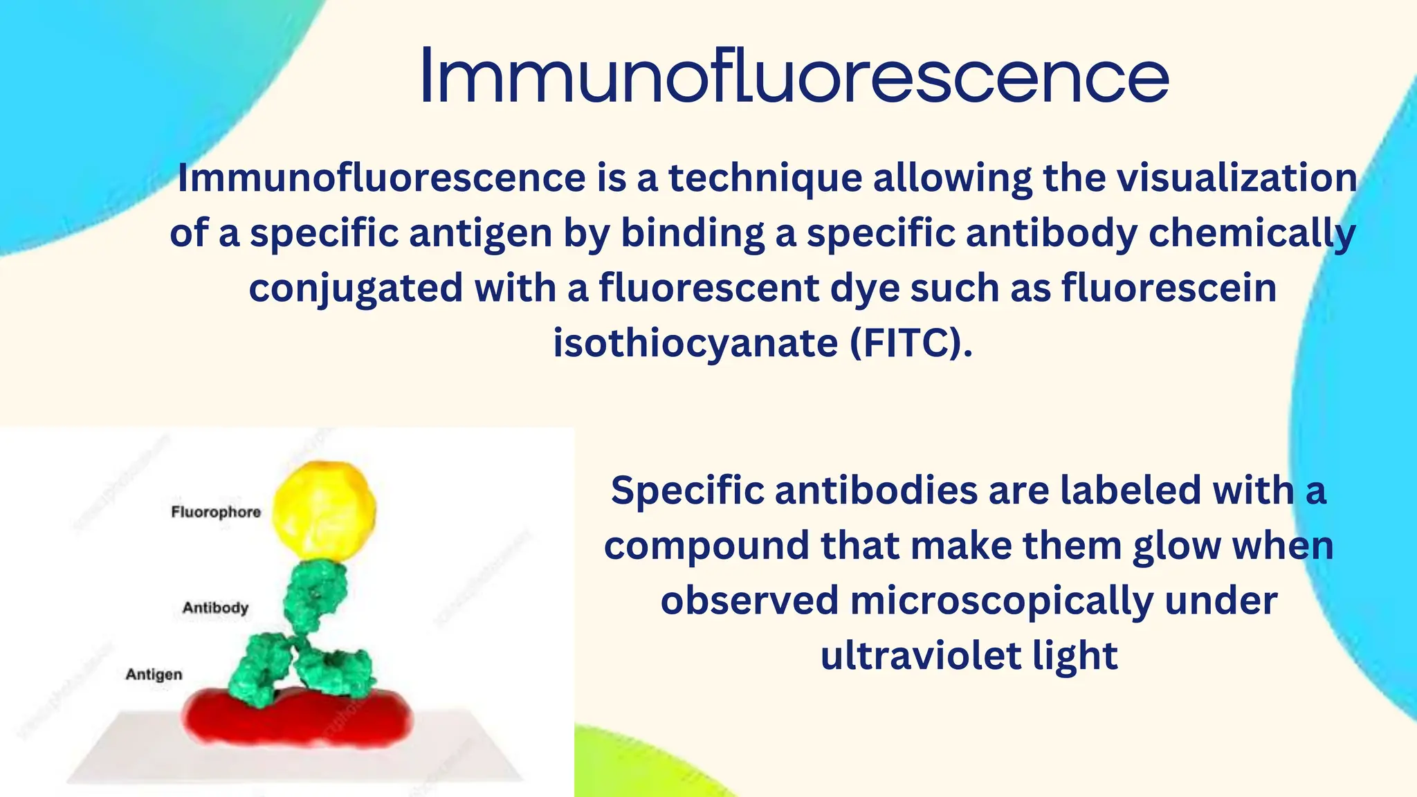
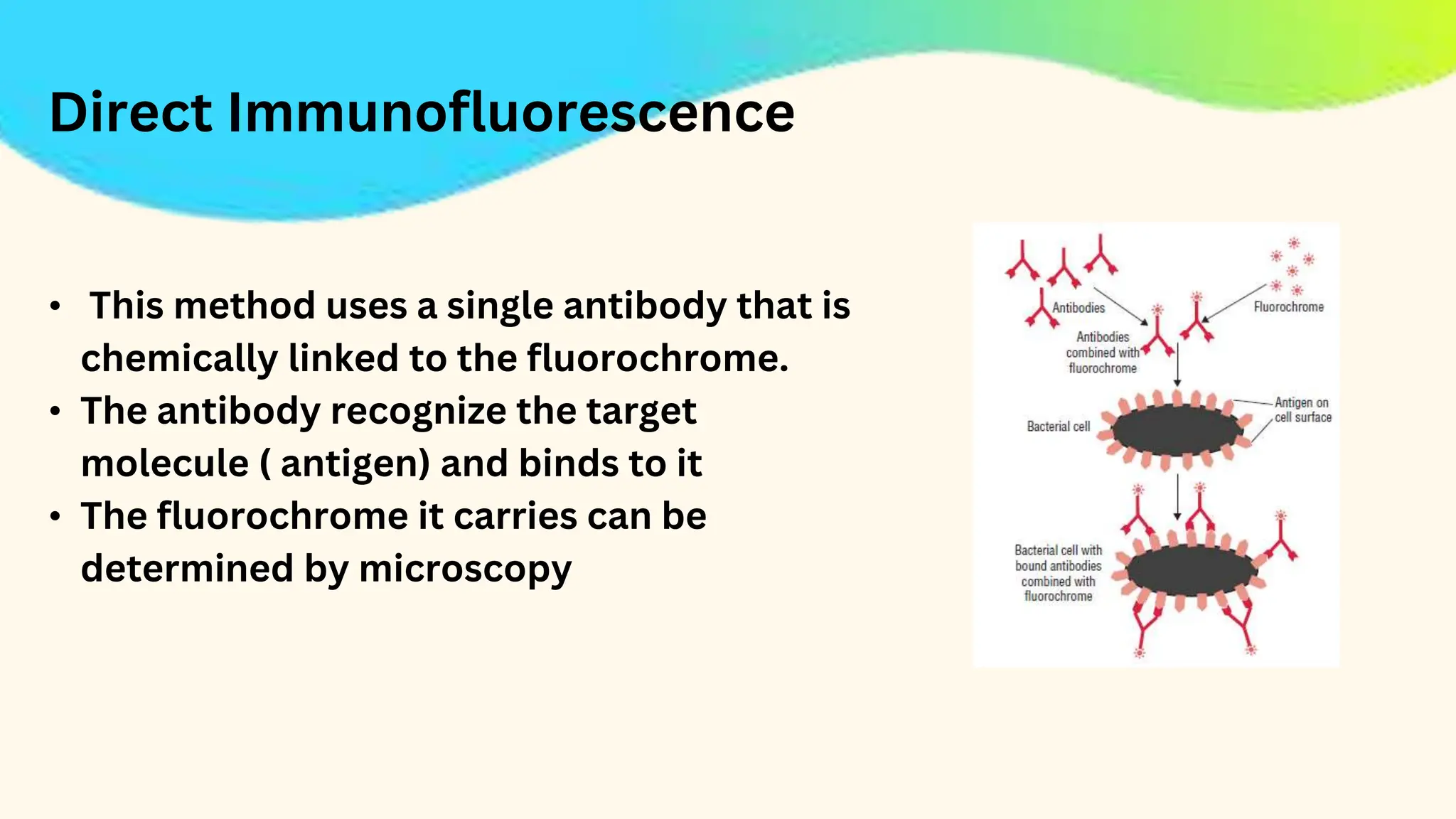
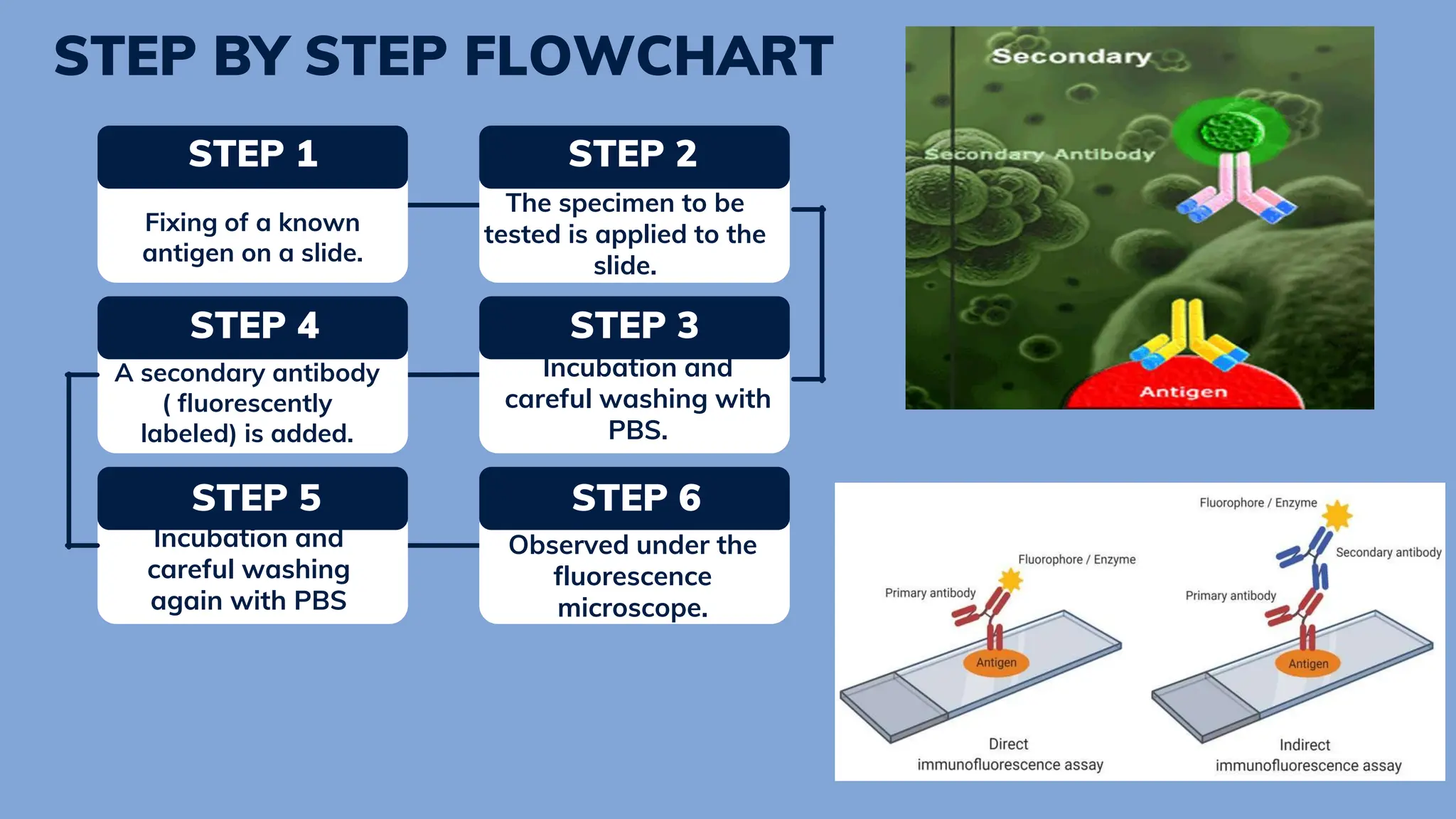
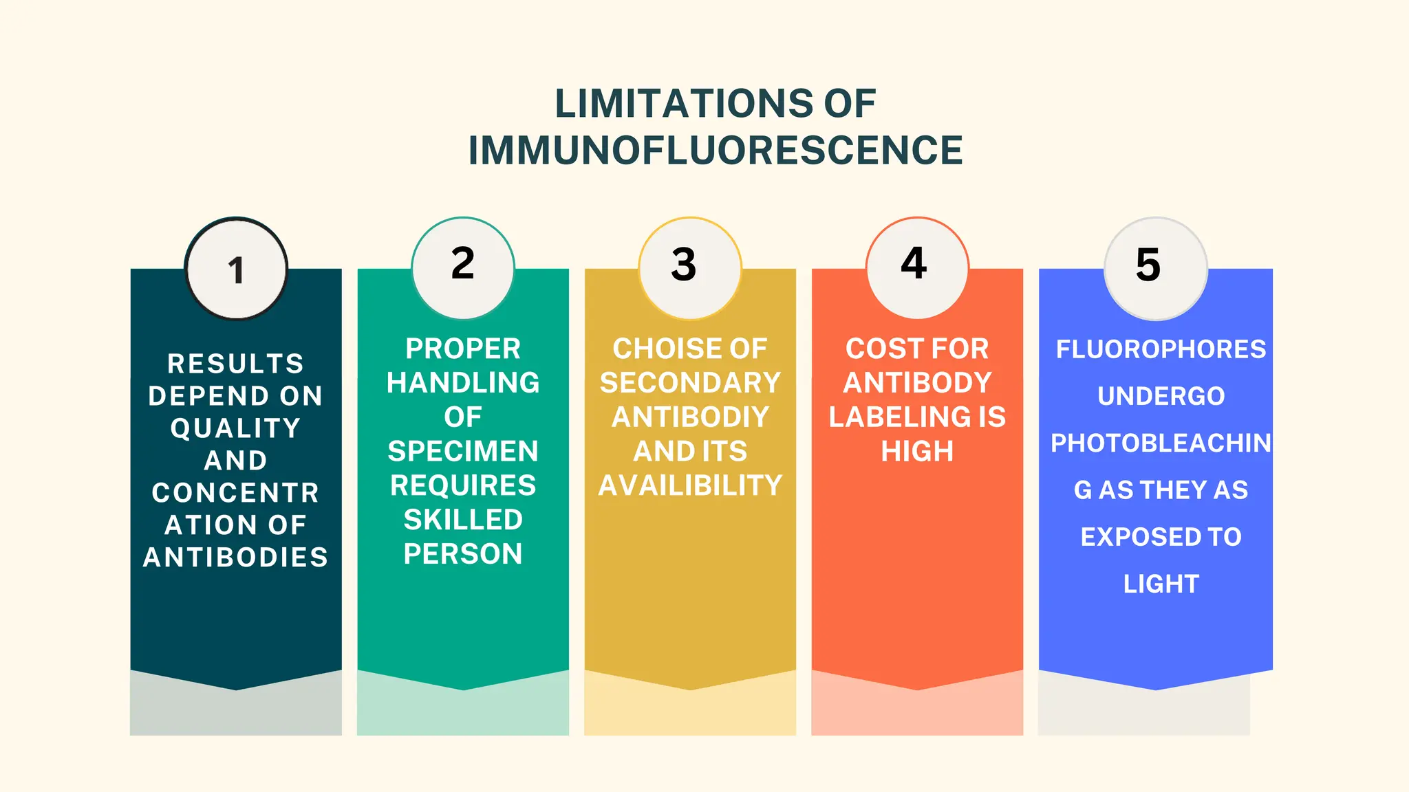
FLUORESCENT
ANTIBODY
and its
applications
by: Devraj Kulkarni
Microbiologist
Lets dive into
basics
• Antibodies are proteins produced by the immune system
in response to infection.
• They are an important part of the body's defence system
as they work to destroy disease-causing organisms (such
as viruses or bacteria) and block them from infecting
human cells.
• These antibodies bind with
antigens ( foreign material entered
in body,which can cause infection)
What is
Fluorescence
Fluorescence is a property of a material to
absorb light at one wavelength and emit light of
longer wavelength when it is illuminate by light
The fluorescence can be visualized by
fluorescence microscopy
Immunofluorescence
Immunofluorescence is a technique allowing the visualization
of a specific antigen by binding a specific antibody chemically
conjugated with a fluorescent dye such as fluorescein
isothiocyanate (FITC).
Specific antibodies are labeled with a
compound that make them glow when
observed microscopically under
ultraviolet light
Types of
Immunofluorescence
• Direct
Immunofluorescence
2.Indirect
Immunofluorescence
Direct Immunofluorescence
• Single antibody i.e. primary antibody is used that is
chemically linked to a fluorochrome.
• If the antigen is present, the primary antibody directly
reacts with it and fluorescence can be observed under the
fluorescent microscope
Primary antibody is an
Immunoglobulin(protein) which has affinity
towards antigen
Direct Immunofluorescence
• This method uses a single antibody that is
chemically linked to the fluorochrome.
• The antibody recognize the target
molecule ( antigen) and binds to it
• The fluorochrome it carries can be
determined by microscopy
2. FLUOROCHROME
LABELED ANTIBODIES
ARE THEN ADDED TO THE
SLIDE.
• FIXING OF SPECIMEN
(ANTIGEN) INTO THE
SLIDE.
3. INCUBATION AND CAREFUL
WASHING WITH WASH BUFFERS LIKE
PBS TO REMOVE OTHER COMPONENTS
EXCEPT FOR THE COMPLEX OF
ANTIGEN AND FLUOROCHROME-
LABELED ANTIBODY.
4. OBSERVED UNDER A
FLUORESCENCE
MICROSCOPE.
Steps for
visualization
Indirect Immunofluorescence
• This technique employs two types of antibodies primary
antibody is not labeled, it binds to the desired antigen
• A secondary antibody, which is labeled with fluorochrome is
added. It has affinity towards primary antibody.
• It binds to the Fc region of primary antibody which has already
combined to the antigen.
• Thus, by using fluorescent microscopy antigen can be detected
STEP 1
Fixing of a known
antigen on a slide.
STEP 4
Incubation and
careful washing with
PBS.
STEP 5
Incubation and
careful washing
again with PBS
STEP 2
The specimen to be
tested is applied to the
slide.
STEP 3
A secondary antibody
( fluorescently
labeled) is added.
STEP 6
Observed under the
fluorescence
microscope.
STEP BY STEP FLOWCHART
Used in the detection of autoantibodies that cause
auto immune disorders.
1
Significance of Indirect
immunofluorescence
4
3
2 Single fluorochrome-labeled antibody can be used
for detecting many Ag-Ab interactions
Multiple secondary antibodies can bind to the Fc
region of primary antibody which amplifies the
fluorescence signal
More sensitive than direct immunofluorescence test.
Applications of
immuno-
fluorescence
used on cell
sections to
determine
presence of
different
biological
molecules
visualization of
cytoskeletons
such as
intermediate
filaments.
diagnosis of
syphilis,
amoebiasis,
leptospirosis,
toxoplasmosis,
and other
diseases.
detection of
rabies virus
antigen in
the skin
smear
to study
distribution of
biological/non
biological
molecules in
tissue
For DNA
labeling
(combination
with non
antibody
method)
detection
and
localization
of a wide
variety of
antigens
7
LIMITATIONS OF
IMMUNOFLUORESCENCE
RESULTS
DEPEND ON
QUALITY
AND
CONCENTR
ATION OF
ANTIBODIES
PROPER
HANDLING
OF
SPECIMEN
REQUIRES
SKILLED
PERSON
CHOISE OF
SECONDARY
ANTIBODIY
AND ITS
AVAILIBILITY
COST FOR
ANTIBODY
LABELING IS
HIGH
FLUOROPHORES
UNDERGO
PHOTOBLEACHIN
G AS THEY AS
EXPOSED TO
LIGHT
2 4 5
3
Dyes used for Antibody labeling
Fluorescei
n
Rhodamine AlexaFluor 532-SE
532 nm
573 nm
498 nm
Quantification methods
Once the desired antigen is detected using immunofluorescence, following
methods are employed for their quantification
• Flow cytometry
• fluorescence-activated cell sorter (FACS)
Bibliography
• https://courses.lumenlearning.com/sunymicrobiology/chapter/fluorescent-
antibody-techniques
• https://www.chemspider.com/Chemical-Structure.19813515.html
• https://www.slideshare.net/TapeshwarYadav1/immunofluorescence-
55934842
• https://www.slideshare.net/rajpalchoudharyjat/immunofluorescence-
technique
• https://microbenotes.com/immunofluorescence/
Thank you...!

Immunofluorescence :Fluorescent antibody and its application

  • 1.
  • 2.
    Lets dive into basics •Antibodies are proteins produced by the immune system in response to infection. • They are an important part of the body's defence system as they work to destroy disease-causing organisms (such as viruses or bacteria) and block them from infecting human cells. • These antibodies bind with antigens ( foreign material entered in body,which can cause infection)
  • 3.
    What is Fluorescence Fluorescence isa property of a material to absorb light at one wavelength and emit light of longer wavelength when it is illuminate by light The fluorescence can be visualized by fluorescence microscopy
  • 4.
    Immunofluorescence Immunofluorescence is atechnique allowing the visualization of a specific antigen by binding a specific antibody chemically conjugated with a fluorescent dye such as fluorescein isothiocyanate (FITC). Specific antibodies are labeled with a compound that make them glow when observed microscopically under ultraviolet light
  • 5.
  • 6.
    Direct Immunofluorescence • Singleantibody i.e. primary antibody is used that is chemically linked to a fluorochrome. • If the antigen is present, the primary antibody directly reacts with it and fluorescence can be observed under the fluorescent microscope Primary antibody is an Immunoglobulin(protein) which has affinity towards antigen
  • 7.
    Direct Immunofluorescence • Thismethod uses a single antibody that is chemically linked to the fluorochrome. • The antibody recognize the target molecule ( antigen) and binds to it • The fluorochrome it carries can be determined by microscopy
  • 8.
    2. FLUOROCHROME LABELED ANTIBODIES ARETHEN ADDED TO THE SLIDE. • FIXING OF SPECIMEN (ANTIGEN) INTO THE SLIDE. 3. INCUBATION AND CAREFUL WASHING WITH WASH BUFFERS LIKE PBS TO REMOVE OTHER COMPONENTS EXCEPT FOR THE COMPLEX OF ANTIGEN AND FLUOROCHROME- LABELED ANTIBODY. 4. OBSERVED UNDER A FLUORESCENCE MICROSCOPE. Steps for visualization
  • 9.
    Indirect Immunofluorescence • Thistechnique employs two types of antibodies primary antibody is not labeled, it binds to the desired antigen • A secondary antibody, which is labeled with fluorochrome is added. It has affinity towards primary antibody. • It binds to the Fc region of primary antibody which has already combined to the antigen. • Thus, by using fluorescent microscopy antigen can be detected
  • 10.
    STEP 1 Fixing ofa known antigen on a slide. STEP 4 Incubation and careful washing with PBS. STEP 5 Incubation and careful washing again with PBS STEP 2 The specimen to be tested is applied to the slide. STEP 3 A secondary antibody ( fluorescently labeled) is added. STEP 6 Observed under the fluorescence microscope. STEP BY STEP FLOWCHART
  • 11.
    Used in thedetection of autoantibodies that cause auto immune disorders. 1 Significance of Indirect immunofluorescence 4 3 2 Single fluorochrome-labeled antibody can be used for detecting many Ag-Ab interactions Multiple secondary antibodies can bind to the Fc region of primary antibody which amplifies the fluorescence signal More sensitive than direct immunofluorescence test.
  • 12.
    Applications of immuno- fluorescence used oncell sections to determine presence of different biological molecules visualization of cytoskeletons such as intermediate filaments. diagnosis of syphilis, amoebiasis, leptospirosis, toxoplasmosis, and other diseases. detection of rabies virus antigen in the skin smear to study distribution of biological/non biological molecules in tissue For DNA labeling (combination with non antibody method) detection and localization of a wide variety of antigens 7
  • 13.
    LIMITATIONS OF IMMUNOFLUORESCENCE RESULTS DEPEND ON QUALITY AND CONCENTR ATIONOF ANTIBODIES PROPER HANDLING OF SPECIMEN REQUIRES SKILLED PERSON CHOISE OF SECONDARY ANTIBODIY AND ITS AVAILIBILITY COST FOR ANTIBODY LABELING IS HIGH FLUOROPHORES UNDERGO PHOTOBLEACHIN G AS THEY AS EXPOSED TO LIGHT 2 4 5 3
  • 14.
    Dyes used forAntibody labeling Fluorescei n Rhodamine AlexaFluor 532-SE 532 nm 573 nm 498 nm
  • 15.
    Quantification methods Once thedesired antigen is detected using immunofluorescence, following methods are employed for their quantification • Flow cytometry • fluorescence-activated cell sorter (FACS)
  • 16.
    Bibliography • https://courses.lumenlearning.com/sunymicrobiology/chapter/fluorescent- antibody-techniques • https://www.chemspider.com/Chemical-Structure.19813515.html •https://www.slideshare.net/TapeshwarYadav1/immunofluorescence- 55934842 • https://www.slideshare.net/rajpalchoudharyjat/immunofluorescence- technique • https://microbenotes.com/immunofluorescence/
  • 17.