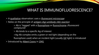
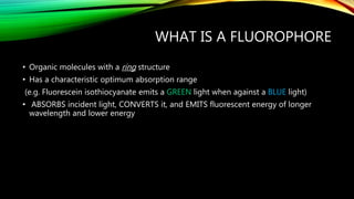
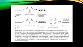
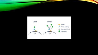
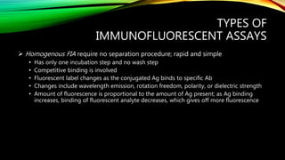
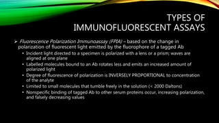
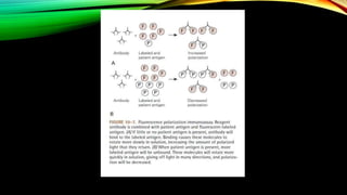
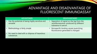
IMMUNOFLUORESCENCE
By GROUP II
WHAT IS IMMUNOFLUORESCENCE?
• A qualitative observation; uses a fluorescent microscope
• Relies on the principle of antigen (Ag)-antibody (Ab) reaction:
> Ab is “tagged” with a fluorophore or fluorochrome (fluorescent
compound)
> Ab binds to a specific Ag of interest
>Ag-Ab complex emits a green or red light (depending on the
fluorophore used) when an incident light (usually UV light) is introduced
• Introduced by Albert Coons in 1941
WHAT IS A FLUOROPHORE
• Organic molecules with a ring structure
• Has a characteristic optimum absorption range
(e.g. Fluorescein isothiocyanate emits a GREEN light when against a BLUE light)
• ABSORBS incident light, CONVERTS it, and EMITS fluorescent energy of longer
wavelength and lower energy
EXAMPLES (FLUOROPHORES)
• Fluorescein isothiocyanate – most common; emits green light
• Tetramethylrhodamine – emits red light
• Phycoerythrin
• Europium (β – naphthyl trifluoroacetone)
• Lucifer Yellow VS
• Acridine Orange
• Lissamine
• Calcofluor White
WORKING PRINCIPLE
• Employs Ag-Ab reaction
• Ab tagged with a fluorophore is introduced to specimen
• Ab binds to specific Ag
• Specimen is viewed under a UV light with a dark background in a fluorescent
microscope
WORKING PRINCIPLE (CONT.)
• Fluorophore absorbs radiant energy and is excited
• Fluorophore goes back to a ground state, emitting light energy of longer
wavelength and lower energy
• Interval of absorption and emission is very short; occurs in nanoseconds
PURPOSE OF
IMMUNOFLUORESCENT ASSAYS
• Rapid identification of microorganisms in cell culture or infected tissue, tumor-
specific Ag on neoplastic tissue, transplantation Ag, and CD Ag on T and B cells (via
Cell Flow Cytometry)
TYPES OF
IMMUNOFLUORESCENT ASSAYS
1. Direct Immunofluorescence Assay (DIFA)
 Tagged Ab is directly added to an unknown Ag fixed to a slide
 Requires incubation and a wash step
 Ag are seen as bright apple green or orange-yellow against a dark backgrounds
 Suitable for detection of specific Ag in tissue or body fluids
e.g. Legionella pneumophila, Pneumocystis carinii, Chlamydia
trachomatis, respiratory syncytial virus (RSV)
TYPES OF
IMMUNOFLUORESCENT ASSAYS
2. Indirect Immunofluorescent Assay
 Specimen with a known Ag is incubated on a solid phase
 Antihuman immunoglobulin (from mouse) tagged with a fluorophore is added
 Specimen Ab binds with antihuman Ab, forming a sandwich and localizing
fluorescence
 Used in Ab identification and detection of Treponema species-specific, antinuclear,
chlamydial, and toxoplasma Ab, and Ab to herpes simplex virus (HSV), Epstein-Barr
virus (EBV), and Cytomegalovirus (CMV)
TYPES OF
IMMUNOFLUORESCENT ASSAYS
ADVANTAGE (over DIFA):
a.) Uses only one Ab conjugate (tagged Ab) for different reactions, eliminating the
need for numerous purified, labeled reagent antibodies)
b.) More sensitive; has increased staining property. Multiple molecules bind to each
primary molecule.
TYPES OF
IMMUNOFLUORESCENT ASSAYS
3. Microimmunofluorescence
 Detects Ab in patient serum
 Has the same working principle as Indirect Immunofluorescence Assay but employs
Teflon slides with many wells dotted with Ag
 Used for:
a. Serodiagnosis of Q fever, Mediterranean Spotted Fever
b. Detection of IgG, IgA, and IgM Ab to Chlamydia, Toxoplasmosis, epidemic
Typhus, etc.
TYPES OF
IMMUNOFLUORESCENT ASSAYS
4. Quantitative Fluorescent Immunoassays (FIAs)
 can be classified as heterogenous or homogenous, depending on the type of
enzymatic immunoassays
 label is fluorescent; can be applied to either Ag or Ab
 Solid-phase heterogenous fluorescent assay is employed for the identification of
Ab to nuclear Ag, Toxoplasma Ag, Rubella Ag, and numerous other virus Ag
 Can also be used to detect important biological compounds
e.g. Cortisol, Progesterone, serum Thyroxine (T4)
TYPES OF
IMMUNOFLUORESCENT ASSAYS
 Homogenous FIA require no separation procedure; rapid and simple
• Has only one incubation step and no wash step
• Competitive binding is involved
• Fluorescent label changes as the conjugated Ag binds to specific Ab
• Changes include wavelength emission, rotation freedom, polarity, or dielectric strength
• Amount of fluorescence is proportional to the amount of Ag present; as Ag binding
increases, binding of fluorescent analyte decreases, which gives off more fluorescence
TYPES OF
IMMUNOFLUORESCENT ASSAYS
 Fluorescence Polarization Immunoassay (FPIA) – based on the change in
polarization of fluorescent light emitted by the fluorophore of a tagged Ab
• Incident light directed to a specimen is polarized with a lens or a prism; waves are
aligned at one plane
• Labelled molecules bound to an Ab rotates less and emits an increased amount of
polarized light
• Degree of fluorescence of polarization is INVERSELY PROPORTIONAL to concentration
of the analyte
• Limited to small molecules that tumble freely in the solution (< 2000 Daltons)
• Nonspecific binding of tagged Ab to other serum proteins occur, increasing polarization,
and falsely decreasing values
TYPES OF
IMMUNOFLUORESCENT ASSAYS
• Used mainly in determining therapeutic drug concentrations and hormone
concentrations
• Requires sophisticated instrumentations; basis for several automated analyzers
ADVANTAGE AND DISADVANTAGE OF
FLUORESCENT IMMUNOASSAY
ADVANTAGES DISADVANTAGES
• Has the potential of being highly sensitive and
versatile
• Separation of signal on the tag from the
autofluorescence produced by different
substance in serum
• Methodology is fairly simple • Nonspecific binding causes quenching;
fluorescence generated is changed
• No need to deal with or dispose of hazardous
substances
ADVANTAGE AND DISADVANTAGE OF
FLUORESCENT IMMUNOASSAY
• FPIA was introduced to overcome some of the problems
• Requires expensive dedicated instrumentation; limits its use in smaller laboratories
REFERENCES:
• Stevens, C. D.. (2010). Clinical Immunology & Serology in Laboratory Medicine, (3rd
ed.). Philadelphia, PA: F.A. Davis Company
• Sridar, R.P.N.. (2006). Immunofluorescence. Retrieved April 11, 2016, from
http://www.microrao.com/micronotes/immunofluorescence.pdf
• Abcam. (n.d.). Direct vs indirect immunofluorescence. Retrieved April 11, 2016, from
http://www.abcam.com/secondary-antibodies/direct-vs-indirect-
immunofluorescence

Immunofluorescence: A Brief Discussion

  • 1.
  • 2.
    WHAT IS IMMUNOFLUORESCENCE? •A qualitative observation; uses a fluorescent microscope • Relies on the principle of antigen (Ag)-antibody (Ab) reaction: > Ab is “tagged” with a fluorophore or fluorochrome (fluorescent compound) > Ab binds to a specific Ag of interest >Ag-Ab complex emits a green or red light (depending on the fluorophore used) when an incident light (usually UV light) is introduced • Introduced by Albert Coons in 1941
  • 3.
    WHAT IS AFLUOROPHORE • Organic molecules with a ring structure • Has a characteristic optimum absorption range (e.g. Fluorescein isothiocyanate emits a GREEN light when against a BLUE light) • ABSORBS incident light, CONVERTS it, and EMITS fluorescent energy of longer wavelength and lower energy
  • 4.
    EXAMPLES (FLUOROPHORES) • Fluoresceinisothiocyanate – most common; emits green light • Tetramethylrhodamine – emits red light • Phycoerythrin • Europium (β – naphthyl trifluoroacetone) • Lucifer Yellow VS • Acridine Orange • Lissamine • Calcofluor White
  • 5.
    WORKING PRINCIPLE • EmploysAg-Ab reaction • Ab tagged with a fluorophore is introduced to specimen • Ab binds to specific Ag • Specimen is viewed under a UV light with a dark background in a fluorescent microscope
  • 6.
    WORKING PRINCIPLE (CONT.) •Fluorophore absorbs radiant energy and is excited • Fluorophore goes back to a ground state, emitting light energy of longer wavelength and lower energy • Interval of absorption and emission is very short; occurs in nanoseconds
  • 7.
    PURPOSE OF IMMUNOFLUORESCENT ASSAYS •Rapid identification of microorganisms in cell culture or infected tissue, tumor- specific Ag on neoplastic tissue, transplantation Ag, and CD Ag on T and B cells (via Cell Flow Cytometry)
  • 10.
    TYPES OF IMMUNOFLUORESCENT ASSAYS 1.Direct Immunofluorescence Assay (DIFA)  Tagged Ab is directly added to an unknown Ag fixed to a slide  Requires incubation and a wash step  Ag are seen as bright apple green or orange-yellow against a dark backgrounds  Suitable for detection of specific Ag in tissue or body fluids e.g. Legionella pneumophila, Pneumocystis carinii, Chlamydia trachomatis, respiratory syncytial virus (RSV)
  • 11.
    TYPES OF IMMUNOFLUORESCENT ASSAYS 2.Indirect Immunofluorescent Assay  Specimen with a known Ag is incubated on a solid phase  Antihuman immunoglobulin (from mouse) tagged with a fluorophore is added  Specimen Ab binds with antihuman Ab, forming a sandwich and localizing fluorescence  Used in Ab identification and detection of Treponema species-specific, antinuclear, chlamydial, and toxoplasma Ab, and Ab to herpes simplex virus (HSV), Epstein-Barr virus (EBV), and Cytomegalovirus (CMV)
  • 12.
    TYPES OF IMMUNOFLUORESCENT ASSAYS ADVANTAGE(over DIFA): a.) Uses only one Ab conjugate (tagged Ab) for different reactions, eliminating the need for numerous purified, labeled reagent antibodies) b.) More sensitive; has increased staining property. Multiple molecules bind to each primary molecule.
  • 13.
    TYPES OF IMMUNOFLUORESCENT ASSAYS 3.Microimmunofluorescence  Detects Ab in patient serum  Has the same working principle as Indirect Immunofluorescence Assay but employs Teflon slides with many wells dotted with Ag  Used for: a. Serodiagnosis of Q fever, Mediterranean Spotted Fever b. Detection of IgG, IgA, and IgM Ab to Chlamydia, Toxoplasmosis, epidemic Typhus, etc.
  • 14.
    TYPES OF IMMUNOFLUORESCENT ASSAYS 4.Quantitative Fluorescent Immunoassays (FIAs)  can be classified as heterogenous or homogenous, depending on the type of enzymatic immunoassays  label is fluorescent; can be applied to either Ag or Ab  Solid-phase heterogenous fluorescent assay is employed for the identification of Ab to nuclear Ag, Toxoplasma Ag, Rubella Ag, and numerous other virus Ag  Can also be used to detect important biological compounds e.g. Cortisol, Progesterone, serum Thyroxine (T4)
  • 15.
    TYPES OF IMMUNOFLUORESCENT ASSAYS Homogenous FIA require no separation procedure; rapid and simple • Has only one incubation step and no wash step • Competitive binding is involved • Fluorescent label changes as the conjugated Ag binds to specific Ab • Changes include wavelength emission, rotation freedom, polarity, or dielectric strength • Amount of fluorescence is proportional to the amount of Ag present; as Ag binding increases, binding of fluorescent analyte decreases, which gives off more fluorescence
  • 16.
    TYPES OF IMMUNOFLUORESCENT ASSAYS Fluorescence Polarization Immunoassay (FPIA) – based on the change in polarization of fluorescent light emitted by the fluorophore of a tagged Ab • Incident light directed to a specimen is polarized with a lens or a prism; waves are aligned at one plane • Labelled molecules bound to an Ab rotates less and emits an increased amount of polarized light • Degree of fluorescence of polarization is INVERSELY PROPORTIONAL to concentration of the analyte • Limited to small molecules that tumble freely in the solution (< 2000 Daltons) • Nonspecific binding of tagged Ab to other serum proteins occur, increasing polarization, and falsely decreasing values
  • 17.
    TYPES OF IMMUNOFLUORESCENT ASSAYS •Used mainly in determining therapeutic drug concentrations and hormone concentrations • Requires sophisticated instrumentations; basis for several automated analyzers
  • 19.
    ADVANTAGE AND DISADVANTAGEOF FLUORESCENT IMMUNOASSAY ADVANTAGES DISADVANTAGES • Has the potential of being highly sensitive and versatile • Separation of signal on the tag from the autofluorescence produced by different substance in serum • Methodology is fairly simple • Nonspecific binding causes quenching; fluorescence generated is changed • No need to deal with or dispose of hazardous substances
  • 20.
    ADVANTAGE AND DISADVANTAGEOF FLUORESCENT IMMUNOASSAY • FPIA was introduced to overcome some of the problems • Requires expensive dedicated instrumentation; limits its use in smaller laboratories
  • 21.
    REFERENCES: • Stevens, C.D.. (2010). Clinical Immunology & Serology in Laboratory Medicine, (3rd ed.). Philadelphia, PA: F.A. Davis Company • Sridar, R.P.N.. (2006). Immunofluorescence. Retrieved April 11, 2016, from http://www.microrao.com/micronotes/immunofluorescence.pdf • Abcam. (n.d.). Direct vs indirect immunofluorescence. Retrieved April 11, 2016, from http://www.abcam.com/secondary-antibodies/direct-vs-indirect- immunofluorescence