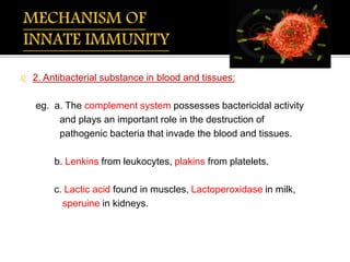
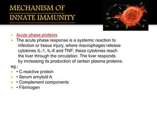
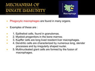
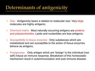
Immunity
Definitions
Components of Immune system
Types
Innate immunity and Mechanism
Adaptive immunity and Mechanism
2. Antigen
Origin of Antigen
Immunogen
3. Antibody- Immunoglobulin
- Structure
- Classification
- Function of each antibody
Immunity is the balanced state of multicellular organisms having adequate biological defenses to fight infection, disease, or other unwanted biological invasion, while having adequate tolerance to avoid allergy, and autoimmune diseases.
presentation includes definition of immunity, its various types, cells of immunity in our body and their working and the various diseases associated immunity deficiency
The term immunity refers to the body’s specific protective response to an invading foreign agent or organism.
The human body has the ability to resist almost all types of organisms or toxins that tend to damage the tissues and organs. The capability is called immunity.
Defence mechanism in finfish and shellfish jassi 2Jaspreet Singh
Farming of fish and shellfish has gained significant grounds in several parts of the world .
Now a days disease is main problem in aquafarming.
In recent years ,lot of attention is being given to health management using various forms of immunoprophylactic techniques such as vaccination and immunostimulation .
To reduce immunoprophylactic application ,it is vital to have insight into specific and non specific defense mechanism of farmed animal .
Through disease process studies ,it is very well known that a pathogen can cause disease only if it can overcome the non specific and specific defense barriers of the host and successfully establish and proliferate
Indian Dental Academy: will be one of the most relevant and exciting training center with best faculty and flexible training programs for dental professionals who wish to advance in their dental practice,Offers certified courses in Dental implants,Orthodontics,Endodontics,Cosmetic Dentistry, Prosthetic Dentistry, Periodontics and General Dentistry.
Flu Vaccine Alert in Bangalore Karnatakaaddon Scans
As flu season approaches, health officials in Bangalore, Karnataka, are urging residents to get their flu vaccinations. The seasonal flu, while common, can lead to severe health complications, particularly for vulnerable populations such as young children, the elderly, and those with underlying health conditions.
Dr. Vidisha Kumari, a leading epidemiologist in Bangalore, emphasizes the importance of getting vaccinated. "The flu vaccine is our best defense against the influenza virus. It not only protects individuals but also helps prevent the spread of the virus in our communities," he says.
This year, the flu season is expected to coincide with a potential increase in other respiratory illnesses. The Karnataka Health Department has launched an awareness campaign highlighting the significance of flu vaccinations. They have set up multiple vaccination centers across Bangalore, making it convenient for residents to receive their shots.
To encourage widespread vaccination, the government is also collaborating with local schools, workplaces, and community centers to facilitate vaccination drives. Special attention is being given to ensuring that the vaccine is accessible to all, including marginalized communities who may have limited access to healthcare.
Residents are reminded that the flu vaccine is safe and effective. Common side effects are mild and may include soreness at the injection site, mild fever, or muscle aches. These side effects are generally short-lived and far less severe than the flu itself.
Healthcare providers are also stressing the importance of continuing COVID-19 precautions. Wearing masks, practicing good hand hygiene, and maintaining social distancing are still crucial, especially in crowded places.
Protect yourself and your loved ones by getting vaccinated. Together, we can help keep Bangalore healthy and safe this flu season. For more information on vaccination centers and schedules, residents can visit the Karnataka Health Department’s official website or follow their social media pages.
Stay informed, stay safe, and get your flu shot today!
The prostate is an exocrine gland of the male mammalian reproductive system
It is a walnut-sized gland that forms part of the male reproductive system and is located in front of the rectum and just below the urinary bladder
Function is to store and secrete a clear, slightly alkaline fluid that constitutes 10-30% of the volume of the seminal fluid that along with the spermatozoa, constitutes semen
A healthy human prostate measures (4cm-vertical, by 3cm-horizontal, 2cm ant-post ).
It surrounds the urethra just below the urinary bladder. It has anterior, median, posterior and two lateral lobes
It’s work is regulated by androgens which are responsible for male sex characteristics
Generalised disease of the prostate due to hormonal derangement which leads to non malignant enlargement of the gland (increase in the number of epithelial cells and stromal tissue)to cause compression of the urethra leading to symptoms (LUTS
Lung Cancer: Artificial Intelligence, Synergetics, Complex System Analysis, S...Oleg Kshivets
RESULTS: Overall life span (LS) was 2252.1±1742.5 days and cumulative 5-year survival (5YS) reached 73.2%, 10 years – 64.8%, 20 years – 42.5%. 513 LCP lived more than 5 years (LS=3124.6±1525.6 days), 148 LCP – more than 10 years (LS=5054.4±1504.1 days).199 LCP died because of LC (LS=562.7±374.5 days). 5YS of LCP after bi/lobectomies was significantly superior in comparison with LCP after pneumonectomies (78.1% vs.63.7%, P=0.00001 by log-rank test). AT significantly improved 5YS (66.3% vs. 34.8%) (P=0.00000 by log-rank test) only for LCP with N1-2. Cox modeling displayed that 5YS of LCP significantly depended on: phase transition (PT) early-invasive LC in terms of synergetics, PT N0—N12, cell ratio factors (ratio between cancer cells- CC and blood cells subpopulations), G1-3, histology, glucose, AT, blood cell circuit, prothrombin index, heparin tolerance, recalcification time (P=0.000-0.038). Neural networks, genetic algorithm selection and bootstrap simulation revealed relationships between 5YS and PT early-invasive LC (rank=1), PT N0—N12 (rank=2), thrombocytes/CC (3), erythrocytes/CC (4), eosinophils/CC (5), healthy cells/CC (6), lymphocytes/CC (7), segmented neutrophils/CC (8), stick neutrophils/CC (9), monocytes/CC (10); leucocytes/CC (11). Correct prediction of 5YS was 100% by neural networks computing (area under ROC curve=1.0; error=0.0).
CONCLUSIONS: 5YS of LCP after radical procedures significantly depended on: 1) PT early-invasive cancer; 2) PT N0--N12; 3) cell ratio factors; 4) blood cell circuit; 5) biochemical factors; 6) hemostasis system; 7) AT; 8) LC characteristics; 9) LC cell dynamics; 10) surgery type: lobectomy/pneumonectomy; 11) anthropometric data. Optimal diagnosis and treatment strategies for LC are: 1) screening and early detection of LC; 2) availability of experienced thoracic surgeons because of complexity of radical procedures; 3) aggressive en block surgery and adequate lymph node dissection for completeness; 4) precise prediction; 5) adjuvant chemoimmunoradiotherapy for LCP with unfavorable prognosis.
MANAGEMENT OF ATRIOVENTRICULAR CONDUCTION BLOCK.pdfJim Jacob Roy
Cardiac conduction defects can occur due to various causes.
Atrioventricular conduction blocks ( AV blocks ) are classified into 3 types.
This document describes the acute management of AV block.
These lecture slides, by Dr Sidra Arshad, offer a quick overview of physiological basis of a normal electrocardiogram.
Learning objectives:
1. Define an electrocardiogram (ECG) and electrocardiography
2. Describe how dipoles generated by the heart produce the waveforms of the ECG
3. Describe the components of a normal electrocardiogram of a typical bipolar leads (limb II)
4. Differentiate between intervals and segments
5. Enlist some common indications for obtaining an ECG
Study Resources:
1. Chapter 11, Guyton and Hall Textbook of Medical Physiology, 14th edition
2. Chapter 9, Human Physiology - From Cells to Systems, Lauralee Sherwood, 9th edition
3. Chapter 29, Ganong’s Review of Medical Physiology, 26th edition
4. Electrocardiogram, StatPearls - https://www.ncbi.nlm.nih.gov/books/NBK549803/
5. ECG in Medical Practice by ABM Abdullah, 4th edition
6. ECG Basics, http://www.nataliescasebook.com/tag/e-c-g-basics
Explore natural remedies for syphilis treatment in Singapore. Discover alternative therapies, herbal remedies, and lifestyle changes that may complement conventional treatments. Learn about holistic approaches to managing syphilis symptoms and supporting overall health.
Title: Sense of Smell
Presenter: Dr. Faiza, Assistant Professor of Physiology
Qualifications:
MBBS (Best Graduate, AIMC Lahore)
FCPS Physiology
ICMT, CHPE, DHPE (STMU)
MPH (GC University, Faisalabad)
MBA (Virtual University of Pakistan)
Learning Objectives:
Describe the primary categories of smells and the concept of odor blindness.
Explain the structure and location of the olfactory membrane and mucosa, including the types and roles of cells involved in olfaction.
Describe the pathway and mechanisms of olfactory signal transmission from the olfactory receptors to the brain.
Illustrate the biochemical cascade triggered by odorant binding to olfactory receptors, including the role of G-proteins and second messengers in generating an action potential.
Identify different types of olfactory disorders such as anosmia, hyposmia, hyperosmia, and dysosmia, including their potential causes.
Key Topics:
Olfactory Genes:
3% of the human genome accounts for olfactory genes.
400 genes for odorant receptors.
Olfactory Membrane:
Located in the superior part of the nasal cavity.
Medially: Folds downward along the superior septum.
Laterally: Folds over the superior turbinate and upper surface of the middle turbinate.
Total surface area: 5-10 square centimeters.
Olfactory Mucosa:
Olfactory Cells: Bipolar nerve cells derived from the CNS (100 million), with 4-25 olfactory cilia per cell.
Sustentacular Cells: Produce mucus and maintain ionic and molecular environment.
Basal Cells: Replace worn-out olfactory cells with an average lifespan of 1-2 months.
Bowman’s Gland: Secretes mucus.
Stimulation of Olfactory Cells:
Odorant dissolves in mucus and attaches to receptors on olfactory cilia.
Involves a cascade effect through G-proteins and second messengers, leading to depolarization and action potential generation in the olfactory nerve.
Quality of a Good Odorant:
Small (3-20 Carbon atoms), volatile, water-soluble, and lipid-soluble.
Facilitated by odorant-binding proteins in mucus.
Membrane Potential and Action Potential:
Resting membrane potential: -55mV.
Action potential frequency in the olfactory nerve increases with odorant strength.
Adaptation Towards the Sense of Smell:
Rapid adaptation within the first second, with further slow adaptation.
Psychological adaptation greater than receptor adaptation, involving feedback inhibition from the central nervous system.
Primary Sensations of Smell:
Camphoraceous, Musky, Floral, Pepperminty, Ethereal, Pungent, Putrid.
Odor Detection Threshold:
Examples: Hydrogen sulfide (0.0005 ppm), Methyl-mercaptan (0.002 ppm).
Some toxic substances are odorless at lethal concentrations.
Characteristics of Smell:
Odor blindness for single substances due to lack of appropriate receptor protein.
Behavioral and emotional influences of smell.
Transmission of Olfactory Signals:
From olfactory cells to glomeruli in the olfactory bulb, involving lateral inhibition.
Primitive, less old, and new olfactory systems with different path
Factory Supply Best Quality Pmk Oil CAS 28578–16–7 PMK Powder in Stockrebeccabio
Factory Supply Best Quality Pmk Oil CAS 28578–16–7 PMK Powder in Stock
Telegram: bmksupplier
signal: +85264872720
threema: TUD4A6YC
You can contact me on Telegram or Threema
Communicate promptly and reply
Free of customs clearance, Double Clearance 100% pass delivery to USA, Canada, Spain, Germany, Netherland, Poland, Italy, Sweden, UK, Czech Republic, Australia, Mexico, Russia, Ukraine, Kazakhstan.Door to door service
Hot Selling Organic intermediates
3. Immunology- branch of biology concerned
with the body’s defence reactions.
Derived from the latin word ‘immunis’ meaning ‘free of burden’.
Maintain the integrity of the body – removing the myriad of
threatening micro organisms which could invade from the
environment.
Internally, mutant cells with malignant potential, may also be
attacked by the immune system.
4. 800 B.C. - Sushruta – “Father of surgery” in India, used patient’s
own skin as ‘graft’ in case of severed nose. At this time, basis of
‘Immunity’ was not established.
1798 – Edward Jenner, gave the first description of ‘cell mediated
immune’ response after injecting ‘vaccinia virus’ as vaccine for
‘small pox’. A local erythematous papule developed within 24-72
hrs. He called it as just as ‘Reaction of Immunity’.
5. 1890 - Koch, described an exaggerated cutaneous reaction to
intradermal injection of ‘tubercle bacillus’ which was a paradigm for
delayed hypersensitivity.
1898 - Bordet, described ‘alexine’, Ehrlich, coined the term
‘complement’ for the factors which completes the immune response.
1902 – Paul Ehrlich, described ‘horror autotoxicosis’ which was later
called as ‘autoimmune reaction’.
6. 1902 - Richet, coined the term ‘Anaphylaxis’.
1903 – Wright, described ‘opsonisation’ & Arthus, described ‘arthus
reaction’.
1905 – Von pirquet & Schick, described ‘serum sickness’.
1906 – Von pirquet, coined the term ‘allergy’.
7. 1923 – Coca and Cooke, described ‘atopy’.
1963 - Coombs & Gell, classified hypersensitivity reactions.
1967 - Burnet, described ‘immuno surveillance’
1975 – Kohler & Milstein, gave a large scale production of
monoclonal antibodies, for which they have received ‘noble prize for
medicine’ in 1984.
1980 – Snell, Dausset & Benaceraff, described about Major Histo
Compatibility antigens & its genetic control in immune response.
8. Immunity:
Resistance exhibited by the host towards injury caused by
pathogenic microorganisms and their products.
9. •Recognition
•Antibody production
•Cell mediated reactions
•Memory
BODY DEFENSE
NORMAL
IMMUNNE
DEFICIENCY
DISORDERS
HYPERSENSITIVITY
REACTIONS
AUTOIMMUNE
DISEASES
ABNORMAL
IMMUNOPHYSIOLOGY
IMMUNOPATHOLOGY
10. • NON – SPECIFIC
• SPECIFICINNATE
IMMUNITY
• ACTIVE
• PASSIVEADAPTIVE
IMMUNITY
11. Innate Immunity:
the resistance to infections which an individual posses by virtue of
his genetic and constitutional makeup.
Non-specific : degree of resistance to infection in general (to all
infections) eg. plant pathogens.
Specific : degree of resistance to particular pathogens.
divided into Species immunity, racial immunity and, individual
immunity.
12. Species Immunity: total or relative refractoriness to a
pathogen shown by all members of a species.
eg: All human beings are not susceptible to plant pathogens.
Racial Immunity: within species, different races may show
differences in susceptibility to infections.
eg: Negros are resistant to yellow fever and malaria.
Individual Immunity: exhibited by different individuals in a race.
eg: Homozygous twins exhibit similar degree of resistance to
leprosy and tuberculosis. Such correlations are not seen in
heterozygous twins.
13. 1.Epithelial surfaces:
The intact skin and mucous membrane covering the body
protect considerably against invasion of microorganisms.
Healthy skin possess anti bacterial activity in form of high
concentration salt in the drying sweats, the sebaceous secretion
Mucosa of respiratory tract has several innate mechanisms of
defense (cough reflex, secretion contain mucopolysaccharides
capable of neutralizing the bacterial products).
14. The mouth is constantly bathed in saliva, which has an
inhibitory effect on many microorganisms.
IgA, T-helper and suppressor cell, B-cell of the IgA, and IgM
isotopes and macrophages.
High acidity of the stomach destroys most of the microbes.
Conjunctiva by lacrymal secretion, tears contains the
antibacterial substance lysozyme.
15. 2. Antibacterial substance in blood and tissues:
eg. a. The complement system possesses bactericidal activity
and plays an important role in the destruction of
pathogenic bacteria that invade the blood and tissues.
b. Lenkins from leukocytes, plakins from platelets.
c. Lactic acid found in muscles, Lactoperoxidase in milk,
speruine in kidneys.
16. 3. Fever : physiological process that destroys the infecting
pathogens. Fever stimulates the production of interferon and aids
recovery from viral infections.
5.Acute phase proteins (CRP): Infection or injury leads to production
of CRP, which activate the alternate pathway of complement.
17. Acute phase proteins
The acute phase response is a systemic reaction to
infection or tissue injury, where macrophages release
cytokines IL-1, IL-6 and TNF; these cytokines reach
the liver through the circulation. The liver responds
by increasing its production of certain plasma proteins.
eg.:
• C-reactive protein
• Serum amyloid A
• Complement components
• Fibrinogen
18. Cells of the innate immune system:
Phagocytes:
Phagocytes belong to two major lineage
1. Monocyte / macrophage
2. PMN granulocyte
A. Neutrophil
B. Basophil
C. Eosinophil
19. The mononuclear phagocyte system has two main functions, which
result from the activities of two different types of bone marrow
derived cells.
A. ‘Professional’ phagocytic macrophages, whose main role is to
remove particulate antigens.
B. Antigen presenting cells, whose role is to take up, process and
present antigenic peptide to T cells.
20. The term macrophages is generally applied to the cells found free in
body cavities while histiocytes is applied to cells found fixed in
tissues.
They possess:
CR1, CR3, CR4, C5aR receptors and molecules important in
antigen presenting to an MHC class II receptor, CD1.
21. Macrophages secrete molecules, engulf and remove excess
antigen and aid in the induction of immune response of presenting
determinants to T cells.
An organism that successfully penetrates an epithelial surface
encounter phagocyte cells of the monocyte macrophage lineage.
22. Phagocytic macrophages are found in many organs.
Examples of these are :
1. Epitheliod cells, found in granulomas.
2. Myeloid progenitors in the bone marrow.
3. Kupffer cells are long lived resident liver macrophages.
4. Dendritic cells are characterized by numerous long, slender
processes and by irregularly shaped nuclei.
5. Multinucleated giant cells are formed by the fusion of
macrophages.
23. Polymorphonuclear granulocytes:
PMN granulocytes mainly consist of neutrophills and differentiate
completely with in bone marrow (14 days) are released from the
bone marrow at a rate of around 7 million per minute.
They are short lived (2-3 days) relative to monocytes
/macrophages, which many live months or years.
Granulocytes do not show any inherent specificity for antigens, but
they play an important role in acute inflammation, which provides a
primary nonspecific internal defense mechanisms.
24. Chemotactic stimuli result in neutrophil margination and diapedesis.
Neutrophils have a large arsenal of antibiotic proteins stored in two
main types of granules.
The primary granules are lysosomes containing acid hydrolase,
myeloperoxidase and muranidase.
The secondary granules contain lactoferrin and lysozyme.
In addition the granules also contain the antibiotic proteins
defensins, seprocidins, cathelicidins and bacterial permeability
inducing proteins.
25. Eosinophils :
2-5%of blood leukocytes in healthy, non-allergic individuals.
Eosinophils limit inflammatory reactions of antagonizing the effects
of mediators.
Release histaminase and arylsulphatase, which inactivates the
mast cell products histamine and some of the leukotriene.
Eosinophils contain granules with eosinophilic basic protein which
are found to be toxic to certain parasites.
26. Basophils:
Very small numbers in the circulation, accounting for less than 0.2%
of leucocytes.
Contain primarily histamine and leukotrienes which are potent
spasmogenic agents causing constriction of smooth muscle.
Granules of basophils contain heparin, leukotrienes, histamine and
eosinophil chemotactic factor for anaphylaxis.
The stimulus basophil degranulation is often an allergen. Mediators
such as histamine released by degranulation, cause the adverse
symptoms of allergy.
On the positive side, they also play a role in immunity against
parasites by enhancing inflammation.
27. Mast cells: The mast cell, which is not found in the circulation is
indistinguishable from the basophil.
2 kinds a) mucosal mast cell
b) connective tissue mast cell
Important in immediate inflammation -possess receptors for
complement components (C3a and C5a) as well as receptors for
the Fc portion of the antibody molecules IgE and IgG.
Feature prominent cytoplasmic granules, termed lysosymes, which
store inflammatory mediators such as histamine, eosinophil
chemotactic factor, neutrophil chemotactic factor and heparin.
28. Stimulation of these receptors > secretion of vasoactive substances
> increase vascular permeability and dilation (important signs of
anaphylaxis.)
Analphylaxis can be life threatening if it is widespread (systemic)
but it is usually localized and is important in initiating inflammatory
response against local microbial invasion.
Mast cells can synthesize denovo, other inflammatory mediators,
such as slow-reacting substance of anaphylaxis (SRS-A), tumor
necrosis factor (TNF-) and leukotriene .
In addition mast cell interleukin has been shown to enhance
collagenase activity and heparin -may augment bone resorption.
29. Platelets:
Induce immune response and especially in inflammation.
Derived from megakaryocytes in the bone marrow following injury to
endothelial cells, platelets aggregate at, the endothelial surface of
damaged vascular tissue.
Release two types of granules which include serotonin and
fibrinogen > increased permeability, activation of complement
> attraction of leucocytes
30. From a anatomic stand point, the immune system has a
a) Fixed compartment distributed through
Bone marrow
Thymus
Spleen
Lymph nodes
Tonsils
Peyer’s Patches
b) Circulatory compartment represented by
diverse lymphocytes that move via the blood stream
into lymphoid organs and other part of the body where needed,
returning to the blood via the lymphatic channels.
31. Cells, tissues and organs of the immune system:
Lymphoid cells:
Organized into tissues and organs in order to perform their
functions more effectively -lymphoid system.
Comprises lymphocytes, accessory cells (macrophages) and in
some tissues, epithelial cells.
The major lymphoid organs and tissues are classified into either
primary (central) or secondary (peripheral).
32. Primary lymphoid organs:
Major sites of lymphocyte development.
The primary or central lymphoid organs include the
Thymus and Bone marrow.
Secondary lymphoid organs:
Generation of lymphocytes is followed by their migration into
peripheral secondary tissues.
Comprise well organized encapsulated organs, the spleen and
lymphnodes and non-encapsulated accumulations of lymphoid
tissue (Mucosa Associated Lymphiod Tissue).
33. Resistance that an individual acquires during his lifetime, which
may be weak or absent on first exposure but that increases
dramatically with subsequent exposures to same specific
pathogen.
35. Active immunity -Resistance developed by an individual as a
result of an antigenic stimulus.
Involves the active functioning of persons immune apparatus
leading to the synthesis of antibodies and production of
immunologically active cells.
Natural active immunity - acquired due to infection
Artificial active immunity - vaccination
eg. Bacterial vaccines: live (BCG) vaccine
killed (cholera) vaccine
Viral vaccines: live oral polio vaccine - sabine
killed injectable polio vaccine - salk
36. Passive immunity -Resistance that is transmitted to a
recipient in a readymade form.
No antigenic stimulus, instead preformed antibodies
are administered.
Natural passive immunity
A mother’s antibodies pass across the placenta to
the foetus and remain for several months. EG:- IgG
antibody.
Artificial passive immunity
Used when a very rapid immune response is needed
e.g. after infection with tetanus
Antibodies come from blood donors who have
recently had the tetanus vaccination.
37. Combined immunization: a combination of active and passive
method of immunization is employed.
Adoptive immunity: Special type of immunization in which
immunologically competent lymphocytes are injected.
Herd immunity: this refers to overall level of immunity in a
community and is relevant in the control of epidemic diseases.
38. Cells of acquired immune system:
Lymphocytes : lymphocytes includes three types of cells.
T-lymphocytes or T cells, which are derived from the thymus and
play a role in cell-mediated immunity.
B-lymphocytes or B-cells which are derived from liver, spleen and
bone marrow are precursors of plasma cells and play a role in
humoral immunity.
Natural killer (NK) and killer (K) cells.
39. T cells leave the bone marrow via the blood stream and move to
the thymus.
T cell becomes able to differentiate between self and nonself
antigens.
This process involves the immature T cells being presented with
cell surface molecules termed major histocompatibility complex
(MHC) molecules, of which there are two types termed class I
and class II.
40. In the thymus, those T cells which react with very strongly or very
weakly with self MHC molecules are destroyed.
The remaining T cells then mature and are able to recognize foreign
antigens in conjunction with MHC molecules.
The T cells recognize antigen through surface molecules termed the
T cell receptor (TCR).
Each T cell possesses a different TCR to allow it to recognize a
different antigen. After leaving the thymus T cells locate in lymph
nodes and spleen.
41. The T lymphocytes are associated with two types of immunological
functions, effector and regulatory.
The effector functions include activities such as killing of virally
infected cells and tumors.
The regulatory functions are represented by their ability to amplify
or suppress through cytokines or other effector lymphocytes
including B and T cells.
42. T cells are divided into several subsets with different functions.
1. T helper cell/T-inducer cells (Th)
2. T-suppressor cells (Ts)
3. T-cytotoxic cells (Tc)
43. 1.T helper cells/T-inducer cells:
T cells have CD4 surface marker, MHC class II restriction,
stimulates the growth of T cells and macrophages.
TH1 cells with CD4 molecule produce cytokines IL-2 which activate
macrophages, IFN-y -involved in hypersensitivity reactions.
TH2 express CD4 molecule on their surface and function by
secreting cytokines (IL-4, IL-5, IL-6, IL-10) that promote B cells to
produce antibodies.
44. T-suppressor cells:
Suppressor effector T cells bind antigen and release factor that
inactivate T-helper cells.
T-suppressor cells can
(1) suppress delayed –type hypersensitivity reactions
(2) prevent proliferation and antibody secretion by antigen-
binding B cells, and
(3) suppress antibody secretion by having CD8 surface marker
and MHC class I restriction.
45. T-Cytotoxic cells:
These cells recognize certain histocompatibility antigens and are
capable of killing foreign cells (i.e., virus) and altered self-cells (i.e.
tumor antigens).
Important in the cytotoxicity of graft reactions and graft-versus-host
reactions.
46. B lymphocytes are primarily defined by surface immunoglobulins
S(Ig).
Common in areas of antibody production, such as the germinal
centers of the lymph nodes and diffuse lymphoid tissue of mucosal
systems.
B lymphocyte development and maturation results in the formation
of plasma cells, the antibody producing cells of the body.
Bone marrow -major repository of stem cells for B lymphocytes.
47. Mature B lymphocytes are the products of lymphoid stem cells that
undergo a sequence of differentiation.
In mammals the differentiation occurs first in the fetal liver and
subsequently in the bone marrow.
The B cell development can be divided into two stages, antigen
dependent and antigen independent.
On encountering an antigen, the mature B cells are driven to
undergo cell activation, proliferation and differentiation, giving rise to
plasma cells that synthesize and secrete Ig, (Ig M, IgD, IgG, IgA
,IgE ) which requires T cell. However they seldom divide and have a
life span of 2 to 3 days.
48. Separate lineage of B lymphocytes -predominant in fetal and early
neonatal life, express the T cell marker CD5 on their surface -
named as B1 cells.
They secrete low affinity poly reactive IgM antibodies.
Responsible for the T independent natural IgM antibacterial
antibodies which appear in neonates without antigenic stimulus.
CD5+ B cells may be relevant in the casuation of autoimmune
conditions.
49. Circulating lymphocytes are classified by their surface markers into
T and B lymphocytes, about 5 – 10 %of the cells are found to lack
features of either type -called NULL cells.
Large Granular Lymphocytes having heterogenous group of cells
with differences in their functional and surface marker features. The
most important of this group is called NATURAL KILLER CELLS.
NK cells -destroy antibody-coated target cells irrespective of the
presence of MHC molecules, a process known as antibody-
dependent cell mediated cytotoxicity. This occurs because killing is
initiated by cross-linking of receptors for the Fc portion of IgG1 and
IgG3.
NK cells are not clonally restricted, have no memory and are not
very specific in their action.
50. An antigen has been defined as any substance which, when
introduced partially into the body, stimulates the production of an
antibody with which it react specifically.
An antigen introduced into the body reacts only with those
particular immunocytes which carry the specific marker for that
antigen and which produce antibody complimentary to that antigen
only.
The discrete portions of the antigen that bind to the antibodies
antigen combining site are called epitopes.
51. An antigen may possess several epitopes. The containing area on
the antibody molecule corresponding to the epitope is called
paratope.
Epitope consists of four or five amino acids possessing a specific
chemical structure, electric charge and spatial configuration capable
of sensitizing an immunocyte and of reacting with its
complementary site on the specific antibody or T-cell receptor.
52. Complete antigen: Is able to induce antibodies formation and
produce a specific and observable reaction with the antibody
produced.
Haptens: Low molecular weight molecules which by itself do not
elicit immune response until and unless complexed with an
immunogenic carrier, such as protein.
53. Size : Antigenicity bears a relation to molecular size. Very large
molecules are highly antigenic.
Chemical matrix : Most naturally occurring antigens are proteins
and polysaccharides. Lipids and nucleotides are less antigenic.
Susceptibility to tissue enzymes : Only substances which are
metabolized and are susceptible to the action of tissue enzymes
behave as antigens.
Foreignness : Only antigen which are ‘foreign’ to the individual (non
self) induce an immune response. Breakdown of this homeostatic
mechanism result in autoimmunization and auto immune disease.
54. Depending on their ability to induce antibody formation, antigens
are classified.
Antigens
T cell dependent
(TD) antigens
T cell
independent (TI)
antigens
Biological classes of antigens
55. T cell dependent (TD)
antigens
T cell independent (TI)
antigens
STRUCTURALLY SIMPLE STRUCTURALLY COMPLEX
NO. OF EPITOPES LIMITED NO. OF EPITOPES UNLIMITED
IMMUNOLOGICAL MEMORY-DO NOT
SHOW
IMMUNOLOGICAL MEMORY- SHOW
METABOLISEDVERY SLOWLY METOBILISED RAPIDLY
REMAIN IN BODY FOR LONG PERIODS REMAIN IN BODY FOR SHORT
PERIODS
IMMUNE RESPONSE DOSE
DEPENDENT
IMMUNE RESPONSE NOT DOSE
DEPENDENT
ANTIBODY RESPONSE IgM & IgG3 ANTIBODY RESPONSE IgM, IgG, IgA &
IgE
56. Processing involves the internalization and partial degradation of
antigens by antigen-presenting cells (APCs).
The resultant fragments of antigen are transported back to the APC
surface, where they are bound by class II histocompatibility
antigens.
The antigen fragment class II complex is subsequently recognized
by T cells bearing complementary T cell receptors.
57. Antigen presenting cells are
1. Macrophages
2. Dendrites cells
3. B cells during secondary immune response
4. Langerhans cells
58. The genetic basis of immune response, which had been suggested
by many early observations ,was proved by BENACERRAF and
COLLEAGUES, who established that the ability to respond
immunologically to an antigen was conditioned by specific genes
called the immune response genes.
For this work on MHC and genetic control of immune response,
SNELL ,DUSSET and BENACERRAF were awarded the nobel
prize for Medicine in 1980.
59. The major antigens determining histocompatibility in human beings
are alloantigens, characteristically found on the surface of
leucocytes.
Human MHC antigens are therefore synonymous with human
leukocyte antigen(HLA).
60. The HLA complex of genes is located on the short arm of
chromosome 6.
It consists of 3 separate clusters of genes.
1. Class I consisting of A, B and C loci.
Determine histocompatibility -acceptance or rejection of allografts.
2. Class II or the D region consisting of D or DQ and DP loci.
Regulate immune response.
3. Class III or the complement region containing genes for
complement components C2 and C4 of the classical pathway
as well as properdin factor B of alternative pathway.
61. HLA Class I molecules consists of a heavy peptide chain (α
chain) non convalently linked to a much smaller peptide called
β2 microglobulin (β chain). The β chain has a constant
aminoacid sequence and is coded for by a gene on
chromosome 15.
The α chain consists of three globoid domains ( α1, α 2, α3)
which protrude from the cell membrane and a small length of
transmembrane C terminus reaching into the cytoplasm.
The distal domains α1 & α 2 have highly variable amino acid
sequences and are folded to form a cavity or groove between
them.
63. Protein antigens processed by macrophages or dendritic cells to
form small peptides are bound to groove for presentation to CD8 T
cells.
T cell will recognize the antigen only when presented as a complex
with the MHC class I molecule and not otherwise (MHC restriction).
When so presented, the CD8 cytotoxic killer cell destroys the target
cell ( for example, a virus infected cell).
Class II antigens are heterodimers, consisting of an α and β chain.
64. HLA class II antigens are heterodimers, consisting of an alpha and
beta chain .Each chain has 2 domains, the proximal domain being
the constant region and the distal the variable.
The two distal domains constitute the antigen binding site, for
recognition by CD4 lymphocytes, in a fashion similar to the
recognition of the class I antigen peptide complex by CD8 T cells.
HLA class II molecules are primarly responsible for the graft versus
host response and the mixed leucocyte reaction .
HLA class III molecules are heterogenous. They include
complement components linked to the formation of C3 convertases,
heat shock protiens and tumour necrosis factors. They also show
polymorphysim.
66. The host responds to oral bacteria and their products by plasma cell
production of immunoglobulins or antibodies.
In 1964 endorsed by WHO, the generic term, “Immunoglobulin” was
internationally accepted for proteins of animal origin endowed with
known antibody activity and for certain other protein related to them
by chemical structure.
Immunoglobulin provides a structural and chemical concept, while
the term ‘antibody’ is a biological and functional concept.
ALL THE ANTIBODIES ARE IMMUNOGLOBULINS BUT ALL
IMMUNOGLOBULINS MAY NOT BE ANTIBODIES.
67. Antibodies which are glycoproteins, are found in blood, tissue fluids
and secretions and are the effectors of humoral immunity. They are
highly specific and sensitive.
All classes and subsets of immunoglobulins have similar structural
organizations, but they differ according to their biologic properties,
carbohydrate content, weight and amino acid sequences.
Every antibody molecule has a variable region which, because of its
unique amino acid sequence and tertiary structure of its antibody
combining site, allows it to react highly specifically with a particular
antigen.
68. Studies involving the cleavage of the Ig molecule, pioneered by
Porter, Edelman and their colleageus, have led to a detailed
picture of its structure.
Each molecule of Ig is split into one Fc portion (crystallisable),
and two Fab fragments (antigen binding).
Ig constitutes 20-25% of the total serum proteins.
Based on physiochemical and antigenic differences, five classes
of Ig have been recognized - IgG, IgA, IgM, IgD and IgE. (Both Ig
and g are accepted abbreviations for immunoglobulins).
69.
70. All the immunoglobulin constant region domains (C) maintain a
similar basic structure. The variable region domains (V) have a
slightly different structure. The polypeptide strand connecting the V
and C domains is called the hinge and is important to and body
structure because of the flexibility it allows between the V and C
domains. Heavy chains can be divided into three functional regions;
Fd, hing and Fc.
The Fd in combination with a light chain forms the Fab region,
which has all the antigen-binding properties of an intact
immunoglobulin.
The Fc fragment bears the effector functions of the immunoglobulin
molecule
71. Immunoglobulins (antibodies):
IgM is the first antibody to arrive in our body. It initiates the
complement cascade and is the main antibody in response to T
independent antigens.
IgG is the next antibody to arrive; it remains longest.
Having several subclasses, IgG1, IgG2, IgG3 and IgG4 is also the
most predominant antibody. It coats antigens (opsonization) for
destruction by phagocytes, prepares other antigens for destruction
by killer cells.
72. IgA is found in the saliva (secretory IgA) and other areas
where there is mucous membrane. It is the first line of
defense against microorganisms that would invade via the
mucosa, IgA antibodies deter microorganisms from
penetrating epithelial cells. it is composes of two subclasses
IgA1 and IgA2.
IgD is a trace antibody and differentiates B cells. it
disappears after differentiation.
IgE is the reaginc antibody it is in low concentration and
responsible for acute allergic reactions, binds to mast cells
and basophils stimulating the release of vasoactive
substances such as histamine, prostaglandins and
leukotrienes.
73. Type Number of
ag binding
sites
Site of action Functions
IgG 2 •Blood
•Tissue fluid
•CAN CROSS
PLACENTA
•Increase
macrophage
activity
•Antitoxins
•Agglutination
IgM 10 •Blood
•Tissue fluid
Agglutination
IgA 2 or 4 •Secretions (saliva,
tears, small
intestine, vaginal,
prostate, nasal,
breast milk)
•Stop bacteria
adhering to host
cells
•Prevents bacteria
forming colonies
on mucous
membranes
IgE 2 Tissues •Activate mast
cells
HISTAMINE
•Warm response
74. Recent investigations have revealed that not all
strains of a specific microbial species are equal in
their capacity to cause disease and not all hosts are
equal in their susceptibility to disease.
The challenge for the future is to be able to better
identify the more virulent bacterial strains and more
susceptible host.
In this manner it may be possible to predict
accurately the individuals at risk for future disease
and to develope more effective strategies to prevent
the onset and progression of disease.