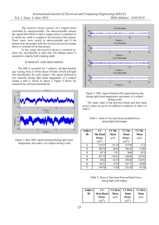
The document describes a brain-computer interface (BCI) system that uses electroencephalography (EEG) to classify motor imagery of the left or right arm and control an assistive device for paralyzed upper limbs. EEG signals are recorded over motor cortex areas during right and left arm imagery tasks. The mu and beta frequency bands are extracted and used to classify intended movement based on features like power and mean. If right arm imagery is classified, a stepper motor attached to the patient's forearm is activated to help lift their arm. The system was tested on 7 subjects with over 10 trials each, achieving classification of intended movement.
![International Journal of Electrical and Computing Engineering (IJECE)
Vol. 1, Issue. 4, June 2015 ISSN (Online): 2349-8218
21
Assistive Device for paralyzed upper limbs using BCI
Ramkumar.B
Department of ECE,
College of Engineering, Guindy
Anna University, Chennai
ramkumar.ssn.eee@gmail.com
Shenbaga Devi.S
Professor,
Department of ECE,
College of Engineering, Guindy
Anna University, Chennai
Abstract: A Human assistive device to lift the forearm of the
patient using Brain-Computer Interface (BCI) was designed.
The electroencephalographic (EEG) activity is used as the
basis for a brain-computer interface (BCI) that could be a new
alternative communication channel forpatients lacking useful
voluntary movement. The intended movement of arms of the
patient during motor imagery is classified by sensory motor
rhythms (Mu and Beta band) in the EEG signals obtained
during the process of imagination of arm movement.
Keywords: Brain Computer Interface, BCI, EEG,
Electroencephalogram, Assistive Device
I INTRODUCTION
A Brain Computer Interface (BCI) is a system
through which a person can control the external world
without relying on muscle activity. BCIs are used for
assisting, augmenting, repairing human cognitive or
sensory-motor functions. An electroencephalogram (EEG)
based Brain-Computer-Interface (BCI) provides a
communication channel between the human brain and a
computer. Patients who are suffering from motor
impairments may use such a BCI system as an alternative
form of communication by mental activity. In this work, the
non-invasive BCI is designed. Non-invasive BCIs aim to
either restore movement in individuals with paralysis or
provide devices to assist them, by the use of computers or
robot arms.
BCI systems are designed for individuals with
motor disabilities to communicate with the outside world.
One of the major factors of stroke is rehabilitation and is
limited with 30 to 60% of patients being unable to use their
more affected limb. Paralysis is the loss of muscle function
in a part of our body. It occurs when something goes wrong
with the way messages pass between human brain and
muscles. Paralysis is most often caused by damage in
nervous system and especially in spinal cord. Paralysis can
be complete or partial. Impairment of motor function, such
as hemiparesis or hemiplegia of the upper limbs and lower
limbs. Recovery of motor function is important to do daily
activities of the patient.
Event Related Desynchronization (ERD) is a
reduction of amplitude in a specific frequency component
and is related to an increase in neural activity. A negative
ERD percentage indicates that there is a decrease in power
with respect to the reference state and a positive value
means there is an increase in power. Event Related
Synchronization (ERS) is an increase in a specific
frequency component and is related to neural suppression.
Activity invoked by right hand movement imagery is most
prominent over electrode location C3 and left hand
movement imagery produces activity most prominent over
electrode location C4.
An EEG based brain-computer interface (BCI)
directly measures brain activity associated with the user’s
intent and translates the recorded brain activity into
corresponding control signals for BCI applications. In this
work the EEG signals are modified by motor imagery and
can be used for automatic classification of left or right hand
movements. If the classification result is right arm, the
assistive device is switched on so that the patient can lift his
arm. The assistive device consists of a support to lift the
forearm of the patient by means of a stepper motor.
II BACKGROUND
In the work by M. Teplan (2002) [1], the author
gives an introduction into EEG Measurements to help with
orientation in EEG field and with building basic knowledge
for performing EEG recordings. The study explained about
the background of the subject, a brief historical overview
and some EEG related research areas and also explains
about EEG recording.
Petia Georgieva et al (2012) [3], developed
electroencephalogram (EEG) based brain machine interface
(BMI). The most successful BMI technologies are
presented and then the protocol for motor imagery
noninvasive BMI for a mobile robot control is discussed.
Source based BMI is a new approach where the idea is to
estimate the strength and the location of the brain most
active zones by some non-invasive technique (for example
EEG). The information (the signal) from the estimated
source is then used in the BMI protocol for discriminating
the user intentions.
Rajesh Kannan Megalingam et al (2012) [4],
developed an EEG acquisition device for a thought
Assistive Device for paralyzed upper limbs using BCI
1
Ramkumar.B, 2
Shenbaga Devi.S
1
M.E, ECE, College of Engineering, Guindy, Anna University, Chennai, INDIA
2
Professor, ECE, College of Engineering, Guindy, Anna University, Chennai, INDIA](https://image.slidesharecdn.com/iisrtramkumar-150705075141-lva1-app6892/85/Iisrt-ramkumar-1-320.jpg)
![International Journal of Electrical and Computing Engineering (IJECE)
Vol. 1, Issue. 4, June 2015 ISSN (Online): 2349-8218
21
Assistive Device for paralyzed upper limbs using BCI
Ramkumar.B
Department of ECE,
College of Engineering, Guindy
Anna University, Chennai
ramkumar.ssn.eee@gmail.com
Shenbaga Devi.S
Professor,
Department of ECE,
College of Engineering, Guindy
Anna University, Chennai
Abstract: A Human assistive device to lift the forearm of the
patient using Brain-Computer Interface (BCI) was designed.
The electroencephalographic (EEG) activity is used as the
basis for a brain-computer interface (BCI) that could be a new
alternative communication channel forpatients lacking useful
voluntary movement. The intended movement of arms of the
patient during motor imagery is classified by sensory motor
rhythms (Mu and Beta band) in the EEG signals obtained
during the process of imagination of arm movement.
Keywords: Brain Computer Interface, BCI, EEG,
Electroencephalogram, Assistive Device
I INTRODUCTION
A Brain Computer Interface (BCI) is a system
through which a person can control the external world
without relying on muscle activity. BCIs are used for
assisting, augmenting, repairing human cognitive or
sensory-motor functions. An electroencephalogram (EEG)
based Brain-Computer-Interface (BCI) provides a
communication channel between the human brain and a
computer. Patients who are suffering from motor
impairments may use such a BCI system as an alternative
form of communication by mental activity. In this work, the
non-invasive BCI is designed. Non-invasive BCIs aim to
either restore movement in individuals with paralysis or
provide devices to assist them, by the use of computers or
robot arms.
BCI systems are designed for individuals with
motor disabilities to communicate with the outside world.
One of the major factors of stroke is rehabilitation and is
limited with 30 to 60% of patients being unable to use their
more affected limb. Paralysis is the loss of muscle function
in a part of our body. It occurs when something goes wrong
with the way messages pass between human brain and
muscles. Paralysis is most often caused by damage in
nervous system and especially in spinal cord. Paralysis can
be complete or partial. Impairment of motor function, such
as hemiparesis or hemiplegia of the upper limbs and lower
limbs. Recovery of motor function is important to do daily
activities of the patient.
Event Related Desynchronization (ERD) is a
reduction of amplitude in a specific frequency component
and is related to an increase in neural activity. A negative
ERD percentage indicates that there is a decrease in power
with respect to the reference state and a positive value
means there is an increase in power. Event Related
Synchronization (ERS) is an increase in a specific
frequency component and is related to neural suppression.
Activity invoked by right hand movement imagery is most
prominent over electrode location C3 and left hand
movement imagery produces activity most prominent over
electrode location C4.
An EEG based brain-computer interface (BCI)
directly measures brain activity associated with the user’s
intent and translates the recorded brain activity into
corresponding control signals for BCI applications. In this
work the EEG signals are modified by motor imagery and
can be used for automatic classification of left or right hand
movements. If the classification result is right arm, the
assistive device is switched on so that the patient can lift his
arm. The assistive device consists of a support to lift the
forearm of the patient by means of a stepper motor.
II BACKGROUND
In the work by M. Teplan (2002) [1], the author
gives an introduction into EEG Measurements to help with
orientation in EEG field and with building basic knowledge
for performing EEG recordings. The study explained about
the background of the subject, a brief historical overview
and some EEG related research areas and also explains
about EEG recording.
Petia Georgieva et al (2012) [3], developed
electroencephalogram (EEG) based brain machine interface
(BMI). The most successful BMI technologies are
presented and then the protocol for motor imagery
noninvasive BMI for a mobile robot control is discussed.
Source based BMI is a new approach where the idea is to
estimate the strength and the location of the brain most
active zones by some non-invasive technique (for example
EEG). The information (the signal) from the estimated
source is then used in the BMI protocol for discriminating
the user intentions.
Rajesh Kannan Megalingam et al (2012) [4],
developed an EEG acquisition device for a thought
Assistive Device for paralyzed upper limbs using BCI
1
Ramkumar.B, 2
Shenbaga Devi.S
1
M.E, ECE, College of Engineering, Guindy, Anna University, Chennai, INDIA
2
Professor, ECE, College of Engineering, Guindy, Anna University, Chennai, INDIA](https://image.slidesharecdn.com/iisrtramkumar-150705075141-lva1-app6892/75/Iisrt-ramkumar-1-2048.jpg)
![International Journal of Electrical and Computing Engineering (IJECE)
Vol. 1, Issue. 4, June 2015 ISSN (Online): 2349-8218
22
controlled robotic arm. The technique lies in the mapping
of the EEG signal of the subject to the 2D cursor. They
designed a low cost and reliable signal acquisition device to
attain the EEG signal and mapped it to cursor control
through signal processing.
J.Arnil et al (2013) [6] developed a BCI-based
assistive robot arm. People who lost their limbs by injury or
congenital missing need prosthesis to replace the missing
body part to assist or enhance the motor ability or for
cosmetic purpose. Brain-computer interface (BCI)
technology is proposed to assist the person with disability
who has no arm. The proposed system includes two BCI
algorithms, i.e. ERD/ERS algorithm and hybrid EEG-EOG
algorithm. Their designed assistive robot arm is light
weight, low power consumption, user friendly and pleasing
aesthetic. The ERD/ERS algorithm can achieve the
accuracy of approximately 66% with 3 commands.
III EXPERIMENTAL SETUP
III A. EEG SIGNAL ACQUISITION
The EEG signal is recorded in 2 channels using
designed acquisition system, one channel measuring
electric potential between electrodes at positions C3 with
respect to Cz and ground electrode A2 in the right ear and
another channel measuring electric potential between
electrodes at positions C4 with respect to Cz and ground
electrode A2 in the right ear. The positions of the electrodes
are shown in figure 1 as shaded region.
Figure 1: Electrode positions for EEG recording
The block diagram of one such channel is shown
in figure 2.
Figure 2: Block diagram of EEG acquisition system in one
channel
The inputs from the electrodes are fed to an
instrumentation amplifier. The output of the
Instrumentation amplifier is fed to a high pass filter having
a cut-off frequency of 0.5 Hz followed by a low pass filter
having a cut-off frequency of 40 Hz. The output is fed to
gain amplifiers. The overall gain of the system varies from
72 dB to 106 dB.
The EEG is recorded for a duration of 15 seconds.
During the time, in the initial first 10 seconds, the patient is
at rest and for the next 5 seconds imagines to move his/her
left/right arm. The raw EEG signal obtained from the
hardware is converted into digital values using NI Data
Acquisition system(DAQ).
III B. CLASSIFICATION
The signals obtained are normalised using
MATLAB. The mu band (8-13 Hz) and beta band (13-30
Hz) rhythms are filtered and extracted. The classification of
the left/right arm is done by the features mean and power of
the signal obtained in the mu band and beta band rhythms.
If the classification result is right arm, then the
assistive device which helps in lifting the right arm is
switched on using NI Data Acquisition system(DAQ).
III C. ASSISTIVE DEVICE
Instrumentation Amplifier
High Pass Filter
Low Pass Filter
Gain Amplifiers
ADC
Computer
Electrodes](https://image.slidesharecdn.com/iisrtramkumar-150705075141-lva1-app6892/85/Iisrt-ramkumar-2-320.jpg)
![International Journal of Electrical and Computing Engineering (IJECE)
Vol. 1, Issue. 4, June 2015 ISSN (Online): 2349-8218
24
1 993.49 -7.607 950.7 16.18
2 1848.5 -34.25 1747 -1.69
3 3880 -29.01 17250 -126.06
4 316.99 44.33 760.69 41.53
5 263.15 10.82 1120 11.88
6 1263.46 26.32 820.15 26.39
7 106.99 -12.45 304.36 -15.64
Table 3: Mean of Mu band Mean and Band Power
during Left hand imagery
Subject
ID
C4
Mu Band
Power
(μV2
)
C4 Mu
Mean
(μV)
C3 Mu
Band
Power
(μV2
)
C3 Mu
Mean
(μV)
1 190.38 -44.37 182.51 42.66
2 137.7 -90.96 175.67 -26.87
3 625.24 -105.76 8420 -471.26
4 425.63 65.42 152.87 12.82
5 398.74 32.4 170.73 36.646
6 170.52 23.65 354.42 -55.74
7 71.9 33.13 323.05 -48.55
Table 4: Mean of Beta band Mean and Band Power during
Left hand imagery
Subject
ID
C4
Beta Band
Power (μV2
)
C4 Beta
Mean
(μV)
C3 Beta
Band
Power
(μV2
)
C3 Beta
Mean
(μV)
1 611.18 -14.52 700.01 -4.27
2 350.2 16.28 347.98 -8.06
3 4200 8.48 22500 83.76
4 1800 13.57 541.37 43.2
5 379.45 18.68 135.22 -0.23
6 482.12 -13.93 803.82 -20.34
7 217.79 3.94 1129.1 55.65
The classification of the left/right arm is done by the
features mean and power of the signal obtained in the mu
band and beta band rhythms. Out of 70 trials, the
classification accuracy for left hand is found out to be
74.28% and the classification accuracy for right hand is
found out to be 80%. The overall classification efficiency is
77.14%. This classified output is given to the assistive
device to activate it.
V CONCLUSION
The classification efficiency can be increased by
extracting more features in the mu and beta band rhythms
and employing neural networks. The time for classification
is a major criteria for the operation for assistive device so
that the assistive device is operated with small delay.
ACKNOWLEDGEMENT
The authors thank Life Sciences Reasearch Board,
DRDO for funding this project. The authors also thank the
Director, DEBEL, Bangalore for the continued support in
this project.
REFERENCES
[1] M. Teplan (2002), “Fundamentals of EEG
Measurement”, Journal of the Measurement Science,
Measurement in Biomedicine Block, Measurement
Science Review, Volume 2, Section 2.
[2] Noor Ashraaf Noorazman and Nor Hidayati (2009),
“Portable EEG Signal Acquisition System”, The
International Journal of College Science in India,
Vol.3.1 February 2009, ISSN: 1939-2648
[3] Petia Georgieva et al (2012), “Brain Machine
Interface – IEETA case Study”, intelligent systems
(IS), 6th
IEEE International Conference 6-8
September 2012, Page 374-379
[4] Rajesh Kannan Megalingam et al (2012), “EEG
Acquisition Device for A Thought Controlled
Robotic Arm” International Journal of Applied
Engineering Research, ISSN 0973-4562 Vol.7 No.11.
[5] Leandro Bueno, Jose Luis Pons and Teodiano Freire
Bastos Filho (2013), “An Embedded System for an
EEG Based BCI”, IEEE Biosignals and Biorobotics
conference (BRC), 18-20 February 2013 ISSNIP,
Page 1-5.
[6] J.Arnil et al (2013) “BCI-based Assistive Robot
Arm”, Medical Information and Communication
Technology (ISMICT), 7th IEEE International
Symposium on 6-8 March 2013, Page 208-212.](https://image.slidesharecdn.com/iisrtramkumar-150705075141-lva1-app6892/85/Iisrt-ramkumar-4-320.jpg)


