Download to read offline
![Ajins et al, International Journal of Advance Research, Ideas and Innovations in Technology.
© 2017, www.IJARIIT.com All Rights Reserved Page | 1114
ISSN: 2454-132X
Impact factor: 4.295
(Volume 3, Issue 6)
Available online at www.ijariit.com
Bionic Arm Using Muscle Sensor V3
Ajins
Robogaph Technologies Pvt. Ltd., Kerala
ajins000@gmail.com
Jaimon Shibu
Robogaph Technologies Pvt. Ltd., Kerala
jaimonshibu07@gmail.com
Jithin Sulfiker
Robogaph Technologies Pvt. Ltd., Kerala
jithinsulfiker@gmail.com
Viswas Eldo
Robogaph Technologies Pvt. Ltd., Kerala
viswaseldo@gmail.com
Abstract: Current motorized limb prostheses provide rudimentary functionality for the application in everyday life. Together with a
poor cosmetic appearance, this is the reason why a large percentage of amputees do not use their prosthetic device regularly. This
paper seeks to present an overview of current state of the art research on neural interfaces. The focus lies on non-invasive
recording with EMG and especially High-Density EMG sensors. Additionally, direct machine learning and pattern recognition
algorithms for the decoding of the recorded signals are discussed. Finally, promising research directions for advanced prosthesis
control will be discussed. The bionic arm uses EMG signals to control each action of the hand. In order to control them, we need to
record the EMG signal for different actions. And compare it with real-time values to move the hand in a different manner. There
are separate servo motors to control the actions of each finger separately. So these are programmed by using microcontrollers.
Keywords: EMG, EEG, Invasive, Non-invasive, Intra-muscular, Prosthetic, Muscle Potential, Muscle Firing Rate.
1. INTRODUCTION
The human arm is kinematically redundant with respect to reaching and grasping tasks in a 3-D dimensional (3D) workspace. As a
result, an upper limb exoskeleton designed for stroke rehabilitation requires a motion control strategy that can render natural arm pos-
tures. Conventionally, motion control has been viewed as a matter of the structure and function of the central nervous system. Studies
from the perspective of neuroanatomy have focused on relating motor functions to different cortical, sub-cortical, and spinal subsys-
tems [2]. From the perspective of neurophysiology, Donders law has been applied to arm postures in reaching movements, yet it is
violated when pointing at a target with the elbow flexed [3]. On the other hand, the equilibrium point (EP) hypothesis specifies physio-
logical variables used by the central nervous system (CNS) as control variables, to address the redundancy problem (i.e., the behavior
of the uncontrolled manifold) in the human motor system at the muscle level [4]. Unlike the neurophysiological perspective, a robotic
viewpoint considers the human body, particularly the musculoskeletal system, as a mechanical system with kinematic and kinetic
properties. The behaviors of this mechanical system are constrained by its physical structures and the laws of physics. The neural sys-
tem does not so much to dictate the movement of this mechanical system as to enhance the compatibility of the system with the envi-
ronment so that the task can be completed according to the requirements and with satisfactory performance [5]. According to Clement,
Bugler and Oliver, 2011, loss of a hand can be devastating. The primary causes of hand loss include; trauma, dysvascularity, and neo-
plasia. 67% of males have upper limb amputees. Upper limb amputations most commonly occur during the productive working years
with sixty percent between the ages of 16 and 54. [1]](https://image.slidesharecdn.com/bionicarmusingmusclesensorv3-180102063033/85/Bionic-arm-using-muscle-sensor-v3-1-320.jpg)
![Ajins et al, International Journal of Advance Research, Ideas and Innovations in Technology.
© 2017, www.IJARIIT.com All Rights Reserved Page | 1114
ISSN: 2454-132X
Impact factor: 4.295
(Volume 3, Issue 6)
Available online at www.ijariit.com
Bionic Arm Using Muscle Sensor V3
Ajins
Robogaph Technologies Pvt. Ltd., Kerala
ajins000@gmail.com
Jaimon Shibu
Robogaph Technologies Pvt. Ltd., Kerala
jaimonshibu07@gmail.com
Jithin Sulfiker
Robogaph Technologies Pvt. Ltd., Kerala
jithinsulfiker@gmail.com
Viswas Eldo
Robogaph Technologies Pvt. Ltd., Kerala
viswaseldo@gmail.com
Abstract: Current motorized limb prostheses provide rudimentary functionality for the application in everyday life. Together with a
poor cosmetic appearance, this is the reason why a large percentage of amputees do not use their prosthetic device regularly. This
paper seeks to present an overview of current state of the art research on neural interfaces. The focus lies on non-invasive
recording with EMG and especially High-Density EMG sensors. Additionally, direct machine learning and pattern recognition
algorithms for the decoding of the recorded signals are discussed. Finally, promising research directions for advanced prosthesis
control will be discussed. The bionic arm uses EMG signals to control each action of the hand. In order to control them, we need to
record the EMG signal for different actions. And compare it with real-time values to move the hand in a different manner. There
are separate servo motors to control the actions of each finger separately. So these are programmed by using microcontrollers.
Keywords: EMG, EEG, Invasive, Non-invasive, Intra-muscular, Prosthetic, Muscle Potential, Muscle Firing Rate.
1. INTRODUCTION
The human arm is kinematically redundant with respect to reaching and grasping tasks in a 3-D dimensional (3D) workspace. As a
result, an upper limb exoskeleton designed for stroke rehabilitation requires a motion control strategy that can render natural arm pos-
tures. Conventionally, motion control has been viewed as a matter of the structure and function of the central nervous system. Studies
from the perspective of neuroanatomy have focused on relating motor functions to different cortical, sub-cortical, and spinal subsys-
tems [2]. From the perspective of neurophysiology, Donders law has been applied to arm postures in reaching movements, yet it is
violated when pointing at a target with the elbow flexed [3]. On the other hand, the equilibrium point (EP) hypothesis specifies physio-
logical variables used by the central nervous system (CNS) as control variables, to address the redundancy problem (i.e., the behavior
of the uncontrolled manifold) in the human motor system at the muscle level [4]. Unlike the neurophysiological perspective, a robotic
viewpoint considers the human body, particularly the musculoskeletal system, as a mechanical system with kinematic and kinetic
properties. The behaviors of this mechanical system are constrained by its physical structures and the laws of physics. The neural sys-
tem does not so much to dictate the movement of this mechanical system as to enhance the compatibility of the system with the envi-
ronment so that the task can be completed according to the requirements and with satisfactory performance [5]. According to Clement,
Bugler and Oliver, 2011, loss of a hand can be devastating. The primary causes of hand loss include; trauma, dysvascularity, and neo-
plasia. 67% of males have upper limb amputees. Upper limb amputations most commonly occur during the productive working years
with sixty percent between the ages of 16 and 54. [1]](https://image.slidesharecdn.com/bionicarmusingmusclesensorv3-180102063033/75/Bionic-arm-using-muscle-sensor-v3-1-2048.jpg)
![Ajins et al, International Journal of Advance Research, Ideas and Innovations in Technology.
© 2017, www.IJARIIT.com All Rights Reserved Page | 1115
2. BASIC CONCEPTS
2.1 BIONICS
An electro-mechanical device that replaces parts of the body and possesses natural human capabilities (WORLDINFO, 2014). Accord-
ing to NATURE, 2014, Bionics also refers to prostheses interconnected with the nervous system and operated by myoelectric control.
[2]
2.2 Prothesis
This refers to an artificial device used to replace a missing body part (”Nature,” 2014). Its functions to restore an amount of normal
functioning to the amputee (”Made How,” 2014). The five generic types of prostheses include • Postoperative, • Initial, preparatory, •
Definitive and • Special-purpose (Michael J. Quigley, 1988). [2, 3]
2.3 Myoelectric Control
This refers to the process of surgically grating an artificial limb onto living bone. The process enables efficient energy transfer and fits
between body and prosthesis (Communication, 2014). [2]
3. METHODS
Here are the methods which are used in controlling Bionic Arm.
3.1 Electroencephalography (EEG) Recording
The following are the fundamental blocks of building an Electroencephalographic capturing system [2, 4]
• Conducting electrodes
• Signal Conditioners with Gain
• Analog to Digital Converters
•Capturing Device
Fig 3.1 Brain Signal Recording
The signals are taken from the scalp by using suitable electrodes. The signals coming from the electrodes are very weak with low am-
plitude and high noise content. In order to manipulate or process this signals, we need to condition these signals by using various digi-
tal or analog signal conditioners. Initially, the low amplitude signals are first amplified to a proper readable gain by using amplifiers
then these are converted into a digital signal so that we can easily read and understand them. [14]
3.2 EMG
EMG or Electromyography is one of the common methods of recording signals used to control bionic devices. It is a non-invasive
method of recording. It does not measure the activity of brain directly instead it measures the electric potential which is generated
from a muscle cell. In this section, we will describe the methods of recording EMG signals and also recent advancement in the re-
search of EMG recording. There are different methods exist to control prosthetic devices using EMG. The most common commercial
application of Bionics use dual-site approach and single site approach the former uses signals of direct muscles for Exon, Pronation
(Extension) and supination and the latter one use only one source of the signal to control both directions by giving different levels of
contraction of muscles in a single site. Signal recording the common signal recording is mainly achieved using monopolar and mono-
deferential setups. In monopolar recording system, the Electrodes senses the muscle signals and referencing electrodes will be kept
away from source whereas mono dierential concept is mainly based on two electrodes one places on muscle ber at an XED
interelectrode distance to record the local dierents. [12, 13]](https://image.slidesharecdn.com/bionicarmusingmusclesensorv3-180102063033/85/Bionic-arm-using-muscle-sensor-v3-2-320.jpg)
![Ajins et al, International Journal of Advance Research, Ideas and Innovations in Technology.
© 2017, www.IJARIIT.com All Rights Reserved Page | 1116
There are two types of recording:
Fig 3.2 Variation of EMG Signals Based on Finger Movements
3.2.1 Invasive Recording
The most efficient and high-quality methodology for acquiring neural signal activities is by placing electrodes below the skull surgi-
cally but these are very dangerous for the patients because in this method the skull is break and electrodes are immersed into it so there
are chances that the patient can be get infected due to the presence of a foreign object so the body may reject it. [7, 9, 15]
In the invasive method of signal acquisition, the electrodes are directly placed on the surface of the brain to record the activities of the
cerebral cortex. This method was discovered at Montreal Neurological Institute in the year 1950.This method was used to treat the
patients with epilepsy to find out the area of cortex which generated an epileptic seizure. The signals are taken by microelectrodes
from local eld potentials. This leads to a resolution (spatial) of one cm and resolution (temporal) five mille seconds. The action poten-
tial of individual neurons can be measured if the electrodes are used with high sampling rate greater than ten kilohertz. The reason for
this massive increase in resolution is, that the potentials are primarily produced by cortical pyramidal cells which lie several layers
below the surface of the cortex.
Fig: Method of Invasive Emg Recording
3.2.2 Non-Invasive Type
In the non-invasive method of EMG recording, the electrodes are placed on the surface of the skin. Usually, we use three electrodes an
active reference and ground electrode to measure the electrical activity in a muscle cell. The electric potential in the electrode is
caused due to the changes in the expansion and contraction of the muscle. When the muscle expands there will be a high potential
compared with muscle contraction. In these methods, the firing angle, the time required for the brain signals to reach muscle is meas-
ured. [6, 8, 17, 18]](https://image.slidesharecdn.com/bionicarmusingmusclesensorv3-180102063033/85/Bionic-arm-using-muscle-sensor-v3-3-320.jpg)
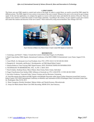

This paper discusses innovations in bionic arms that use muscle sensor technology, specifically focusing on the use of EMG (electromyography) signals to control hand movements. It highlights the importance of advanced neural interfaces and machine learning techniques for improving prosthetic functionality and user acceptance. The research emphasizes both invasive and non-invasive methods for recording muscle signals to enhance the control and performance of prosthetic devices.


