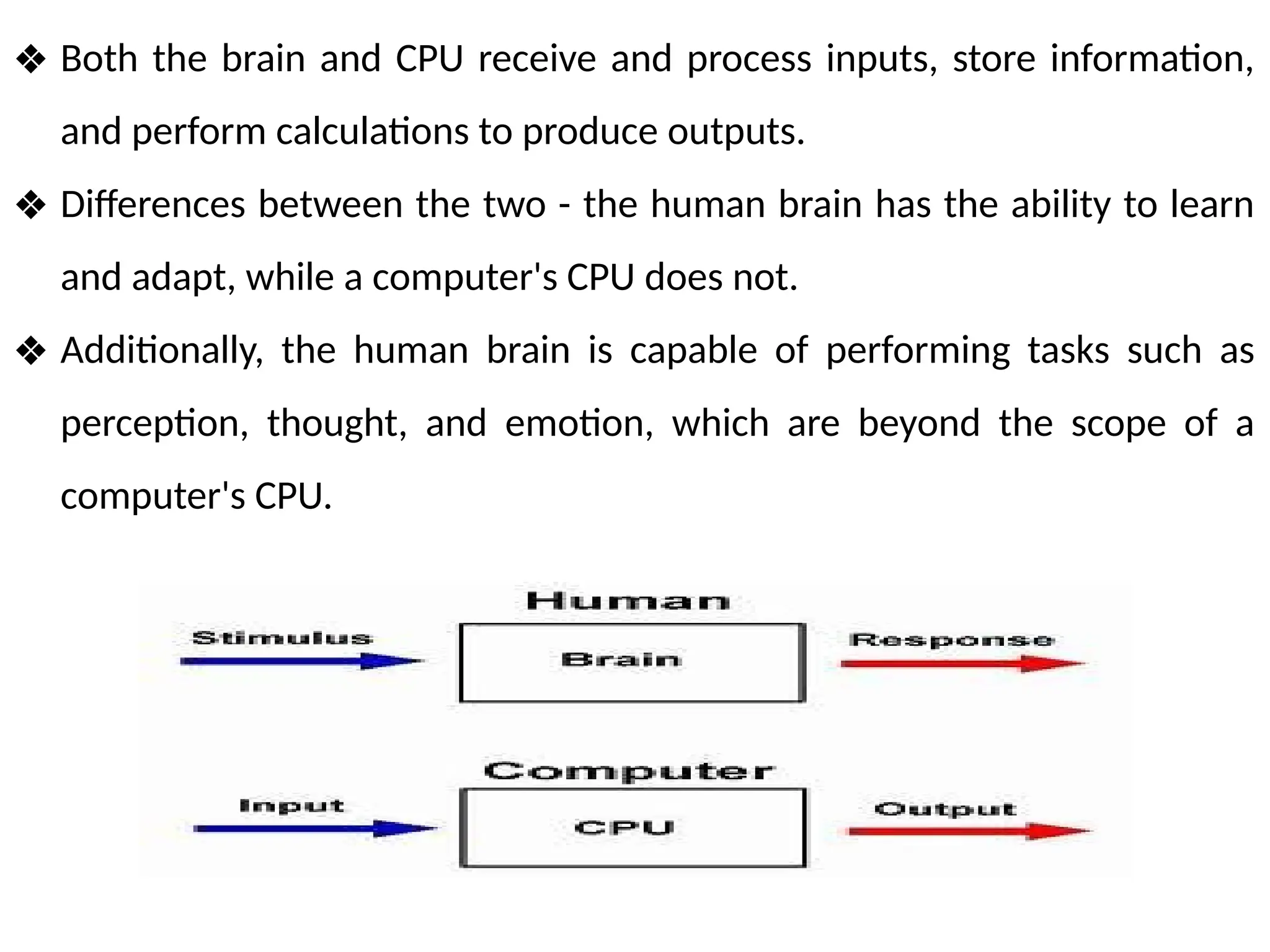
❖ Both thebrain and CPU receive and process inputs, store information,
and perform calculations to produce outputs.
❖ Differences between the two - the human brain has the ability to learn
and adapt, while a computer's CPU does not.
❖ Additionally, the human brain is capable of performing tasks such as
perception, thought, and emotion, which are beyond the scope of a
computer's CPU.
4.
Architecture
• The humanbrain as a CPU system can be compared to that of a parallel
distributed processing system, as opposed to the Von Neumann
architecture of traditional computers.
• In the human brain, information is processed in a distributed manner
across multiple regions, each with specialized functions, rather than
being processed sequentially in a single centralized location.
• Just like how a computer's CPU has an arithmetic logic unit (ALU) to
perform mathematical calculations, the human brain has specialized
regions for processing mathematical and logical operations.
• The prefrontal cortex, for example, is responsible for higher-level
cognitive functions such as decision making and problem solving
5.
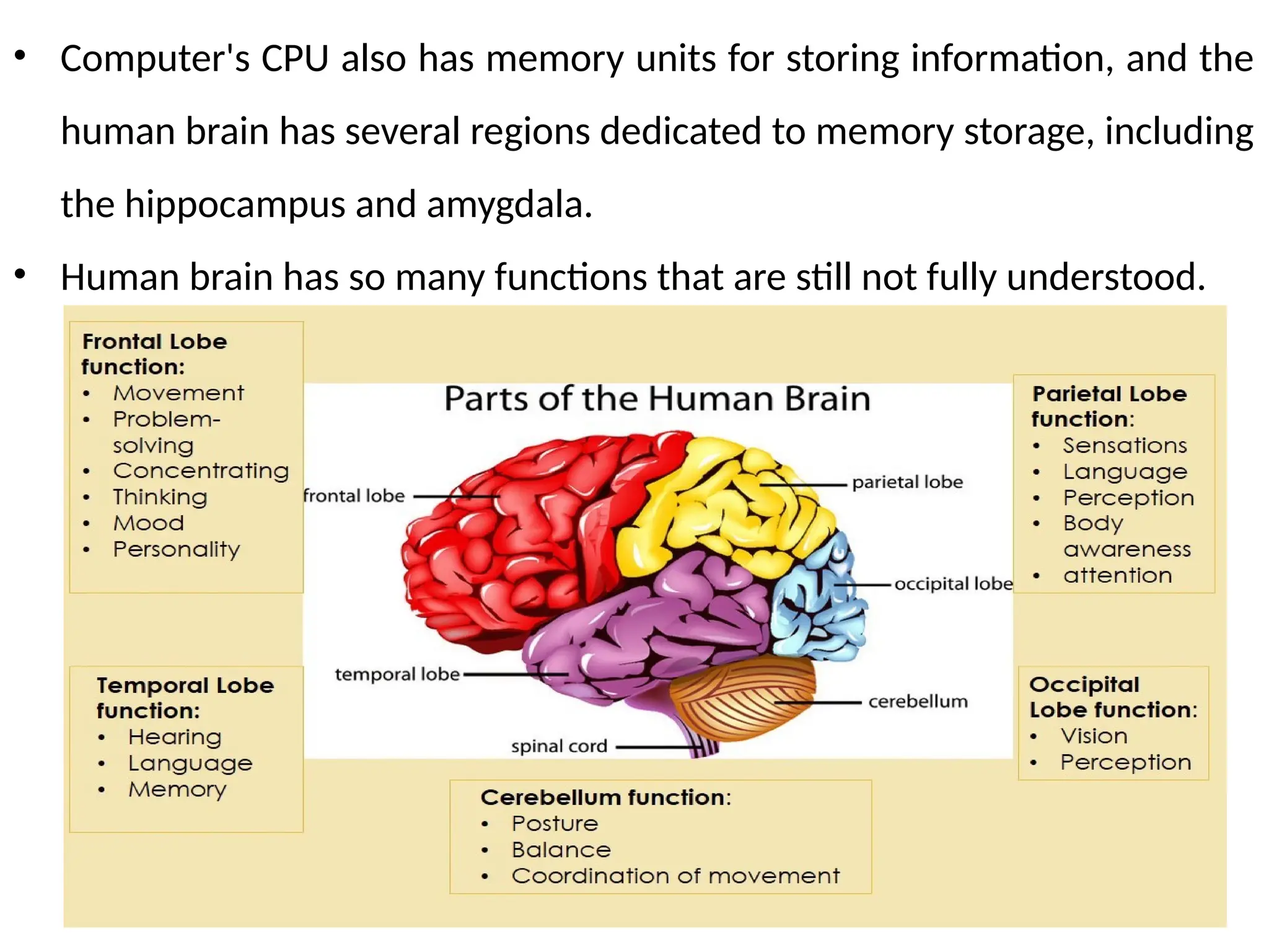
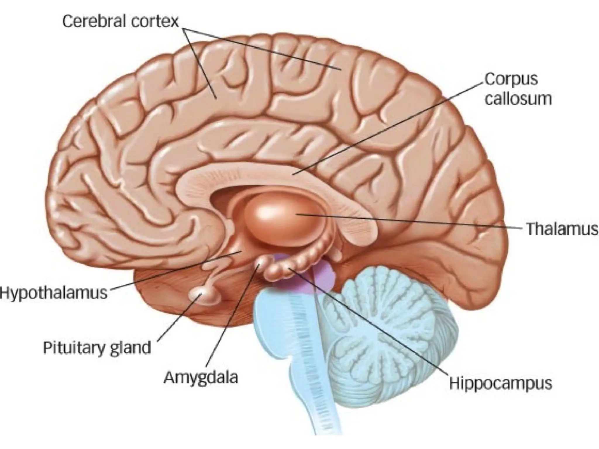
• Computer's CPUalso has memory units for storing information, and the
human brain has several regions dedicated to memory storage, including
the hippocampus and amygdala.
• Human brain has so many functions that are still not fully understood.
7.
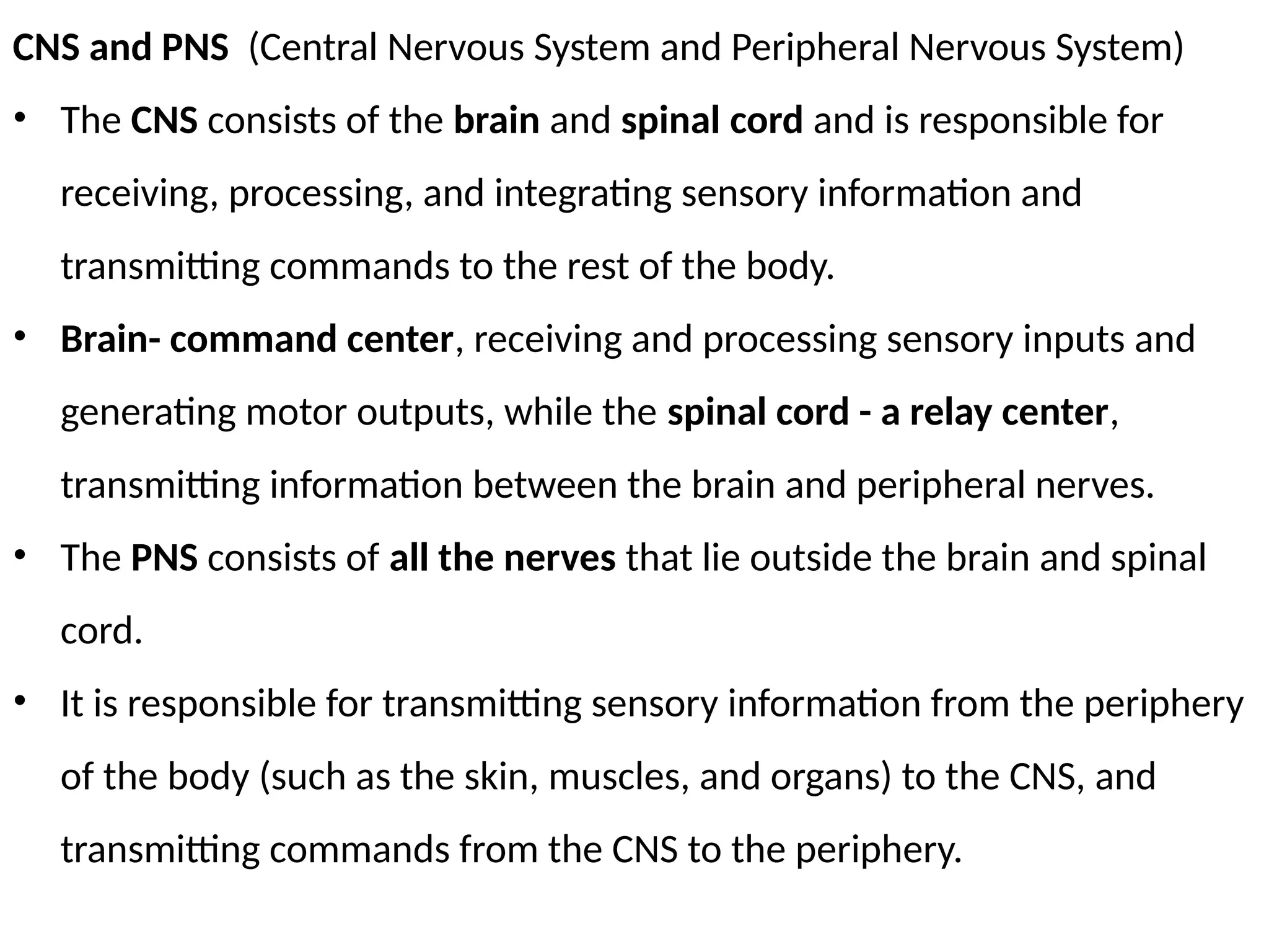
CNS and PNS(Central Nervous System and Peripheral Nervous System)
• The CNS consists of the brain and spinal cord and is responsible for
receiving, processing, and integrating sensory information and
transmitting commands to the rest of the body.
• Brain- command center, receiving and processing sensory inputs and
generating motor outputs, while the spinal cord - a relay center,
transmitting information between the brain and peripheral nerves.
• The PNS consists of all the nerves that lie outside the brain and spinal
cord.
• It is responsible for transmitting sensory information from the periphery
of the body (such as the skin, muscles, and organs) to the CNS, and
transmitting commands from the CNS to the periphery.
8.
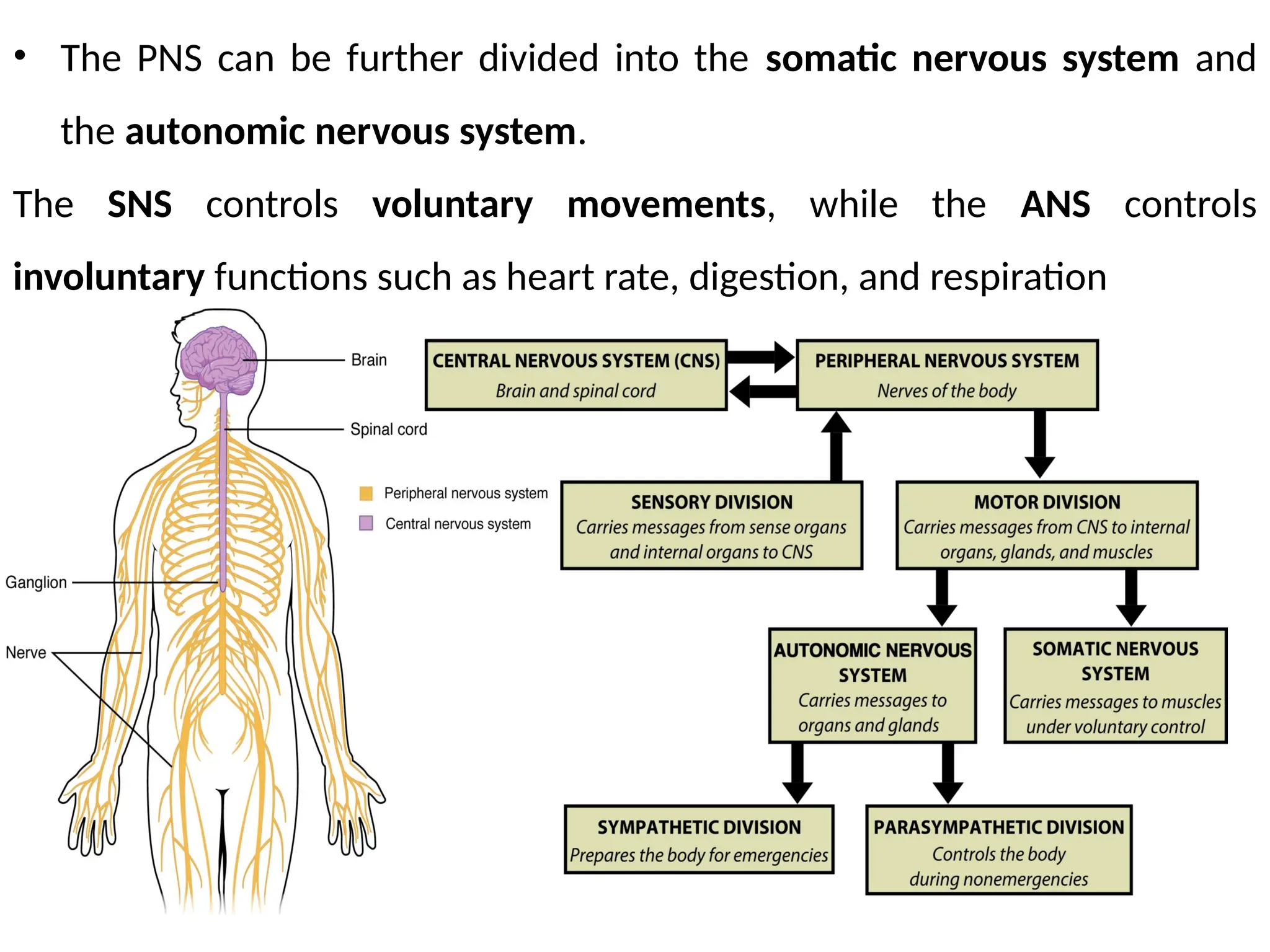
• The PNScan be further divided into the somatic nervous system and
the autonomic nervous system.
The SNS controls voluntary movements, while the ANS controls
involuntary functions such as heart rate, digestion, and respiration
9.
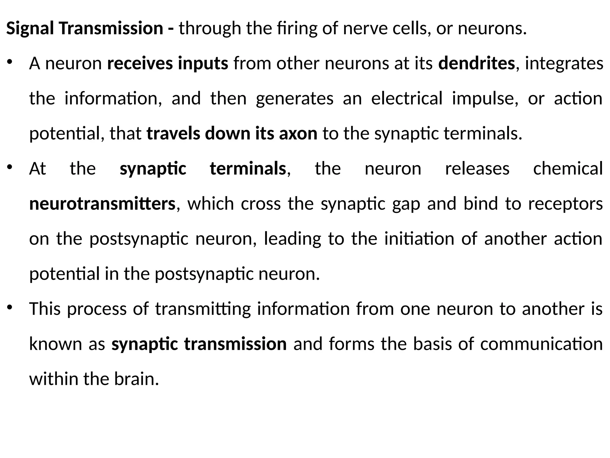
Signal Transmission -through the firing of nerve cells, or neurons.
• A neuron receives inputs from other neurons at its dendrites, integrates
the information, and then generates an electrical impulse, or action
potential, that travels down its axon to the synaptic terminals.
• At the synaptic terminals, the neuron releases chemical
neurotransmitters, which cross the synaptic gap and bind to receptors
on the postsynaptic neuron, leading to the initiation of another action
potential in the postsynaptic neuron.
• This process of transmitting information from one neuron to another is
known as synaptic transmission and forms the basis of communication
within the brain.
10.
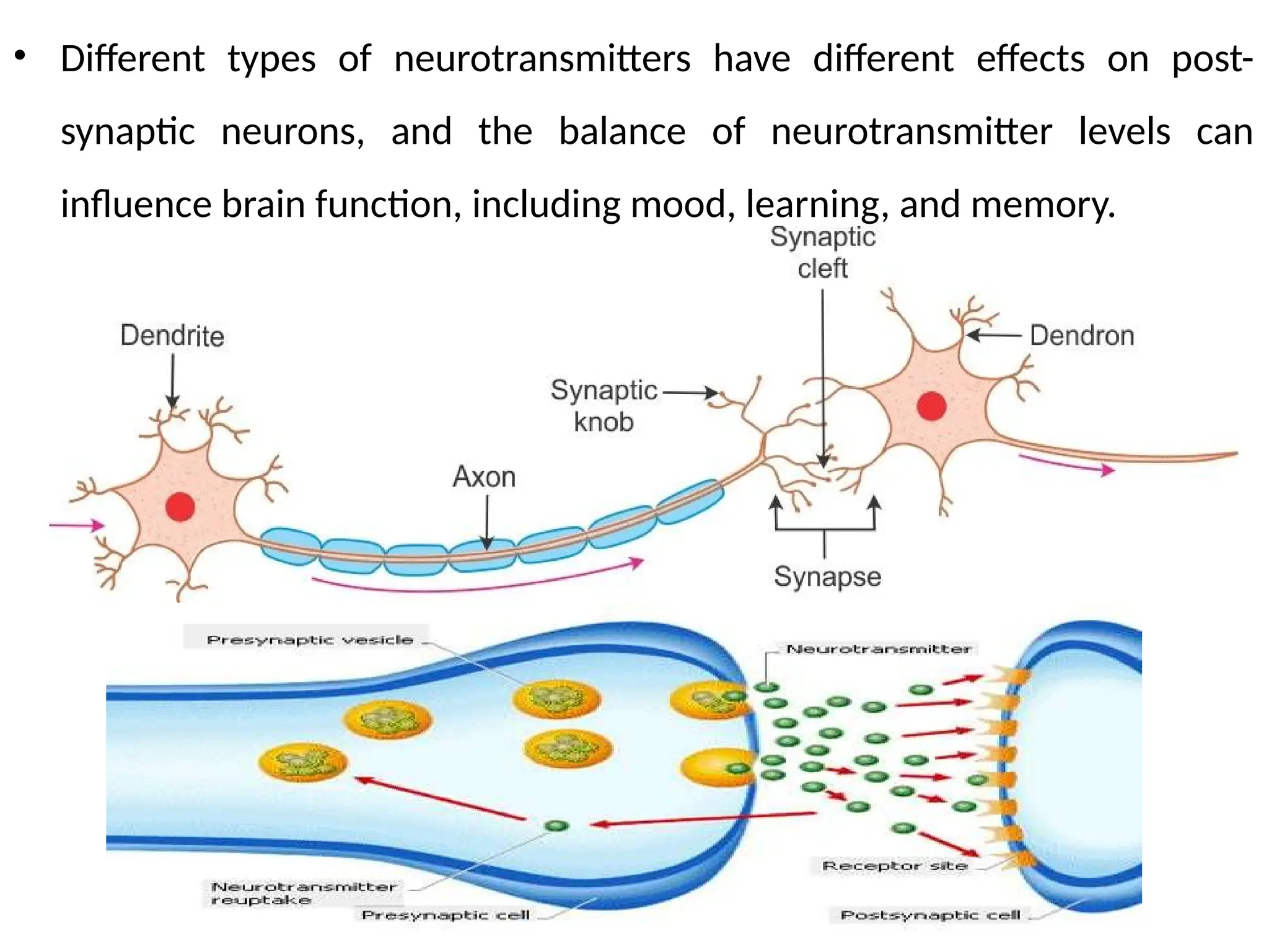
• Different typesof neurotransmitters have different effects on post-
synaptic neurons, and the balance of neurotransmitter levels can
influence brain function, including mood, learning, and memory.
11.
EEG (electroencephalography)
• Non-invasivemethod for measuring and recording of the electrical
activity of the brain.
• The signals are recorded through electrodes placed on the scalp and the
resulting EEG pattern provides information about the synchronized
electrical activity of large populations of neurons.
12.
Applications of EEG
1.Diagnosis of Epilepsy: and other seizure disorders. It can detect
abnormal electrical activity in the brain - diagnosis and determine the
location of the seizure focus.
2. Sleep Studies: to evaluate sleep patterns and diagnose sleep disorders.
3. Brain-Computer Interfaces (BCI): EEG can be used to control external
devices such as prosthetic limbs or computer software. This is done by
detecting specific brain waves associated with a particular mental state,
such as concentration or relaxation.
4. Research on Brain Function: during various activities such as reading,
problem-solving, and decision-making. And also be used to investigate
how the brain responds to stimuli such as light, sound, and touch.
13.
5. Diagnosis ofBrain Disorders: including dementia, Parkinson's disease,
and traumatic brain injury.
6. Anesthesia Monitoring: during surgery to ensure that the patient
remains in a safe and comfortable state.
7. Monitoring Brain Activity during Coma: to determine the level of brain
function and assess the likelihood of recovery.
14.
EEG Signals andTypes of Brain Activity
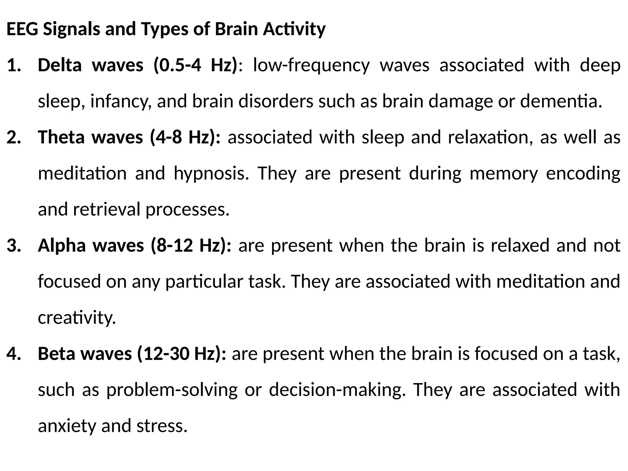
1. Delta waves (0.5-4 Hz): low-frequency waves associated with deep
sleep, infancy, and brain disorders such as brain damage or dementia.
2. Theta waves (4-8 Hz): associated with sleep and relaxation, as well as
meditation and hypnosis. They are present during memory encoding
and retrieval processes.
3. Alpha waves (8-12 Hz): are present when the brain is relaxed and not
focused on any particular task. They are associated with meditation and
creativity.
4. Beta waves (12-30 Hz): are present when the brain is focused on a task,
such as problem-solving or decision-making. They are associated with
anxiety and stress.
15.
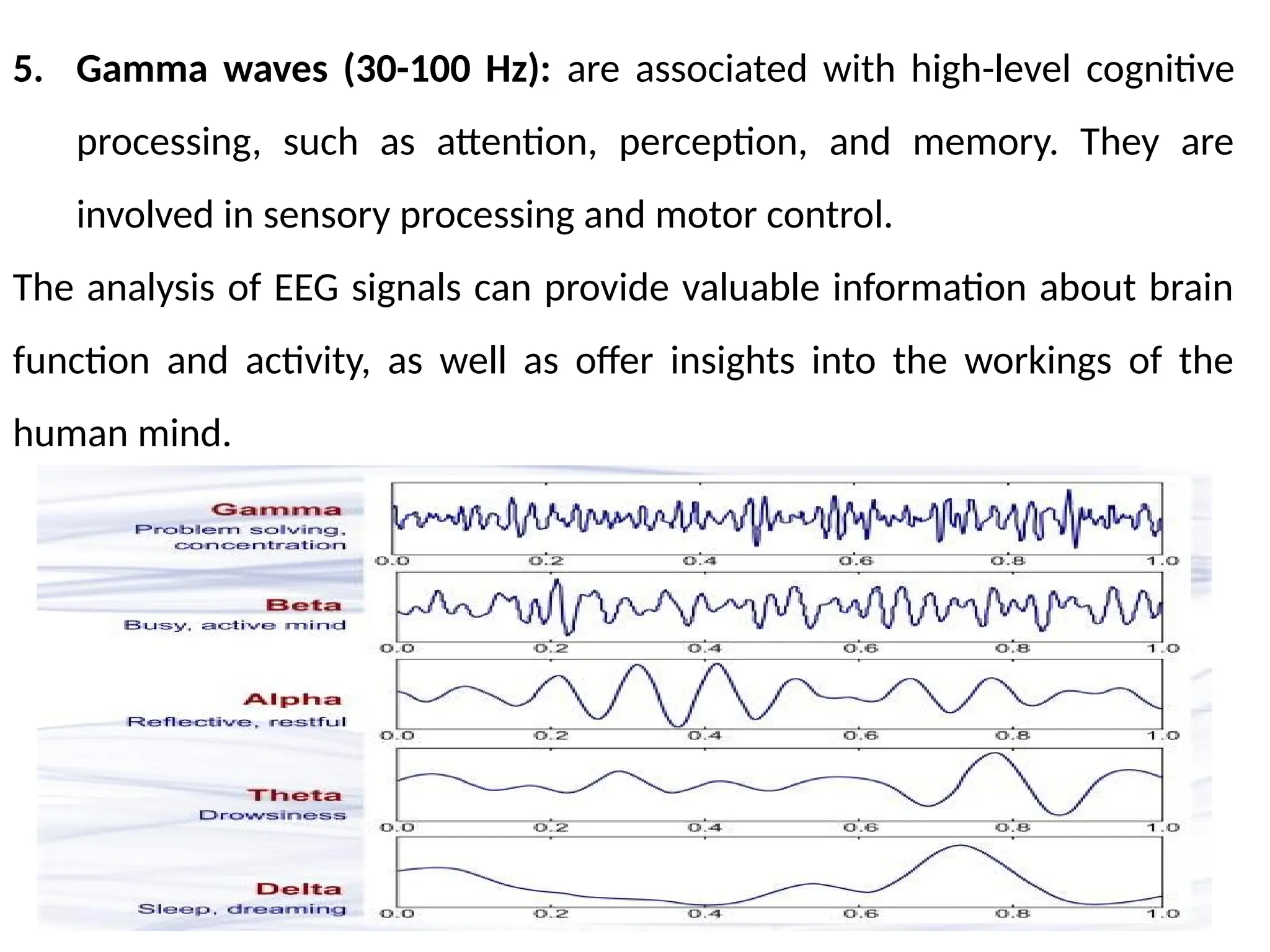
5. Gamma waves(30-100 Hz): are associated with high-level cognitive
processing, such as attention, perception, and memory. They are
involved in sensory processing and motor control.
The analysis of EEG signals can provide valuable information about brain
function and activity, as well as offer insights into the workings of the
human mind.
16.
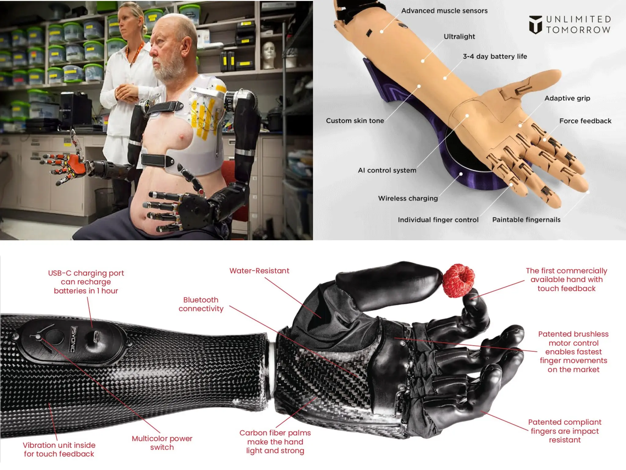
Robotic Arms forProsthetics
• The devices that use robotics technology to restore functionality to
individuals with upper limb amputations.
• These devices typically use motors, actuators, and sensors to mimic the
movements of a human arm and hand, allowing the wearer to perform
tasks such as reaching, grasping, and manipulating objects.
• It can be controlled in a variety of ways, including direct control through
muscle signals (myoelectric control) or brain-machine interfaces, which
use electrodes implanted in the brain or placed on the scalp to detect
and interpret brain activity.
• Some prosthetic arms also incorporate machine learning algorithms to
improve their performance and adapt to the user's needs over time.
18.
Robotic Arm ProstheticDirect Control through Muscle Signals
(myoelectric control)
• It involves using the electrical signals generated by the wearer's remaining
muscles to control the movement of the prosthetic.
• The electrodes placed on the skin over the remaining muscle that are used
to detect and interpret the electrical signals generated by the muscle
contractions.
• When the wearer contracts their muscles, the electrodes detect the
electrical signals and send them to a control unit, which interprets the
signals and uses them to control the movement of the robotic arm.
• Depending on the specific design, the control unit may use pattern
recognition algorithms (movement intending to perform) . or a combination
of muscle signals (specific degrees of freedom ).
19.
• Myoelectric controlhas the advantage of being directly controlled by
the user, allowing for a more intuitive and natural interaction with the
prosthetic.
• It can also provide a high level of control and precision, as the electrical
signals generated by the muscles are unique to each individual and can
be used to perform a wide range of movements.
• However, myoelectric control systems can be complex and may require
extensive rehabilitation and training to use effectively, as well as
ongoing maintenance to ensure proper function.
• Additionally, the system may not be suitable for individuals with muscle
weakness or other conditions that affect the ability to generate strong
electrical signals.
20.
Robotic Arm Prostheticby Brain-Machine Interfaces (BMIs)
• Brain-machine interfaces are a type of technology that allows a user to
control a robotic arm prosthetic directly with their brain activity.
• The system typically involves electrodes placed on the scalp or
implanted directly into the brain to detect and interpret the user's brain
signals.
• When the user thinks about moving the prosthetic arm, the electrodes
detect the corresponding brain activity and send the signals to a control
unit, which uses algorithms to interpret the signals and control the
movement of the prosthetic.
• The user can then control the movement of the prosthetic in real-time
by thinking about the desired movement.
21.
• BMIs havethe advantage of providing a direct and intuitive connection
between the user's brain and the prosthetic, allowing for a high level of
control and precision.
• BMIs can be used to provide sensory feedback to the user, allowing
them to experience the sensation of touch through the prosthetic.
• BMIs can be complex and invasive systems, requiring surgical
implantation and ongoing maintenance to ensure proper function.
• They may not be suitable for individuals with conditions that affect
brain activity or who are unable to generate strong enough brain signals
to control the prosthetic effectively.
Ongoing research and development is aimed to improving the
performance and accessibility and increasing their ease of use and
reliability.
22.
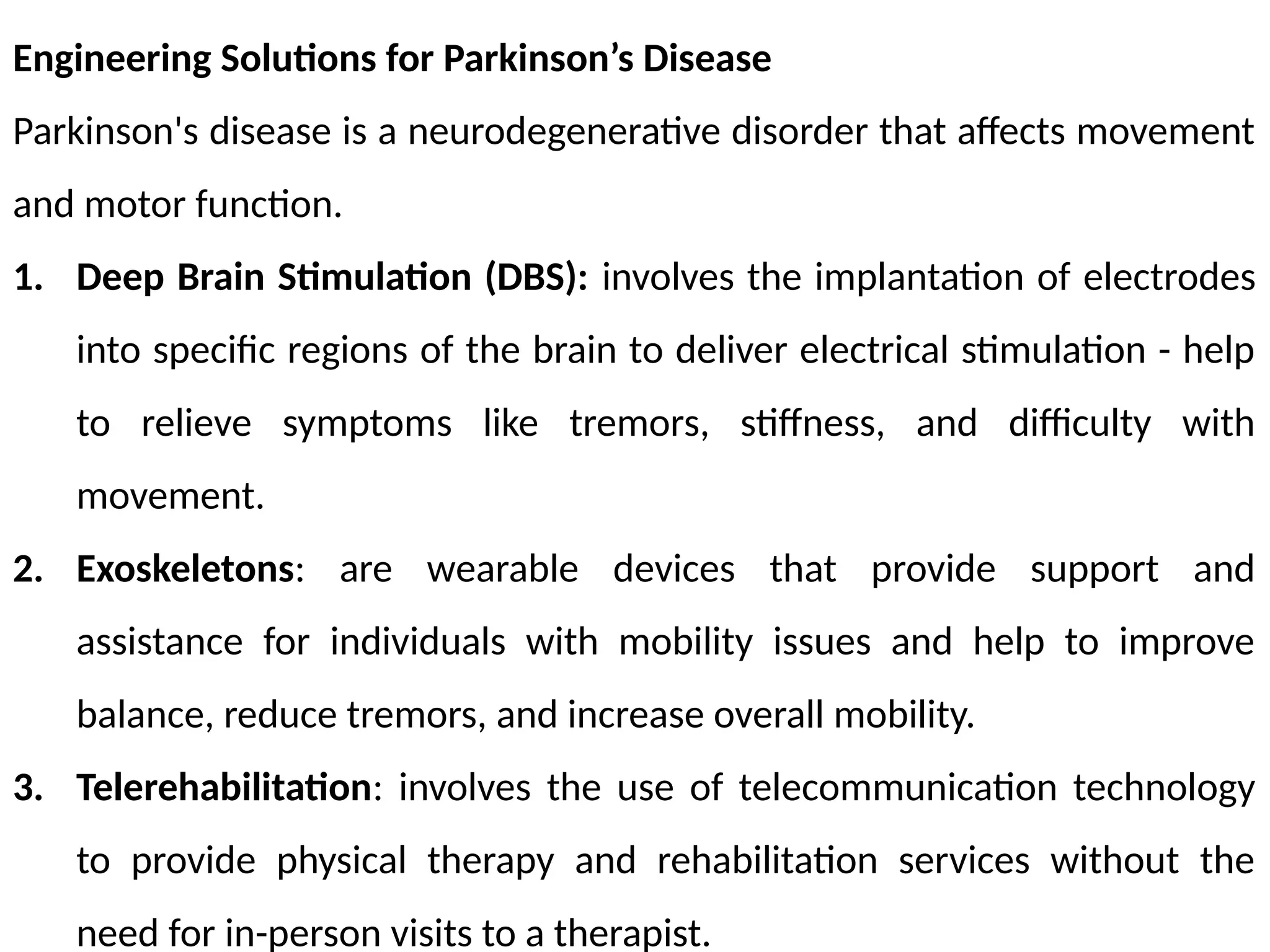
Engineering Solutions forParkinson’s Disease
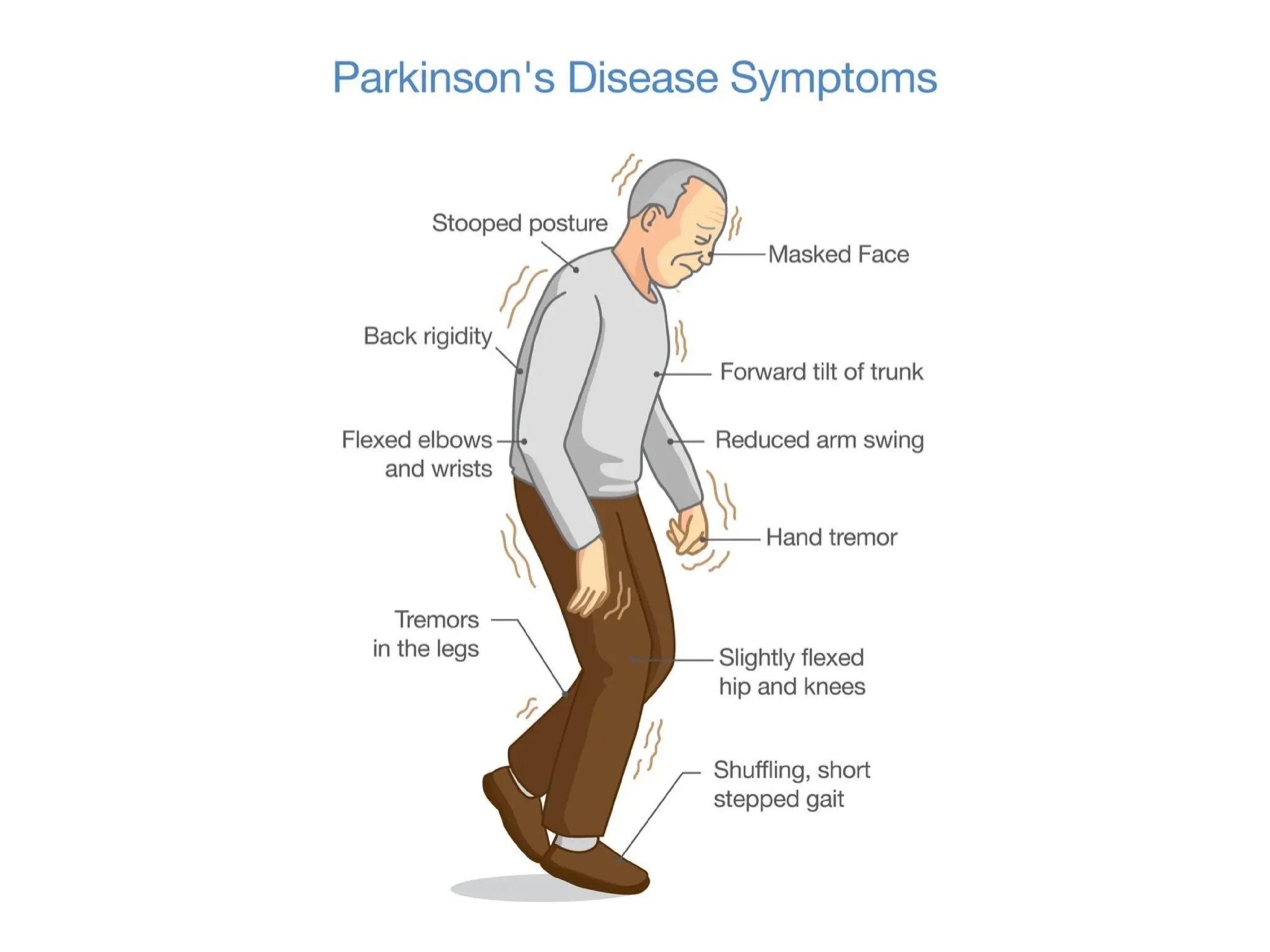
Parkinson's disease is a neurodegenerative disorder that affects movement
and motor function.
1. Deep Brain Stimulation (DBS): involves the implantation of electrodes
into specific regions of the brain to deliver electrical stimulation - help
to relieve symptoms like tremors, stiffness, and difficulty with
movement.
2. Exoskeletons: are wearable devices that provide support and
assistance for individuals with mobility issues and help to improve
balance, reduce tremors, and increase overall mobility.
3. Telerehabilitation: involves the use of telecommunication technology
to provide physical therapy and rehabilitation services without the
need for in-person visits to a therapist.
23.
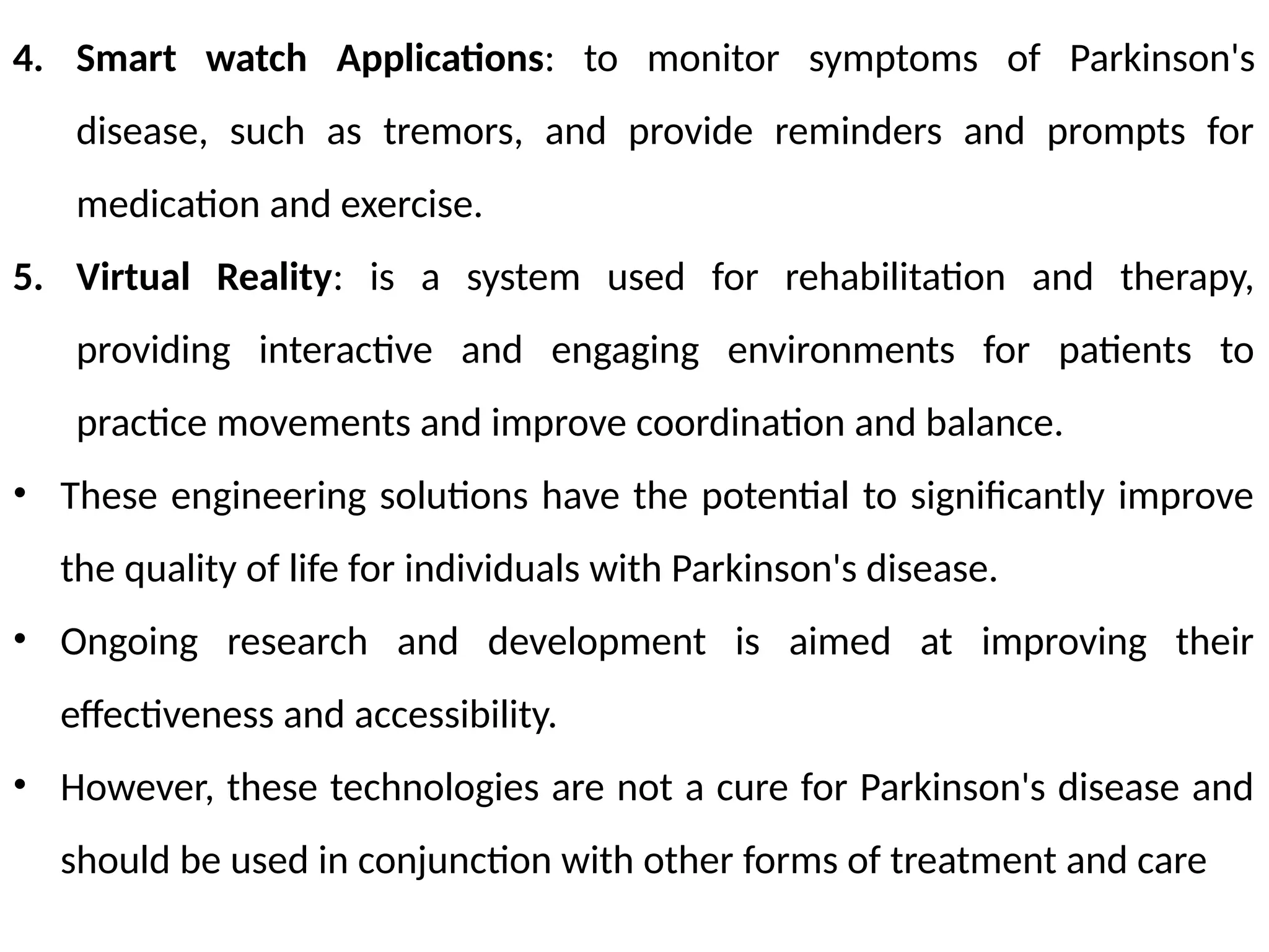
4. Smart watchApplications: to monitor symptoms of Parkinson's
disease, such as tremors, and provide reminders and prompts for
medication and exercise.
5. Virtual Reality: is a system used for rehabilitation and therapy,
providing interactive and engaging environments for patients to
practice movements and improve coordination and balance.
• These engineering solutions have the potential to significantly improve
the quality of life for individuals with Parkinson's disease.
• Ongoing research and development is aimed at improving their
effectiveness and accessibility.
• However, these technologies are not a cure for Parkinson's disease and
should be used in conjunction with other forms of treatment and care
25.
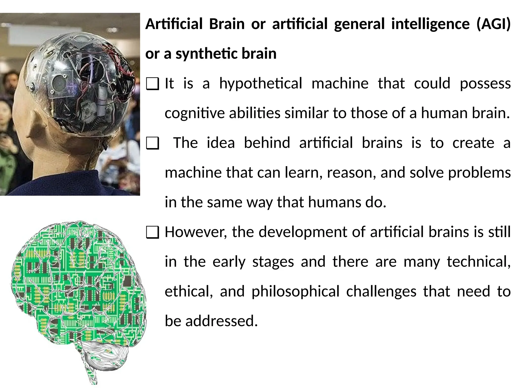
Artificial Brain orartificial general intelligence (AGI)
or a synthetic brain
❑ It is a hypothetical machine that could possess
cognitive abilities similar to those of a human brain.
❑ The idea behind artificial brains is to create a
machine that can learn, reason, and solve problems
in the same way that humans do.
❑ However, the development of artificial brains is still
in the early stages and there are many technical,
ethical, and philosophical challenges that need to
be addressed.
26.
o Currently, artificialintelligence (AI) systems are designed to perform
specific tasks, such as image recognition, speech recognition, or decision
making, but they are not capable of general intelligence
o This is because AI systems are designed to operate within a narrow
domain and lack the ability to learn from new experiences, generalize
from past experiences, or reason about the world in the same way that
humans do.
o The development of artificial brains requires a deep understanding of
the human brain and its functions, as well as advanced computer
science and engineering skills.
o Researchers are working on creating artificial brain models that can
simulate the complex processes of human cognition and adapt to new
situations.
27.
o Despite thesignificant challenges, some experts believe that artificial
brains are a realistic possibility and that they have the potential to
revolutionize the field of AI and bring about new technological
advancements.
o However, others argue that it is unlikely that we will ever be able to
recreate the human brain in a machine, due to the complexity and
intricacy of the brain's structure and functions.
In conclusion, the development of artificial brains is an exciting and
rapidly advancing field of research that has the potential to change the
world in many ways. However, it is important to approach this research
with caution and to consider the ethical and philosophical implications of
creating a machine that can think like a human.
• The humaneye can be analogized to a camera system, as both the eye
and a camera capture light and convert it into an image.
• The main components of the eye that correspond to a camera system
include:
1. The Cornea: This transparent outer layer of the eye functions like a
camera lens, bending light to focus it onto the retina.
2. The Iris: The iris functions like the diaphragm in a camera, controlling
the amount of light that enters the eye.
3. The Pupil: The pupil functions like the aperture in a camera, adjusting
the size to control the amount of light entering the eye.
4. The Retina: The retina functions like the camera film or sensor,
capturing the light and converting it into electrical signals that are sent
to the brain.
30.
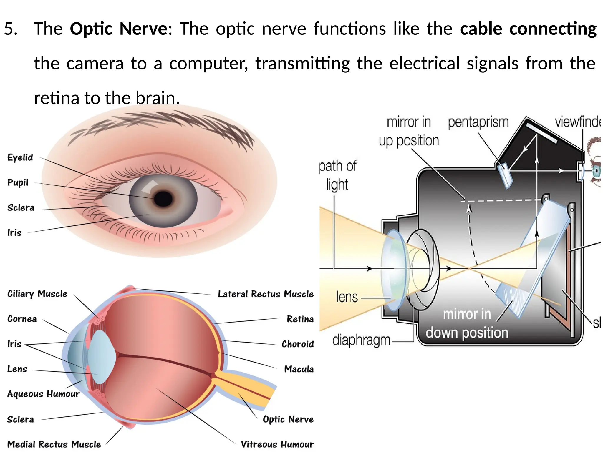
5. The OpticNerve: The optic nerve functions like the cable connecting
the camera to a computer, transmitting the electrical signals from the
retina to the brain.
31.
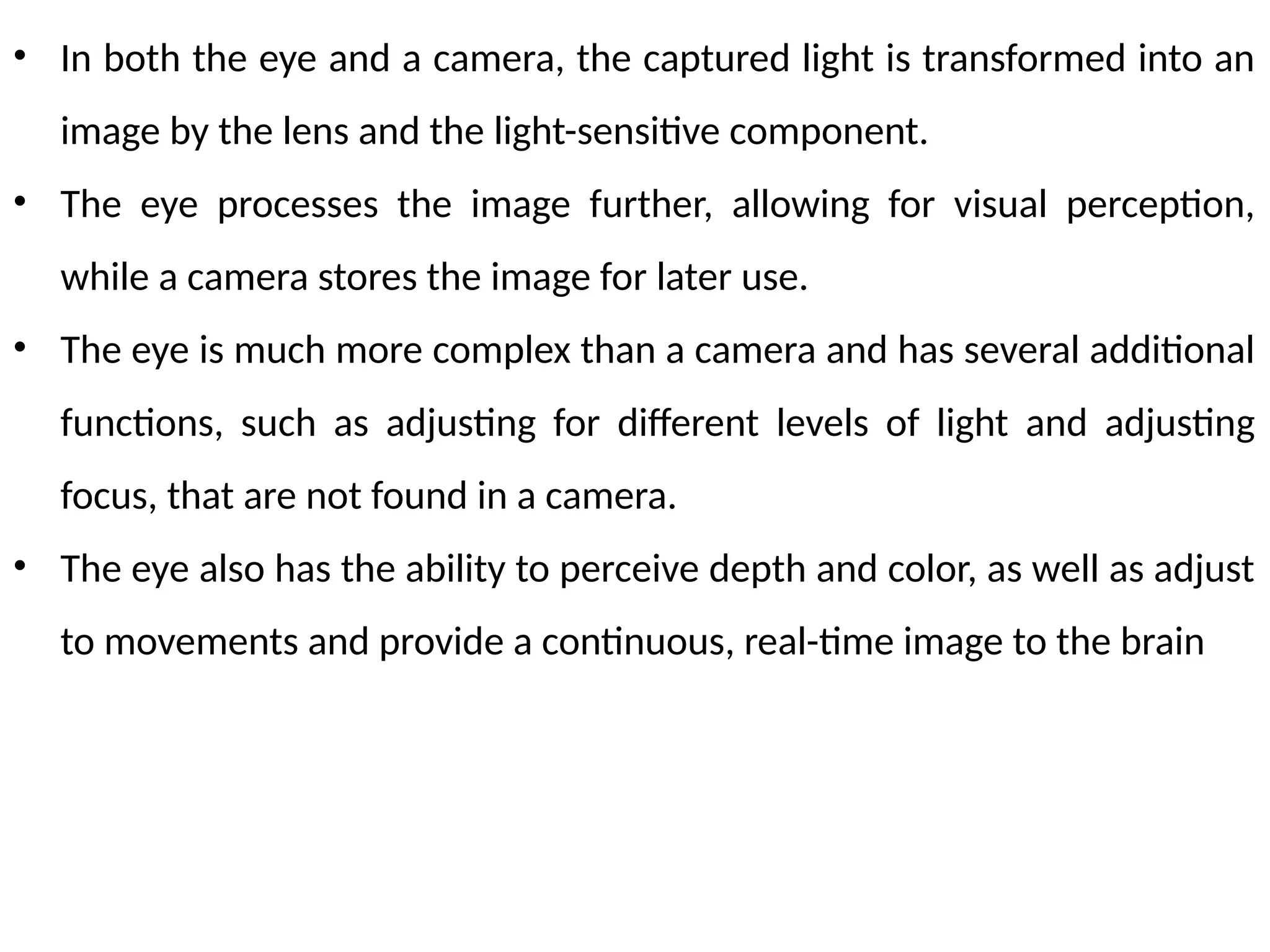
• In boththe eye and a camera, the captured light is transformed into an
image by the lens and the light-sensitive component.
• The eye processes the image further, allowing for visual perception,
while a camera stores the image for later use.
• The eye is much more complex than a camera and has several additional
functions, such as adjusting for different levels of light and adjusting
focus, that are not found in a camera.
• The eye also has the ability to perceive depth and color, as well as adjust
to movements and provide a continuous, real-time image to the brain
32.
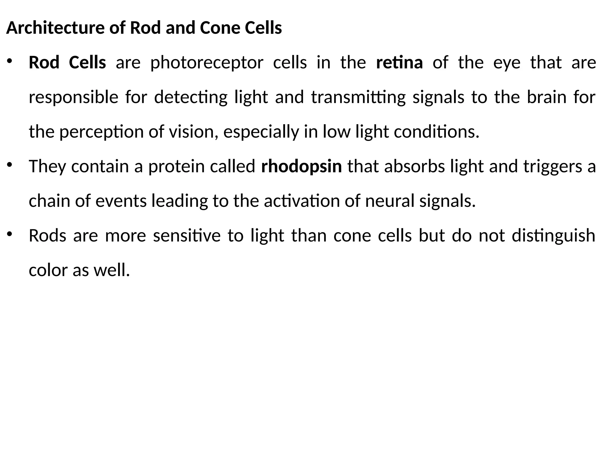
Architecture of Rodand Cone Cells
• Rod Cells are photoreceptor cells in the retina of the eye that are
responsible for detecting light and transmitting signals to the brain for
the perception of vision, especially in low light conditions.
• They contain a protein called rhodopsin that absorbs light and triggers a
chain of events leading to the activation of neural signals.
• Rods are more sensitive to light than cone cells but do not distinguish
color as well.
33.
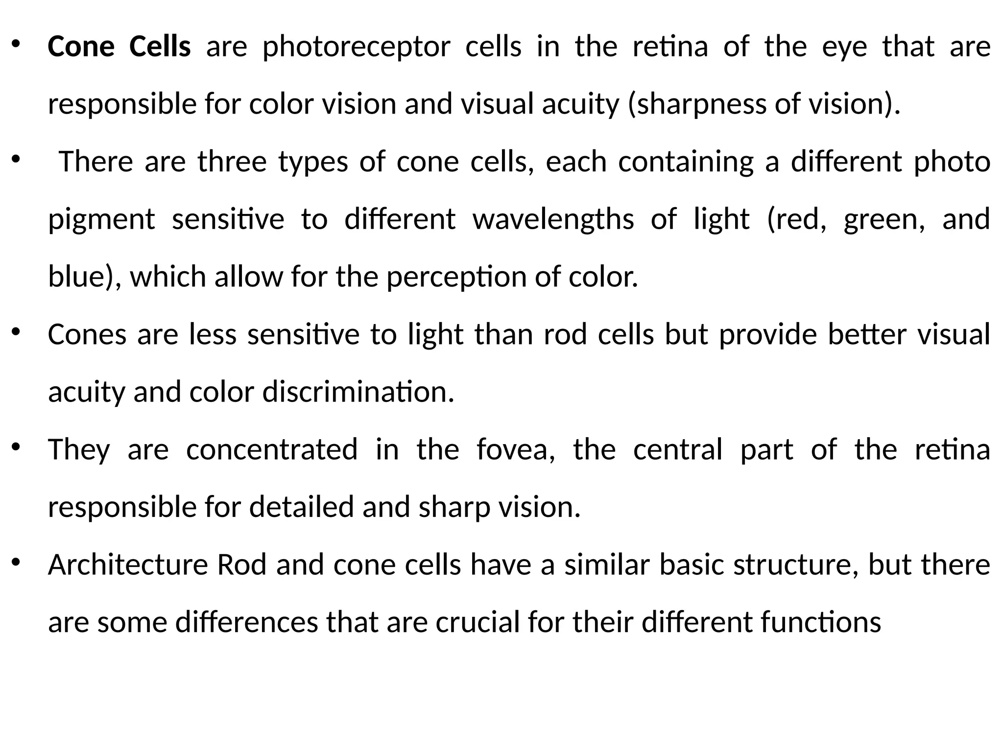
• Cone Cellsare photoreceptor cells in the retina of the eye that are
responsible for color vision and visual acuity (sharpness of vision).
• There are three types of cone cells, each containing a different photo
pigment sensitive to different wavelengths of light (red, green, and
blue), which allow for the perception of color.
• Cones are less sensitive to light than rod cells but provide better visual
acuity and color discrimination.
• They are concentrated in the fovea, the central part of the retina
responsible for detailed and sharp vision.
• Architecture Rod and cone cells have a similar basic structure, but there
are some differences that are crucial for their different functions
34.
• Rod cellsmake synapses with one bipolar cell, while cone cells synapse
with one of several bipolar cells.
• This difference in synapse distribution is critical for the different
functions of rod and cone cells in vision.
35.
Optical Corrections
• Opticalcorrections refer to devices or techniques used to improve or
correct vision problems caused by a refractive error in the eye.
• Refractive errors occur when light entering the eye is not properly
focused on the retina, leading to blurred vision.
There are several types of refractive errors, including:
1. Myopia (nearsightedness): Light is focused in front of the retina,
making distant objects appear blurry.
2. Hyperopia (farsightedness): Light is focused behind the retina, making
near objects appear blurry.
3. Astigmatism: Light is not focused evenly on the retina, leading to
blurred or distorted vision.
36.
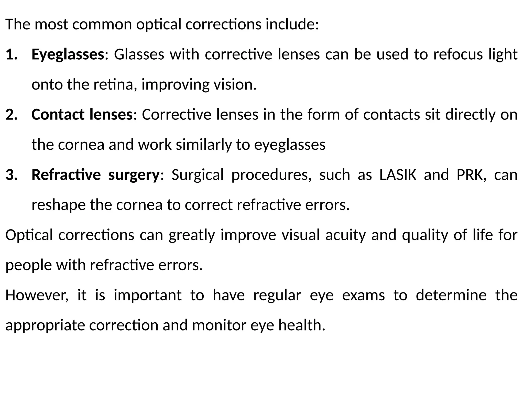
The most commonoptical corrections include:
1. Eyeglasses: Glasses with corrective lenses can be used to refocus light
onto the retina, improving vision.
2. Contact lenses: Corrective lenses in the form of contacts sit directly on
the cornea and work similarly to eyeglasses
3. Refractive surgery: Surgical procedures, such as LASIK and PRK, can
reshape the cornea to correct refractive errors.
Optical corrections can greatly improve visual acuity and quality of life for
people with refractive errors.
However, it is important to have regular eye exams to determine the
appropriate correction and monitor eye health.
37.
Cataract :
• Acataract is a clouding of the lens of the eye that affects vision.
• The lens, located behind the iris and pupil, normally allows light to pass
through to the retina and produces clear, sharp images.
• However, as we age or due to other factors, the proteins in the lens can
clump together and cause the lens to become opaque, leading to vision
problems.
• Symptoms of a cataract include blurred or hazy vision, increased
sensitivity to glare and bright lights, faded or yellowed colors, and
double vision in one eye.
• Cataracts can also cause frequent changes in prescription for eyeglasses
or contacts.
38.
• Cataract surgeryis a common and safe procedure to remove the cloudy
lens and replace it with an artificial lens.
• The surgery is typically performed on an outpatient basis and most
people experience improved vision within a few days after the
procedure.
In conclusion, cataracts can significantly affect vision, but surgical removal
and replacement with an artificial lens can restore clear vision and improve
quality of life.
Regular eye exams can help detect cataracts early and prevent vision loss
39.
Lens Materials
The artificiallenses used in cataract surgery or for vision correction can be
made of a variety of materials, each with its own unique properties and
benefits.
The most common lens materials include:
1. Polymethyl methacrylate (PMMA): PMMA is a type of plastic that has
been used for many years in artificial lenses. It is a durable and
affordable material, but does not have the ability to flex and adjust
focus like the natural lens.
2. Silicone: Silicone is a soft, flexible material that is resistant to cracking
and breaking. It is often used in phakic intraocular lenses (IOLs), which
are implanted in front of the natural lens.
40.
2. Acrylic: Acrylicis a lightweight, clear material that is similar in
properties to PMMA. It is often used in foldable IOLs, which can be
inserted through a smaller incision.
3. Hydrophobic acrylic: Hydrophobic acrylic is a type of acrylic material
that has a special surface treatment that helps to reduce glare and
halos around lights.
4. Hydrophilic acrylic: Hydrophilic acrylic is a type of acrylic material that
is designed to be more compatible with the natural fluid in the eye,
reducing the risk of vision-threatening complications.
The choice of lens material will depend on several factors, including the
patient's individual needs, the surgeon's preference, and the potential risks
and benefits of each material. – suggested eye doctor
41.
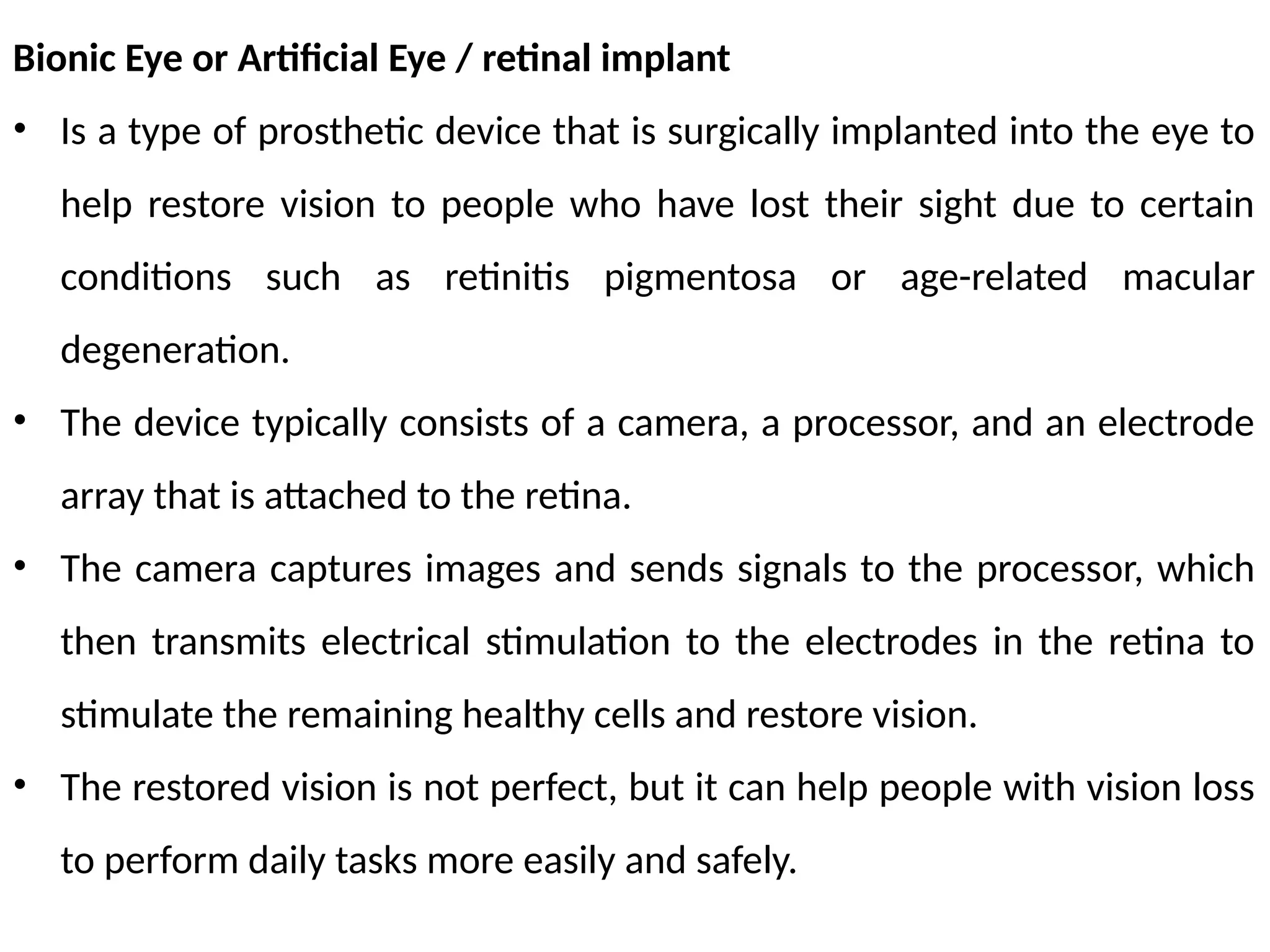
Bionic Eye orArtificial Eye / retinal implant
• Is a type of prosthetic device that is surgically implanted into the eye to
help restore vision to people who have lost their sight due to certain
conditions such as retinitis pigmentosa or age-related macular
degeneration.
• The device typically consists of a camera, a processor, and an electrode
array that is attached to the retina.
• The camera captures images and sends signals to the processor, which
then transmits electrical stimulation to the electrodes in the retina to
stimulate the remaining healthy cells and restore vision.
• The restored vision is not perfect, but it can help people with vision loss
to perform daily tasks more easily and safely.
42.
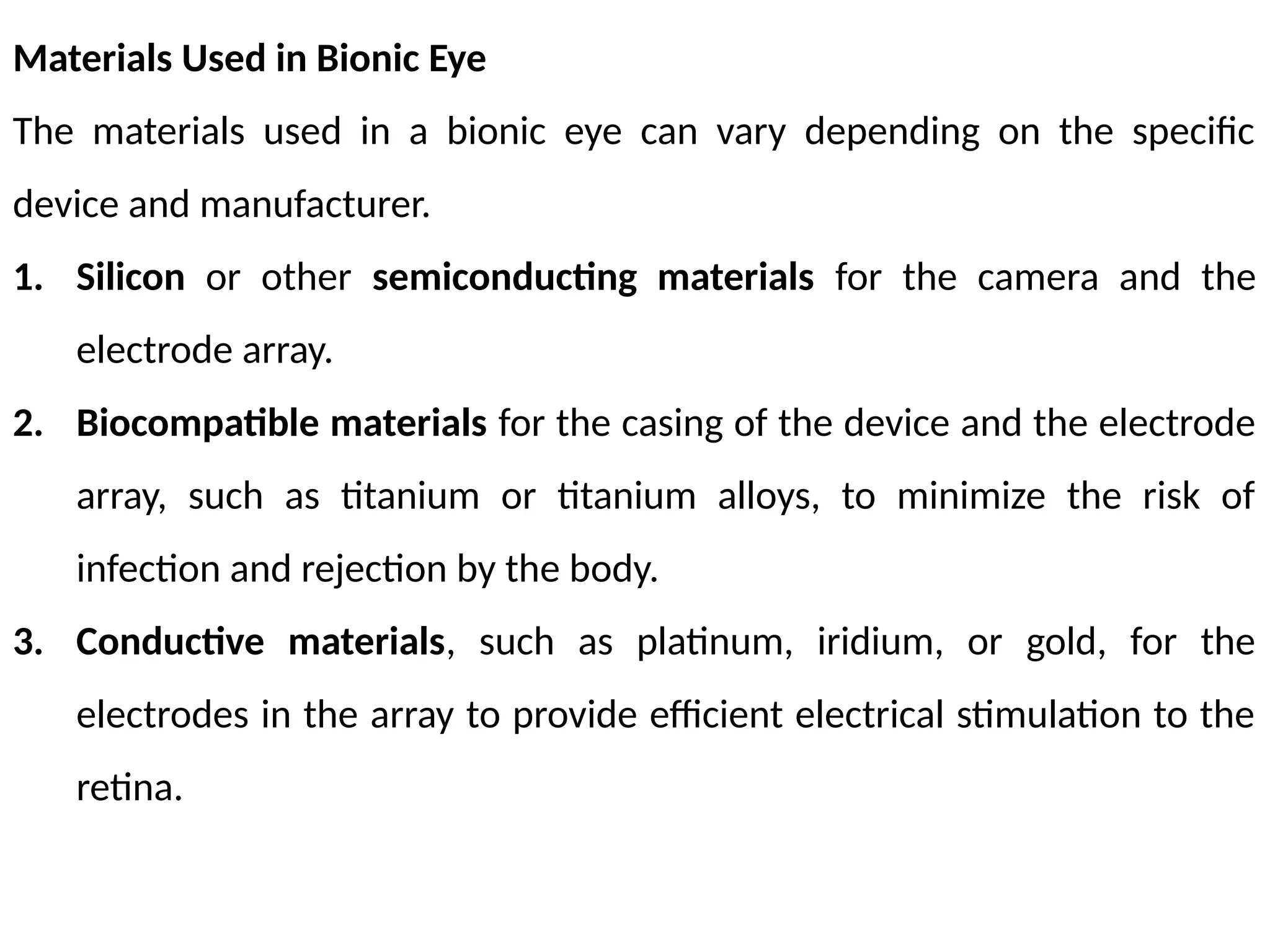
Materials Used inBionic Eye
The materials used in a bionic eye can vary depending on the specific
device and manufacturer.
1. Silicon or other semiconducting materials for the camera and the
electrode array.
2. Biocompatible materials for the casing of the device and the electrode
array, such as titanium or titanium alloys, to minimize the risk of
infection and rejection by the body.
3. Conductive materials, such as platinum, iridium, or gold, for the
electrodes in the array to provide efficient electrical stimulation to the
retina.
43.
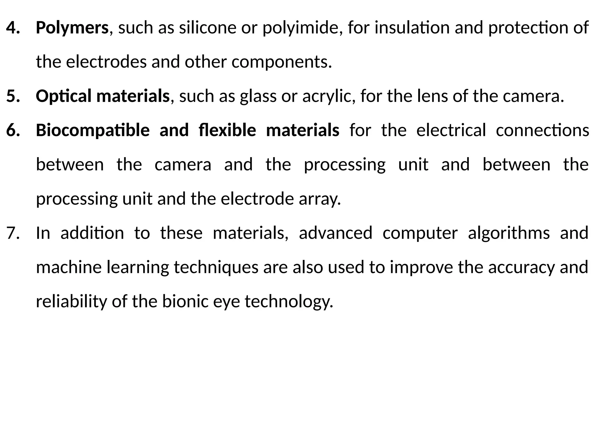
4. Polymers, suchas silicone or polyimide, for insulation and protection of
the electrodes and other components.
5. Optical materials, such as glass or acrylic, for the lens of the camera.
6. Biocompatible and flexible materials for the electrical connections
between the camera and the processing unit and between the
processing unit and the electrode array.
7. In addition to these materials, advanced computer algorithms and
machine learning techniques are also used to improve the accuracy and
reliability of the bionic eye technology.
44.
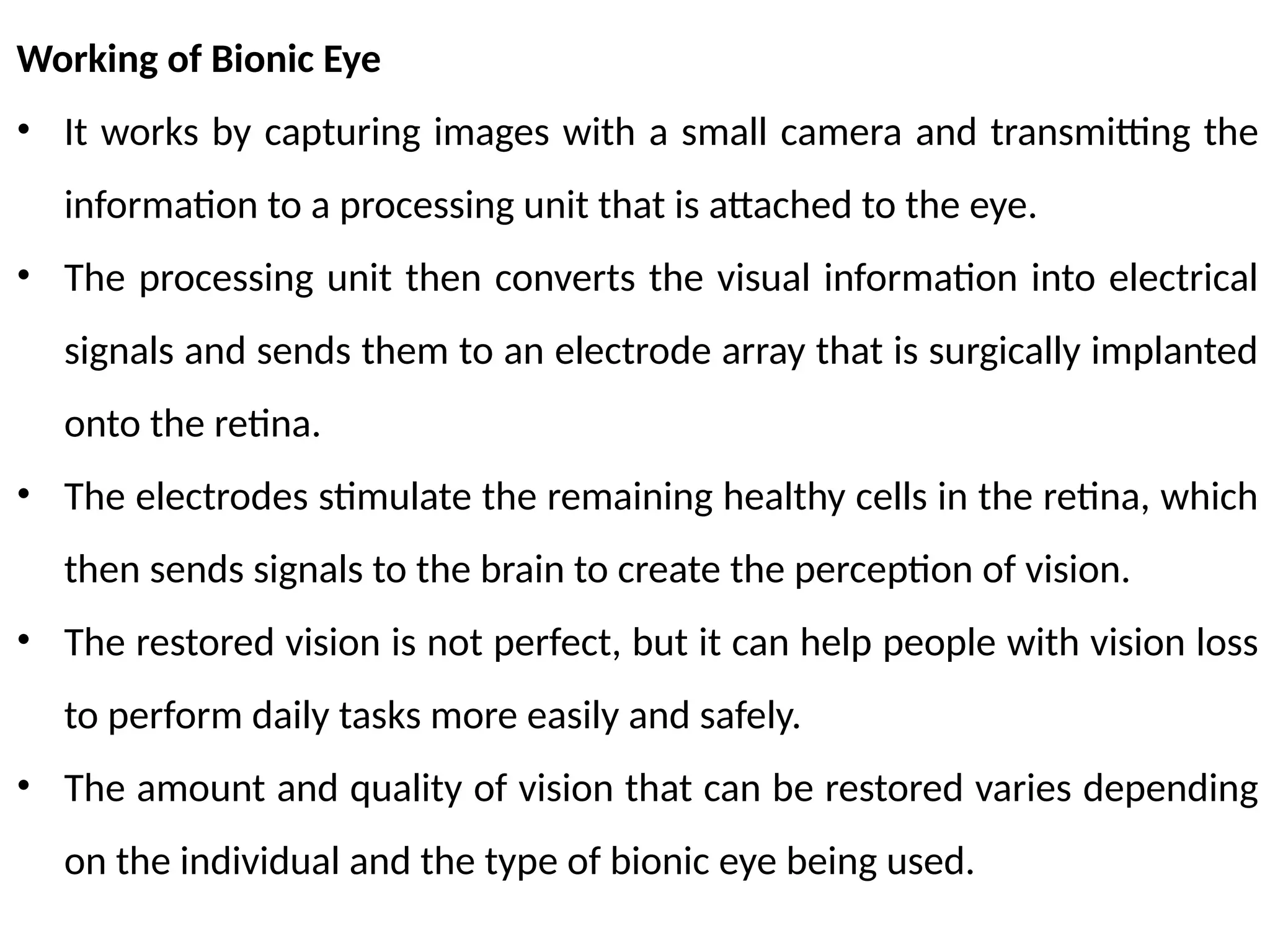
Working of BionicEye
• It works by capturing images with a small camera and transmitting the
information to a processing unit that is attached to the eye.
• The processing unit then converts the visual information into electrical
signals and sends them to an electrode array that is surgically implanted
onto the retina.
• The electrodes stimulate the remaining healthy cells in the retina, which
then sends signals to the brain to create the perception of vision.
• The restored vision is not perfect, but it can help people with vision loss
to perform daily tasks more easily and safely.
• The amount and quality of vision that can be restored varies depending
on the individual and the type of bionic eye being used.
45.
• Some bioniceyes only restore basic visual shapes and patterns, while
others can provide more detailed vision.
• The bionic eye is powered by a battery that is typically implanted behind
the ear.
• The battery is recharged through a device that is held near the eye,
which transmits power wirelessly to the battery.
• The device is typically rechargeable and can be used for several years
before it needs to be replaced.
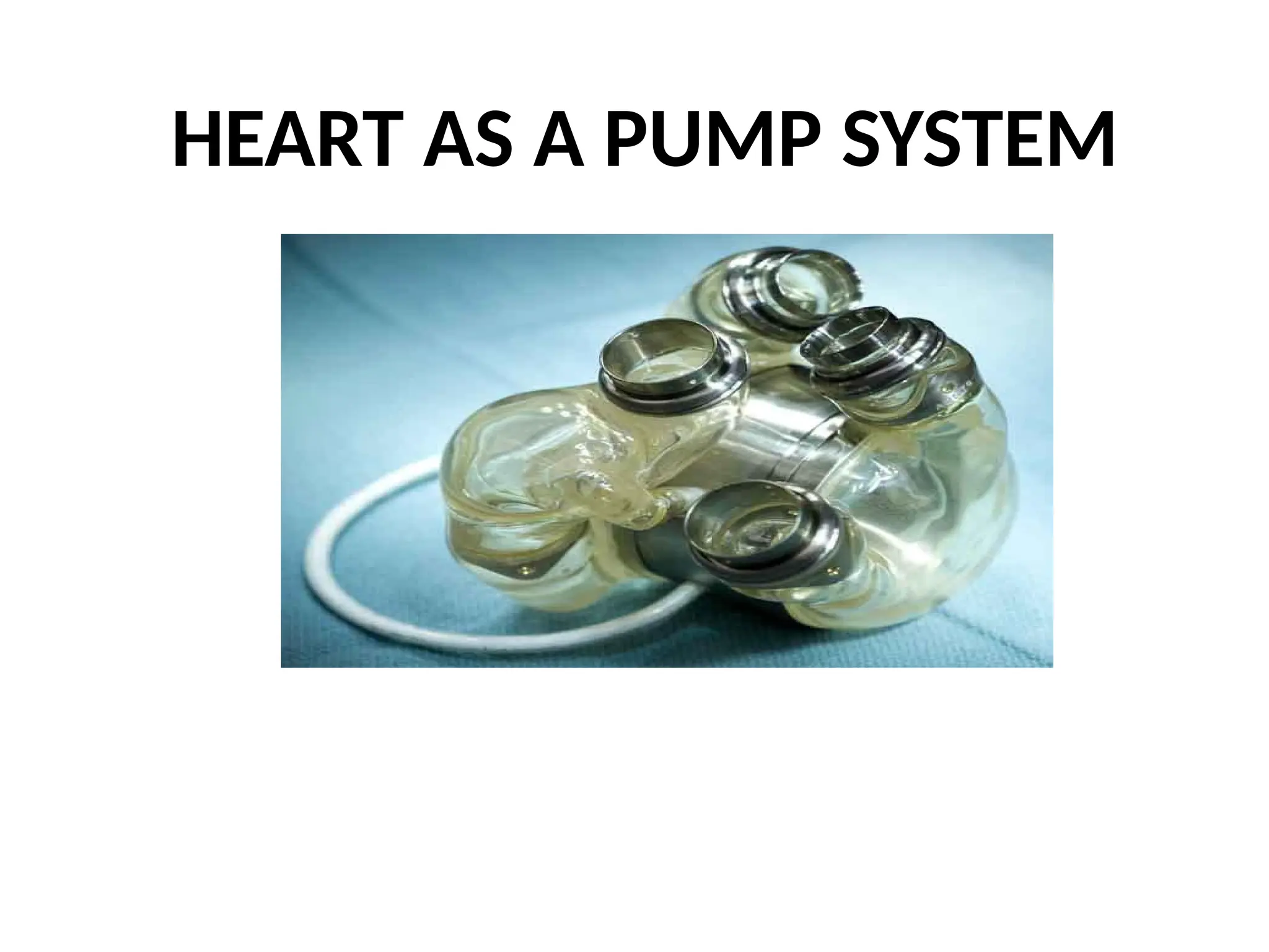
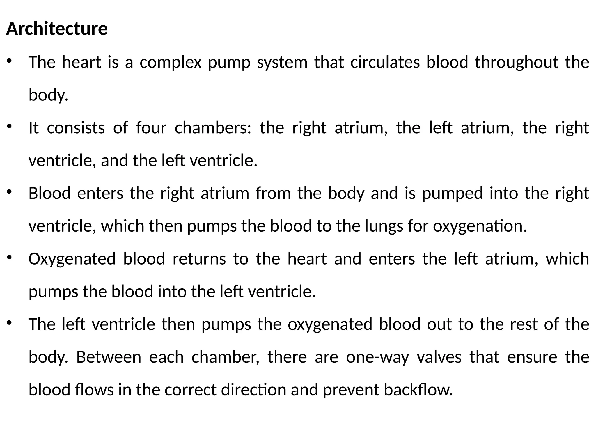
Architecture
• The heartis a complex pump system that circulates blood throughout the
body.
• It consists of four chambers: the right atrium, the left atrium, the right
ventricle, and the left ventricle.
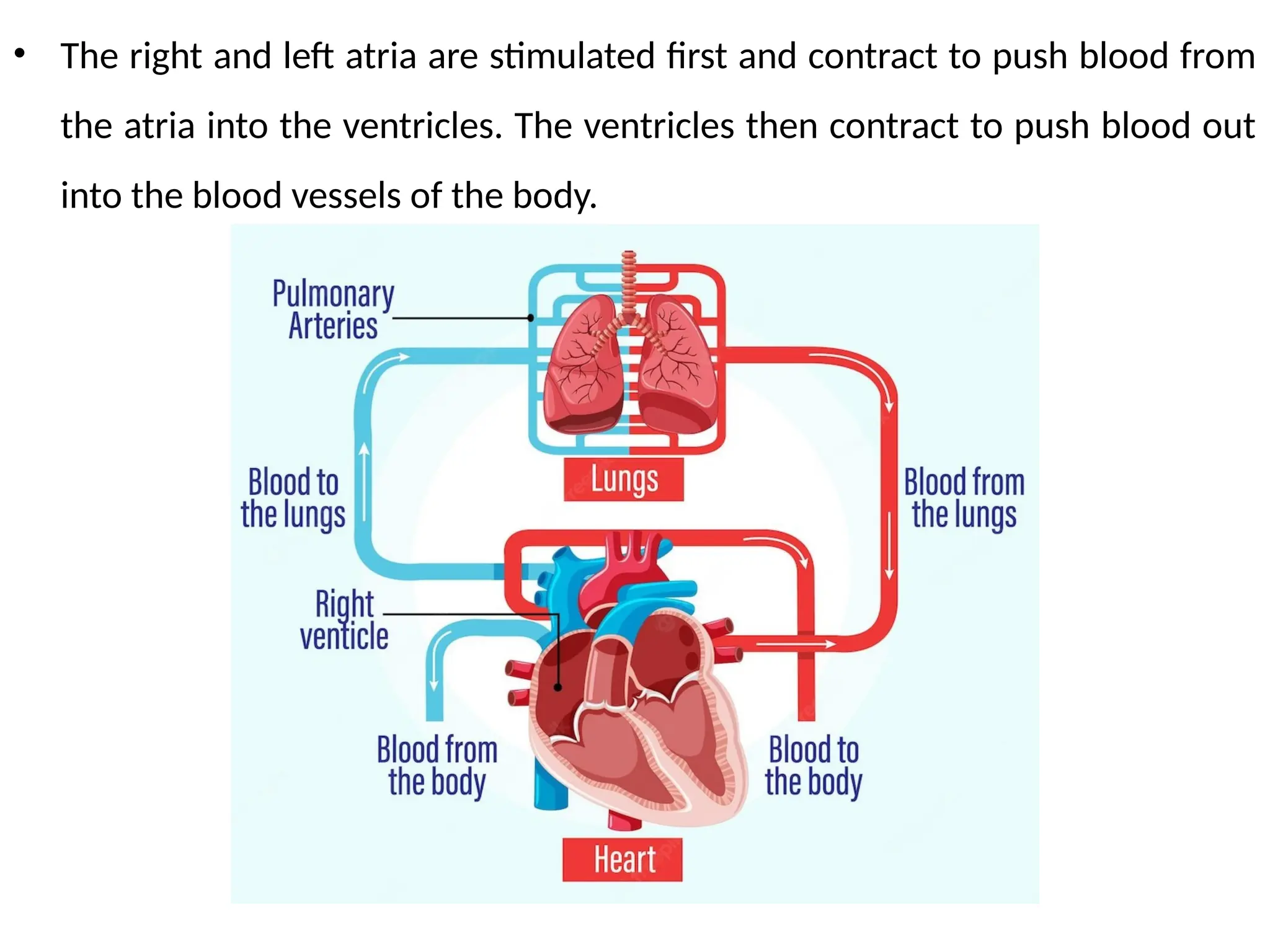
• Blood enters the right atrium from the body and is pumped into the right
ventricle, which then pumps the blood to the lungs for oxygenation.
• Oxygenated blood returns to the heart and enters the left atrium, which
pumps the blood into the left ventricle.
• The left ventricle then pumps the oxygenated blood out to the rest of the
body. Between each chamber, there are one-way valves that ensure the
blood flows in the correct direction and prevent backflow.
48.
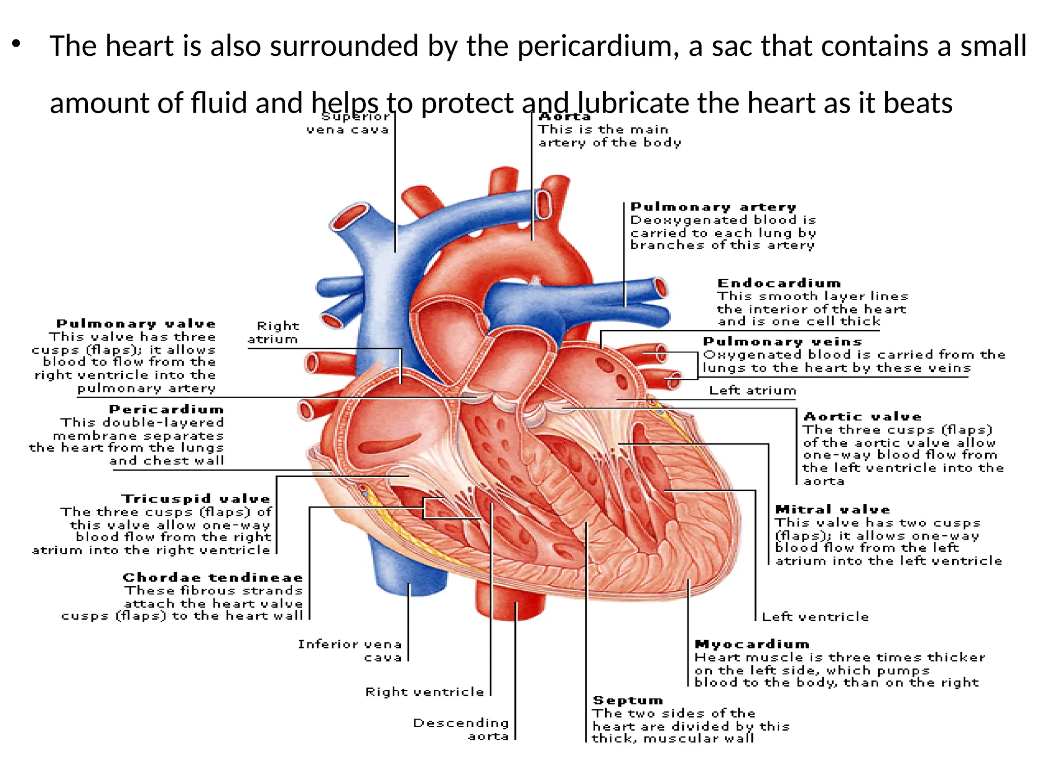
• The heartis also surrounded by the pericardium, a sac that contains a small
amount of fluid and helps to protect and lubricate the heart as it beats
49.
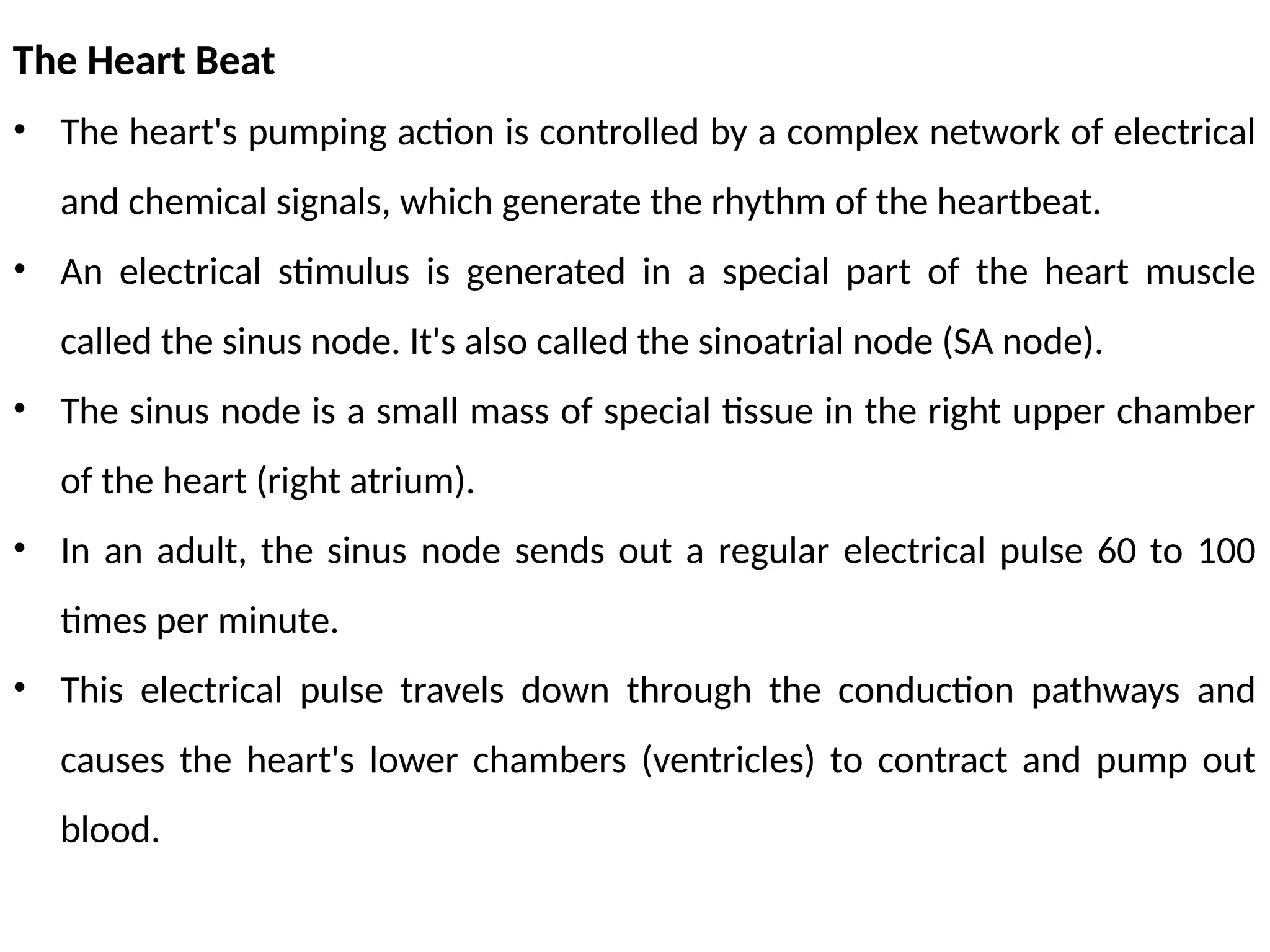
The Heart Beat
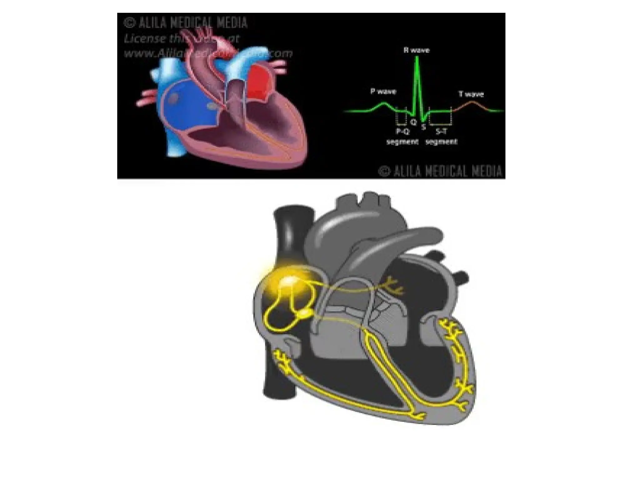
•The heart's pumping action is controlled by a complex network of electrical
and chemical signals, which generate the rhythm of the heartbeat.
• An electrical stimulus is generated in a special part of the heart muscle
called the sinus node. It's also called the sinoatrial node (SA node).
• The sinus node is a small mass of special tissue in the right upper chamber
of the heart (right atrium).
• In an adult, the sinus node sends out a regular electrical pulse 60 to 100
times per minute.
• This electrical pulse travels down through the conduction pathways and
causes the heart's lower chambers (ventricles) to contract and pump out
blood.
50.
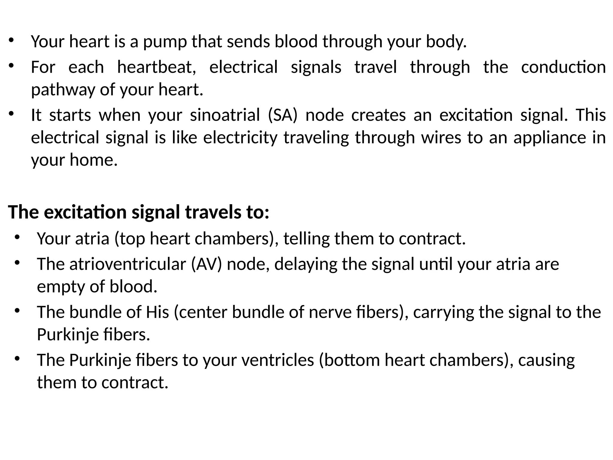
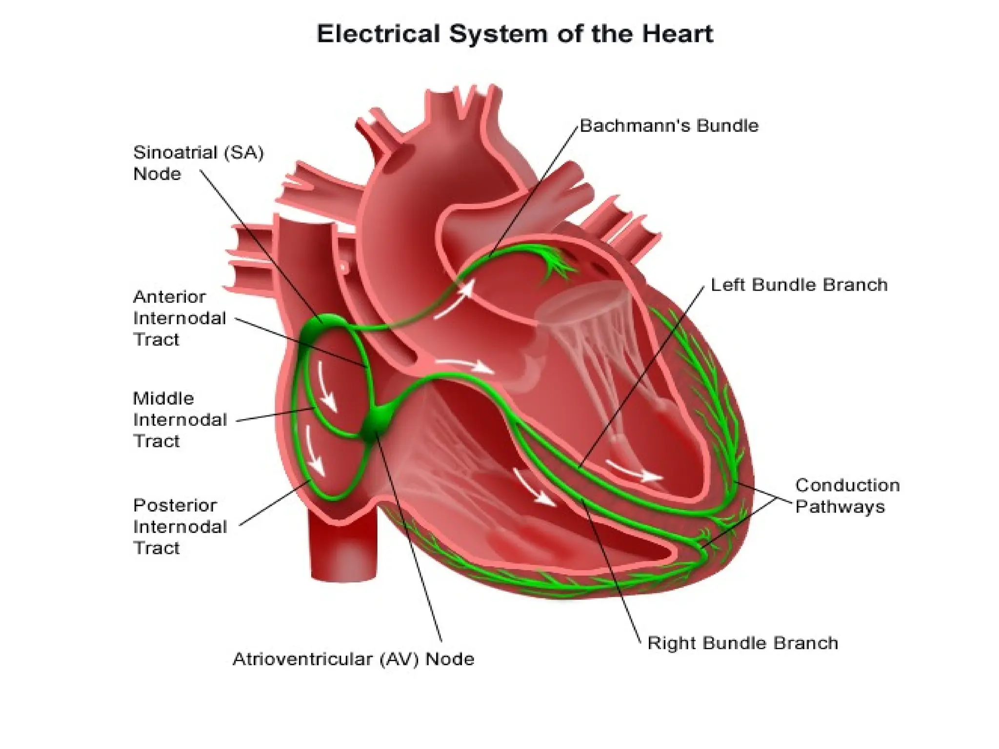
• Your heartis a pump that sends blood through your body.
• For each heartbeat, electrical signals travel through the conduction
pathway of your heart.
• It starts when your sinoatrial (SA) node creates an excitation signal. This
electrical signal is like electricity traveling through wires to an appliance in
your home.
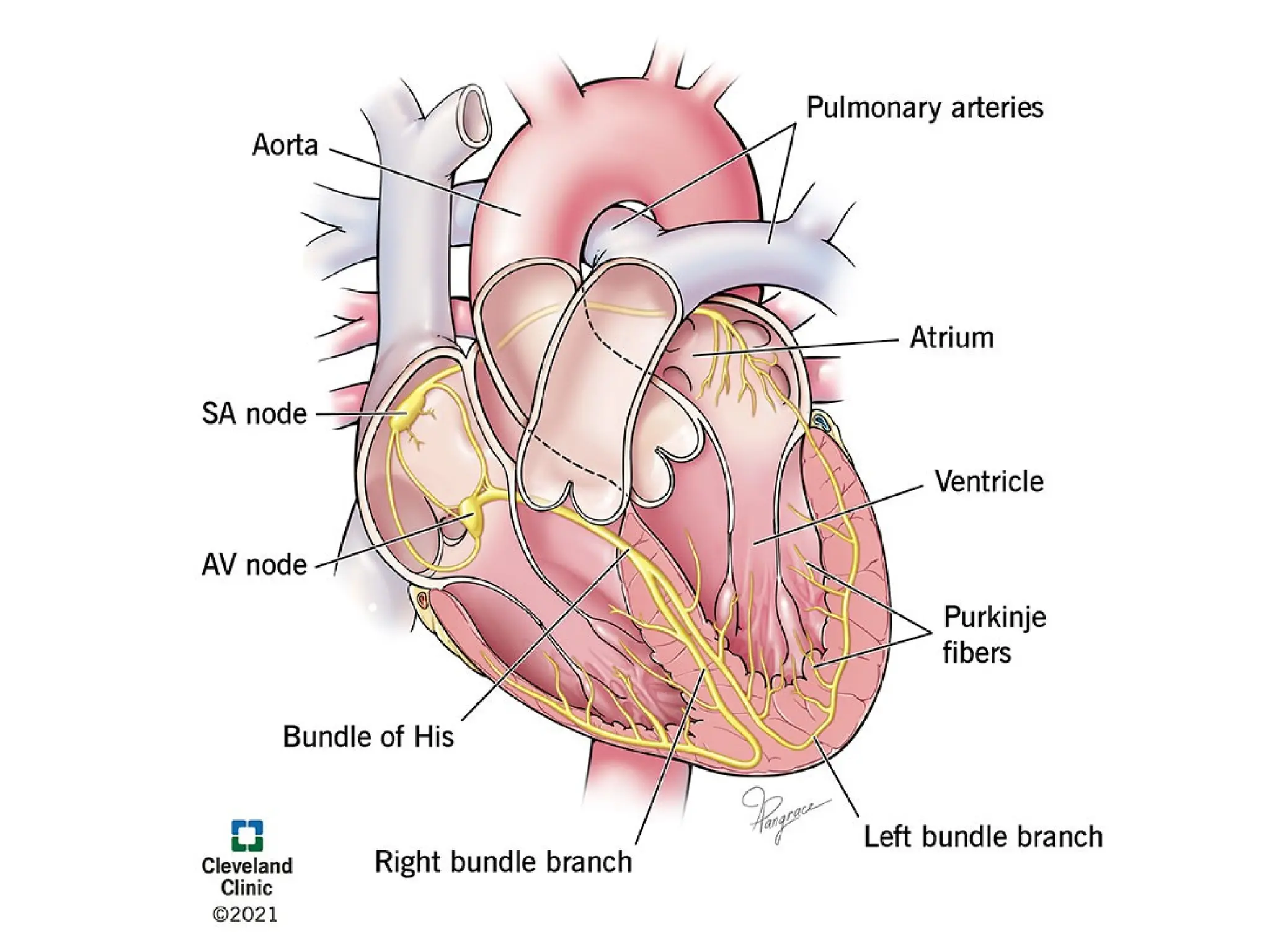
The excitation signal travels to:
• Your atria (top heart chambers), telling them to contract.
• The atrioventricular (AV) node, delaying the signal until your atria are
empty of blood.
• The bundle of His (center bundle of nerve fibers), carrying the signal to the
Purkinje fibers.
• The Purkinje fibers to your ventricles (bottom heart chambers), causing
them to contract.
53.
• The rightand left atria are stimulated first and contract to push blood from
the atria into the ventricles. The ventricles then contract to push blood out
into the blood vessels of the body.
55.
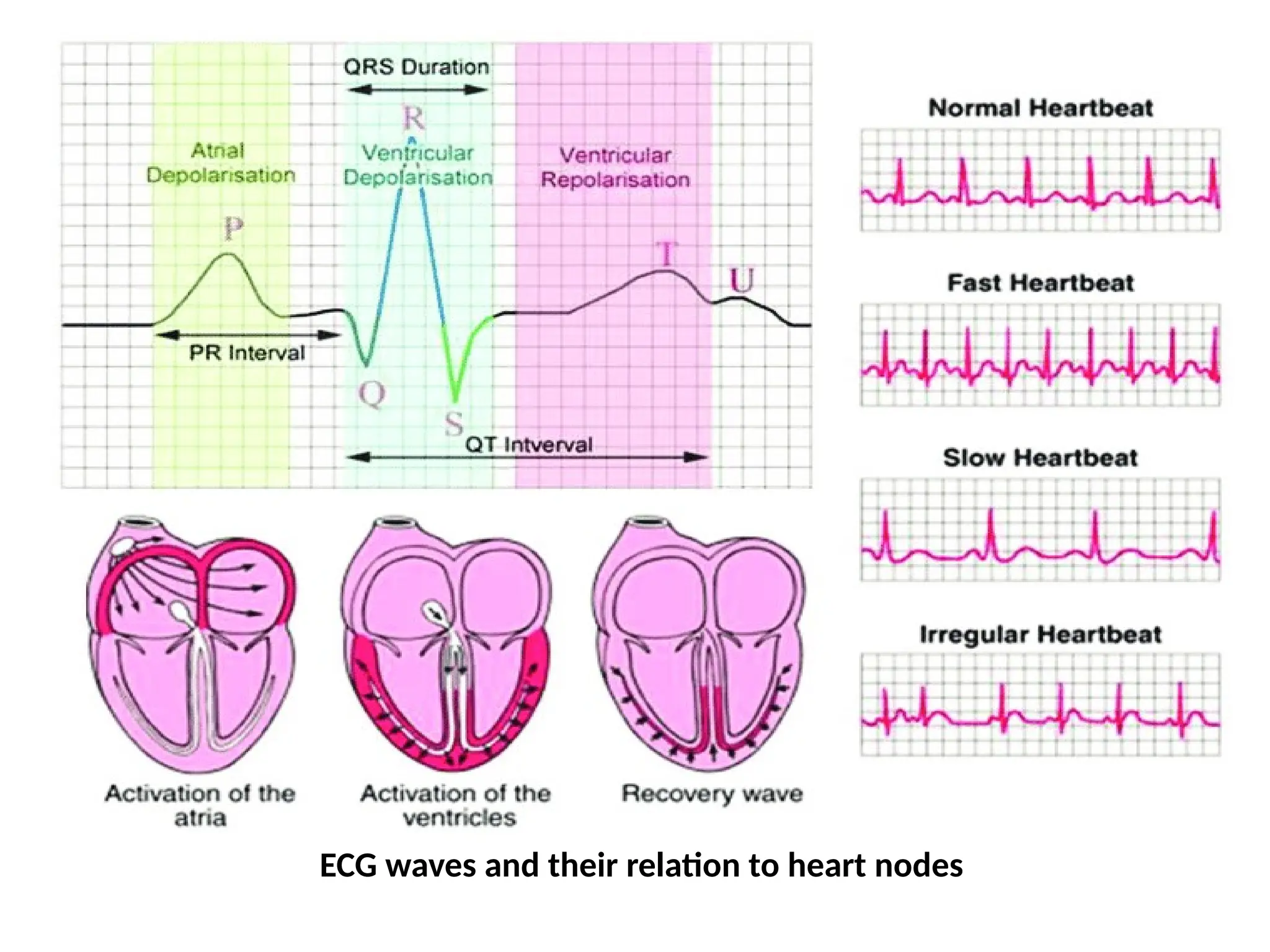
Electrical Signalling –ECG Monitoring and Heart Related Issues
• The heart's pumping action is controlled by electrical signaling, which
generates the rhythm of the heartbeat.
• This electrical signaling can be monitored using an electrocardiogram (ECG),
which records the electrical activity of the heart and provides important
information about the heart's function.
• An ECG measures the electrical signals produced by the heart as it beats
and generates a trace or waveform that reflects the electrical activity of the
heart.
• This trace can be used to diagnose heart conditions and monitor the heart's
function.
56.
Some common heart-relatedissues that can be diagnosed or monitored using
an ECG include:
1. Arrhythmias: Abnormalities in the heart's rhythm and rate can be detected
using an ECG.
2. Heart disease: Changes in the heart's electrical activity can indicate the
presence of heart disease, such as coronary artery disease or heart attacks.
3. Heart attack: An ECG can help diagnose a heart attack by detecting
changes in the heart's electrical activity that indicate a lack of blood flow to
the heart.
Overall, the ECG is a useful tool for diagnosing and monitoring heart-related
issues and helps to provide important information about the heart's function
and health.
Reasons for Blockagesof Blood Vessels / arterial blockages or atherosclerosis
1. High cholesterol levels: Excessive amounts of low-density lipoprotein (LDL)
cholesterol in the blood can lead to the formation of plaque in the blood
vessels, which can narrow or block them.
2. High blood pressure: Over time, high blood pressure can cause damage to
the blood vessels, leading to the formation of plaque and blockages.
3. Smoking: Smoking can damage the inner walls of blood vessels and
promote the buildup of plaque, leading to blockages.
4. Diabetes: People with uncontrolled diabetes are at a higher risk of
developing blockages in their blood vessels, due to damage to the blood
vessels from high levels of glucose.
59.
5. Age: Aspeople age, the blood vessels can become stiff and less flexible,
increasing the risk of blockages
6. Genetics: Some people may be predisposed to developing blockages in
their blood vessels due to genetic factors.
7. Poor diet: A diet high in saturated fats, trans fats, and cholesterol can
increase the risk of developing blockages in the blood vessels.
• The blockages in blood vessels can have serious health consequences, such
as heart attacks and stroke.
• Maintaining a healthy lifestyle, including eating a healthy diet, exercising
regularly, and avoiding smoking, can help reduce the risk of developing
blockages in blood vessels.
60.
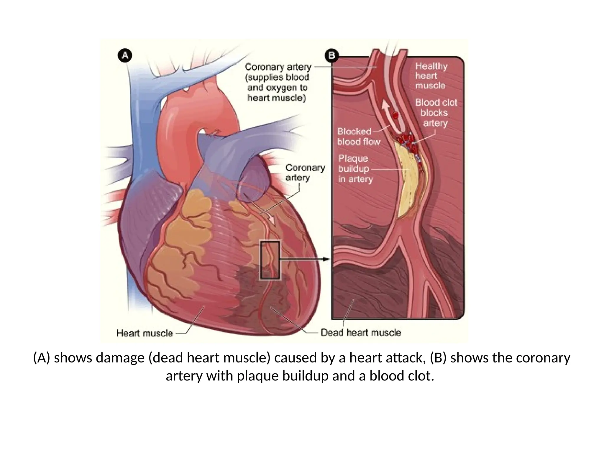
(A) shows damage(dead heart muscle) caused by a heart attack, (B) shows the coronary
artery with plaque buildup and a blood clot.
61.
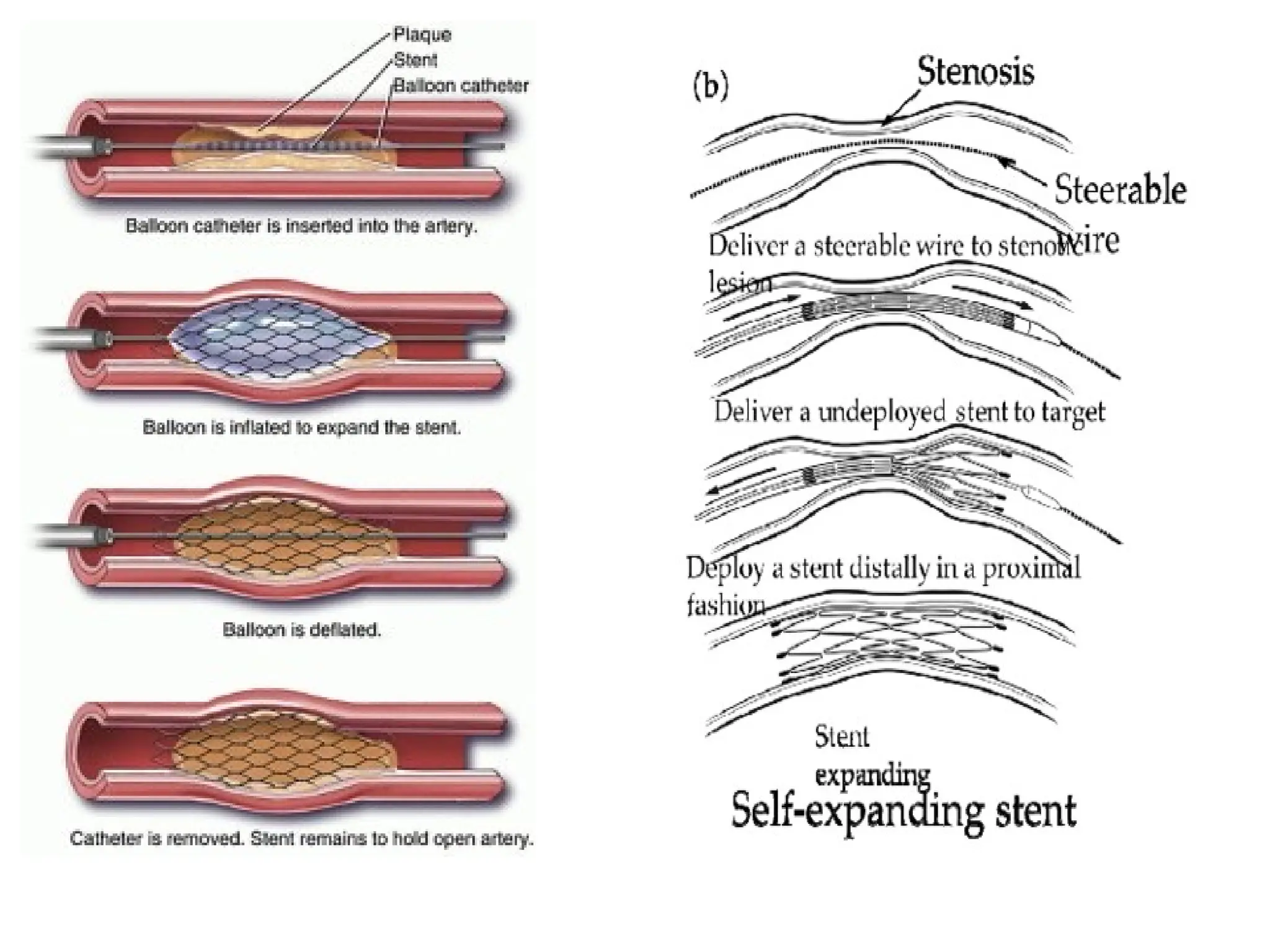
Design of Stents
•Stents are small, metal mesh devices that are used to treat blockages in
blood vessels.
• They are typically used in procedures such as angioplasty, where a balloon
catheter is used to open up a blocked blood vessel and a stent is placed to
keep it open.
• The design of stents can vary depending on the type of stent and the
specific medical condition it is used to treat.
Some common design features of stents include:
1. Shape: Stents can be designed in a variety of shapes, including cylindrical,
helical, and spiraled, to match the shape of the blood vessel and provide
adequate support.
62.
2. Material: Stentscan be made of different materials, including stainless
steel, cobaltchromium, and nitinol (a type of metal that is flexible and can
return to its original shape after being expanded).
3. Coating: Stents can be coated with different materials to prevent blood
clots from forming and reduce the risk of restenosis (recurrent blockage of
the blood vessel).
4. Expansion mechanism: Stents can be designed to expand in different ways,
such as by balloon inflation or self-expansion, depending on the type of
stent and the specific medical condition it is used to treat.
Overall, the design of stents plays an important role in their effectiveness and
safety. Stents must be designed to provide adequate support to the blood
vessel, prevent restenosis, and minimize the risk of complications such as
blood clots.
64.
Pace Makers
• Apacemaker is a small device that is surgically implanted in the chest to
regulate the heartbeat.
• It is used to treat heart rhythm disorders, such as bradycardia (a slow
heartbeat) or arrhythmias (abnormal heart rhythms), by delivering
electrical impulses to the heart to regulate its rhythm.
The basic design of a pacemaker consists of:
1. Generator: The generator is the main component of the pacemaker and
contains a battery and electronic circuitry to generate and control the
electrical impulses.
2. Leads: Leads are thin wires that connect the generator to the heart and
carry the electrical impulses from the generator to the heart.
65.
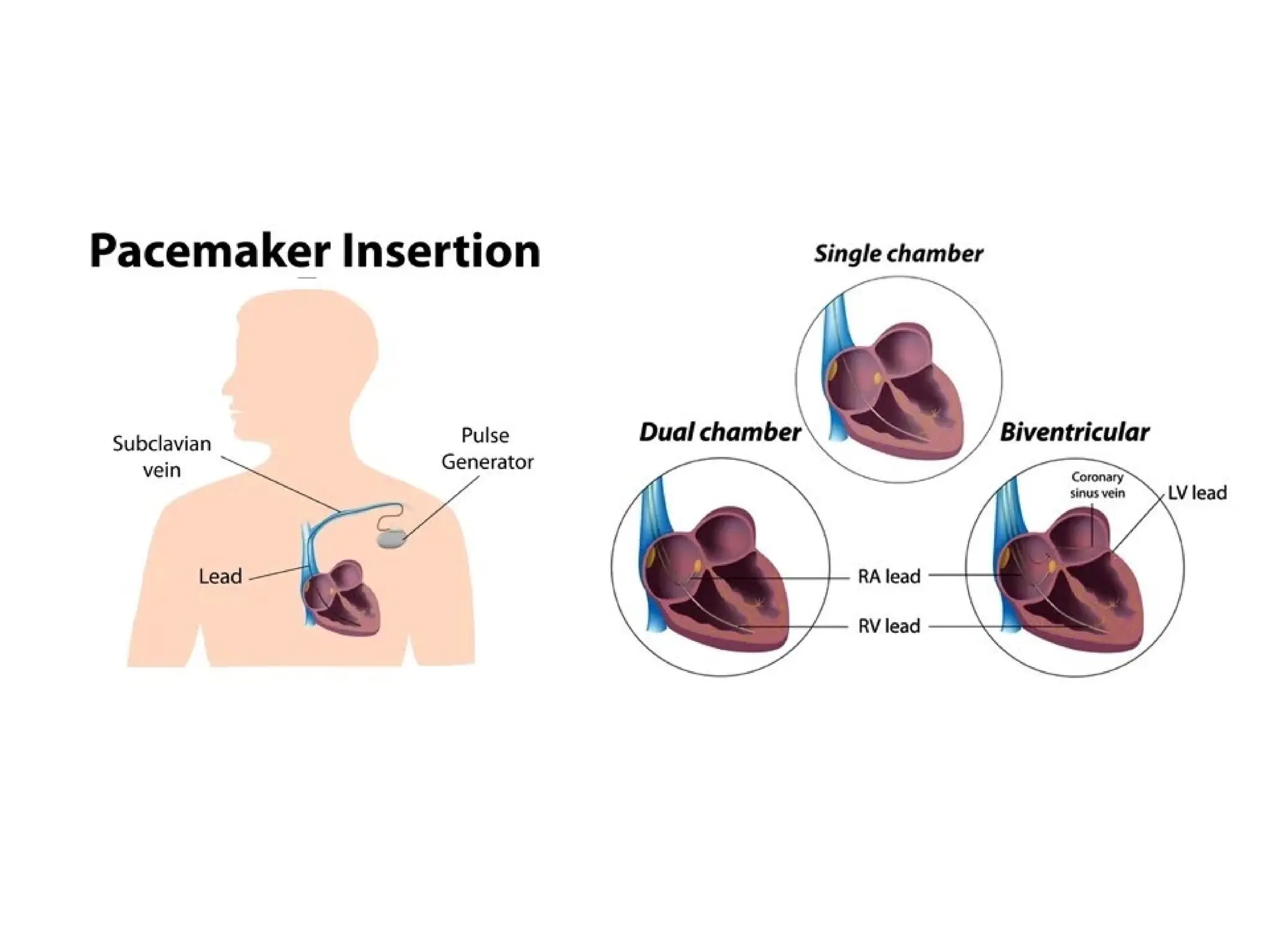
3. Electrodes: Theelectrodes are located at the end of the leads and are used
to deliver the electrical impulses to the heart.
Pacemakers can be designed to work in different ways, including:
4. Single-chamber pacemaker: A single-chamber pacemaker delivers
electrical impulses to either the right atrium or the right ventricle of the
heart to regulate its rhythm.
5. Dual-chamber pacemaker: A dual-chamber pacemaker delivers electrical
impulses to both the right atrium and the right ventricle of the heart to
regulate its rhythm.
6. Biventricular pacemaker: A biventricular pacemaker delivers electrical
impulses to both ventricles of the heart to coordinate their contractions
and improve heart function in people with heart failure.
67.
Construction of aPacemaker
• It involves the use of high-quality materials and specialized manufacturing
processes to ensure their safety and reliability.
Materials used in the construction of pacemakers include:
1. Medical-grade plastics: Medical-grade plastics, such as polycarbonate, are
used to construct the exterior of the device and to provide insulation and
protection for the internal components.
2. Metals: Metals, such as stainless steel and titanium, are used in the
construction of the leads and electrodes to ensure their durability and
long-lasting performance.
3. Electronic components: Electronic components, such as microprocessors,
batteries, and capacitors, are used to control the delivery of the electrical
impulses and to provide power to the device.
68.
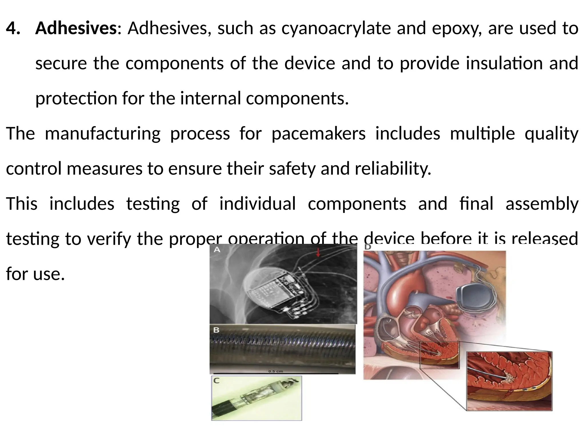
4. Adhesives: Adhesives,such as cyanoacrylate and epoxy, are used to
secure the components of the device and to provide insulation and
protection for the internal components.
The manufacturing process for pacemakers includes multiple quality
control measures to ensure their safety and reliability.
This includes testing of individual components and final assembly
testing to verify the proper operation of the device before it is released
for use.
69.
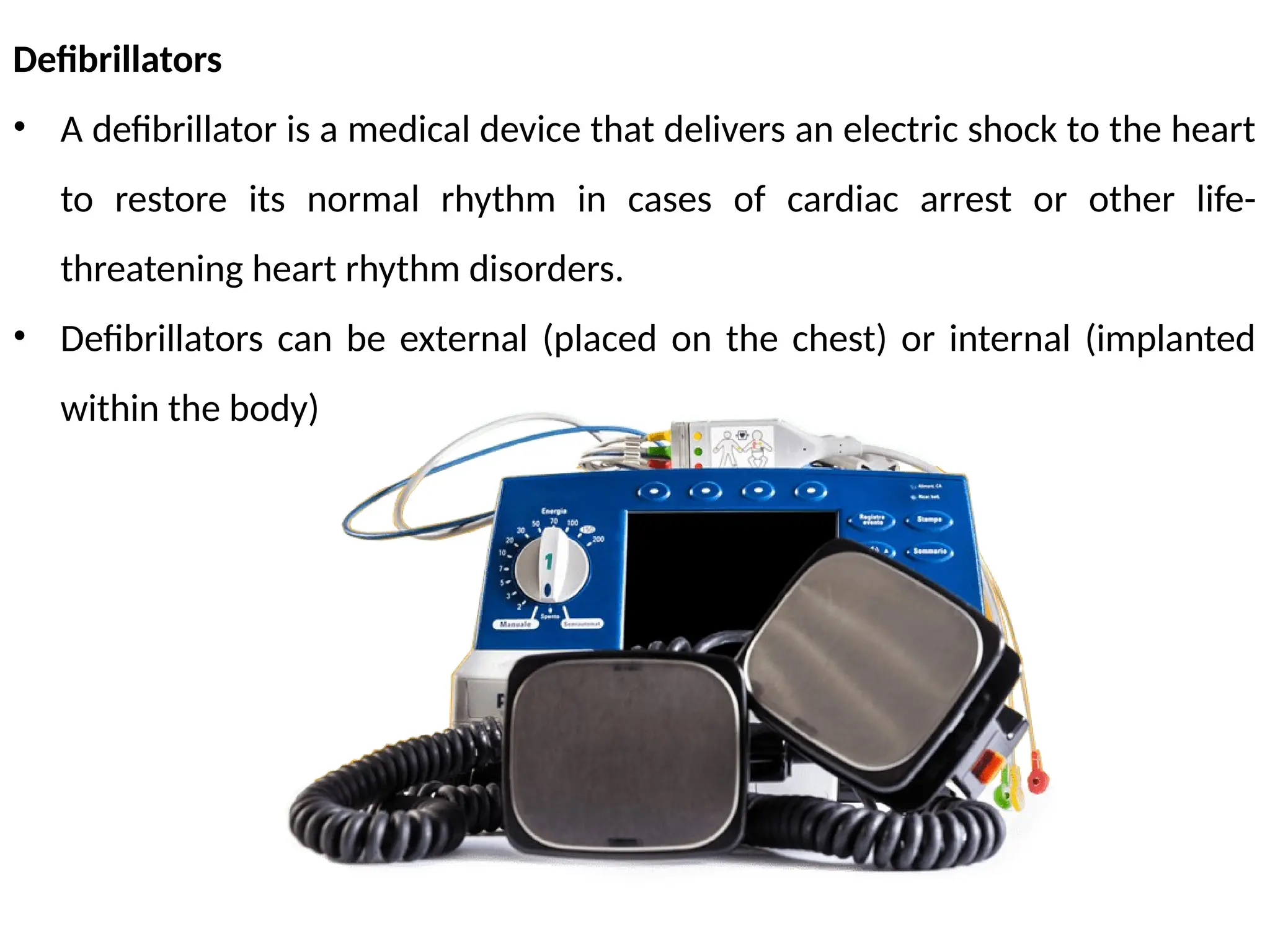
Defibrillators
• A defibrillatoris a medical device that delivers an electric shock to the heart
to restore its normal rhythm in cases of cardiac arrest or other life-
threatening heart rhythm disorders.
• Defibrillators can be external (placed on the chest) or internal (implanted
within the body)
70.
The basic designof a defibrillator consists of:
1. Power source: The power source, typically a battery, provides
energy to deliver the electric shock to the heart.
2. Electrodes: The electrodes are placed on the chest and deliver the
electric shock to the heart.
3. Circuitry: The circuitry in the defibrillator controls the delivery of the
electric shock, including the timing, strength, and duration of the
shock.
4. Display: A display on the defibrillator provides information about the
heart rhythm, battery life, and other relevant information.
71.
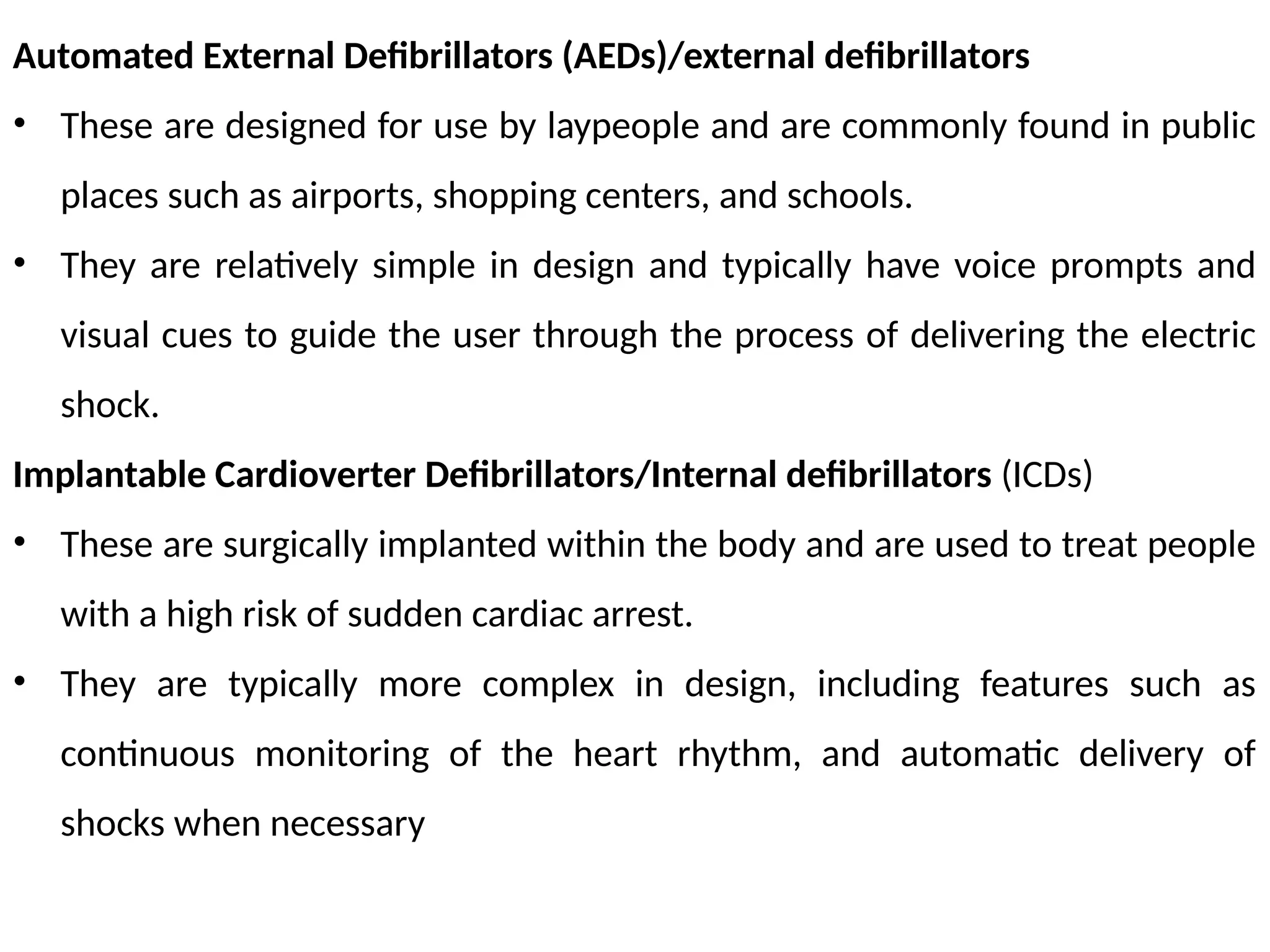
Automated External Defibrillators(AEDs)/external defibrillators
• These are designed for use by laypeople and are commonly found in public
places such as airports, shopping centers, and schools.
• They are relatively simple in design and typically have voice prompts and
visual cues to guide the user through the process of delivering the electric
shock.
Implantable Cardioverter Defibrillators/Internal defibrillators (ICDs)
• These are surgically implanted within the body and are used to treat people
with a high risk of sudden cardiac arrest.
• They are typically more complex in design, including features such as
continuous monitoring of the heart rhythm, and automatic delivery of
shocks when necessary
72.
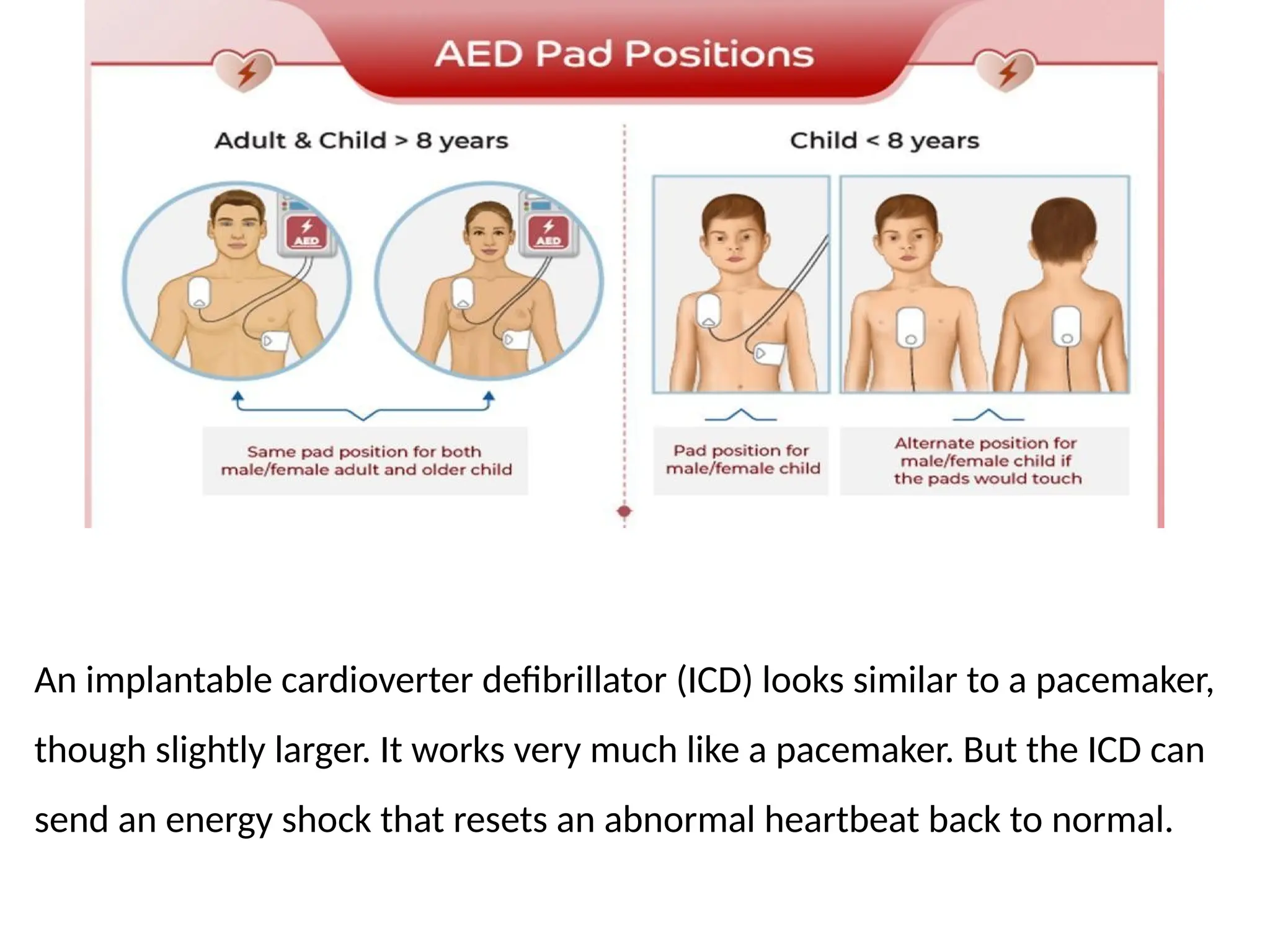
An implantable cardioverterdefibrillator (ICD) looks similar to a pacemaker,
though slightly larger. It works very much like a pacemaker. But the ICD can
send an energy shock that resets an abnormal heartbeat back to normal.
73.
Construction of defibrillators
Theconstruction of defibrillators involves the use of high-quality materials and
specialized manufacturing processes to ensure their safety and reliability.
Materials Used Materials used in the construction of defibrillators include:
1. Medical-grade plastics: such as polycarbonate, are used to construct the
exterior of the device and to provide insulation and protection for the
internal components.
2. Metals: such as stainless steel and titanium, are used in the construction of
the leads and electrodes to ensure their durability and long-lasting
performance.
3. Electronic components: such as microprocessors, batteries, capacitors, and
high-voltage transformers, are used to control the delivery of the electrical
impulses and to provide power to the device.
74.
4. Adhesives: suchas cyanoacrylate and epoxy, are used to secure the
components of the device and to provide insulation and protection for the
internal components
• The manufacturing process for defibrillators includes multiple quality
control measures to ensure their safety and reliability.
• This includes testing of individual components and final assembly testing to
verify the proper operation of the device before it is released for use.
75.
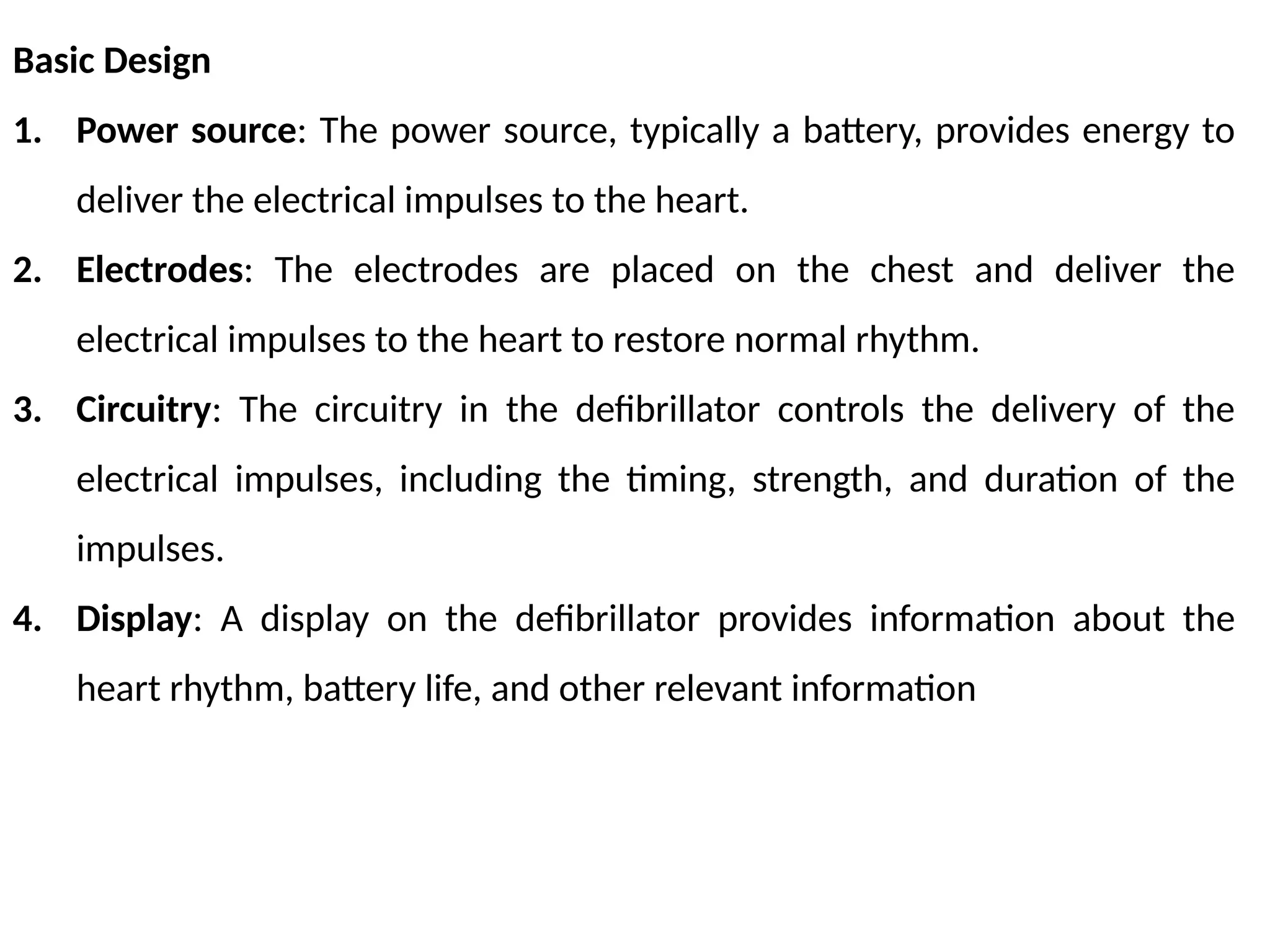
Basic Design
1. Powersource: The power source, typically a battery, provides energy to
deliver the electrical impulses to the heart.
2. Electrodes: The electrodes are placed on the chest and deliver the
electrical impulses to the heart to restore normal rhythm.
3. Circuitry: The circuitry in the defibrillator controls the delivery of the
electrical impulses, including the timing, strength, and duration of the
impulses.
4. Display: A display on the defibrillator provides information about the
heart rhythm, battery life, and other relevant information
76.
Artificial Heart
An artificialheart is a device that is designed to replace the functions of a
damaged or failing heart.
• It can be used as a temporary measure to support a patient while they are
waiting for a heart transplant, or as a permanent solution for people who
are not eligible for a heart transplant.
There are two main types of artificial hearts: total artificial hearts and heart
assist devices.
• A total artificial heart is a self-contained device that completely replaces
the functions of the natural heart. It is used as a bridge to transplant, it
provides temporary support to a patient while they are waiting for a heart
transplant.
77.
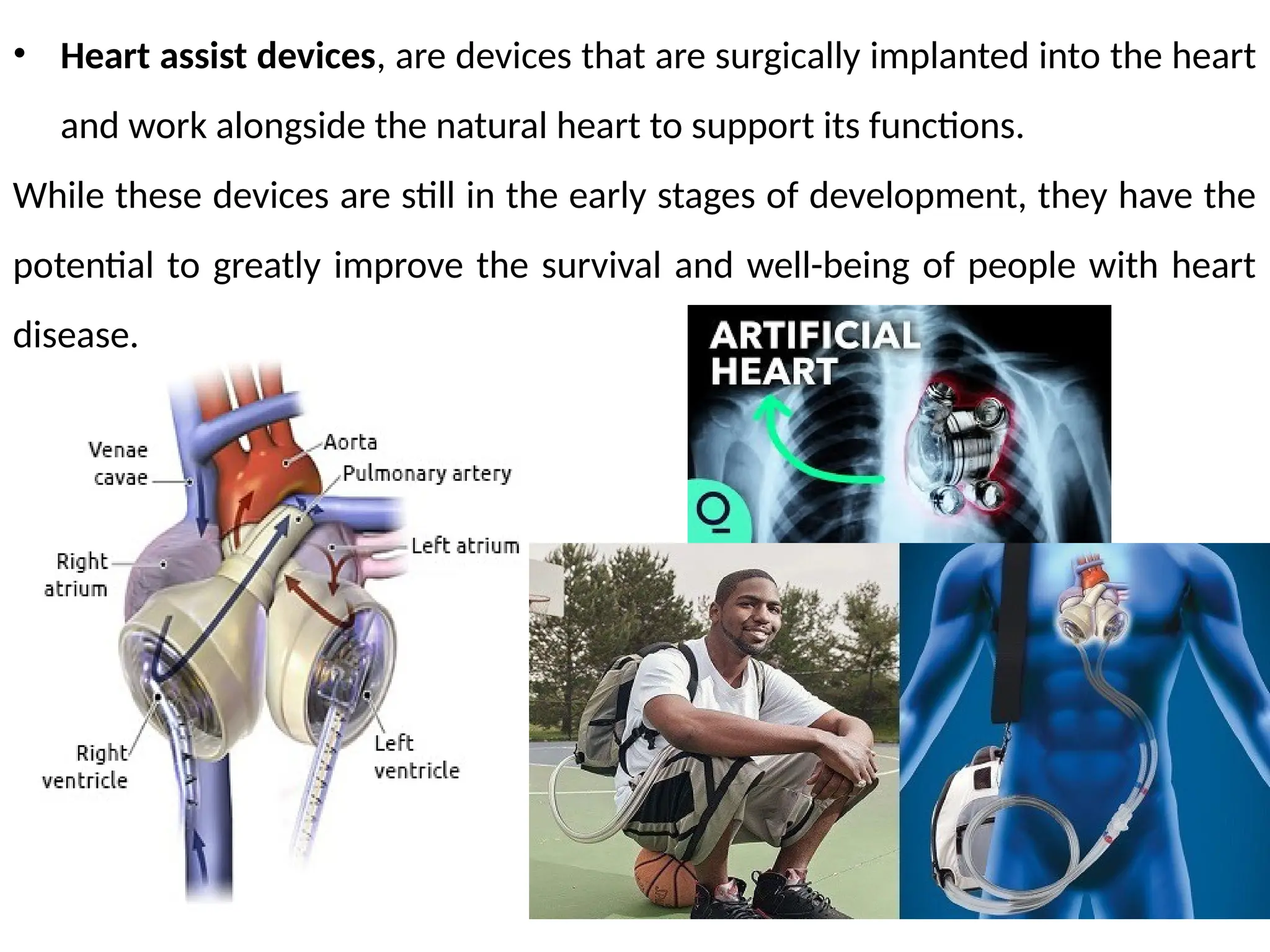
• Heart assistdevices, are devices that are surgically implanted into the heart
and work alongside the natural heart to support its functions.
While these devices are still in the early stages of development, they have the
potential to greatly improve the survival and well-being of people with heart
disease.
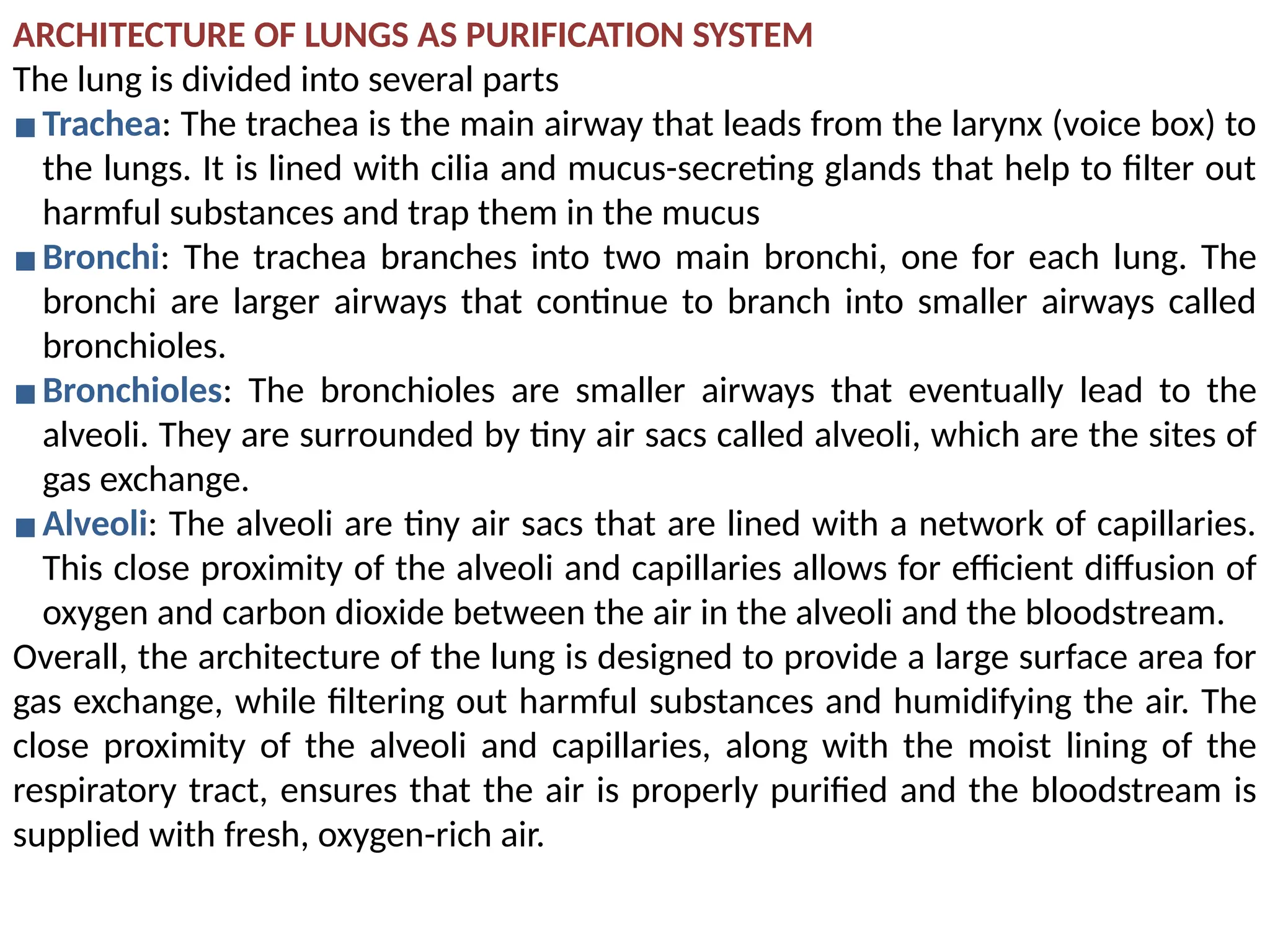
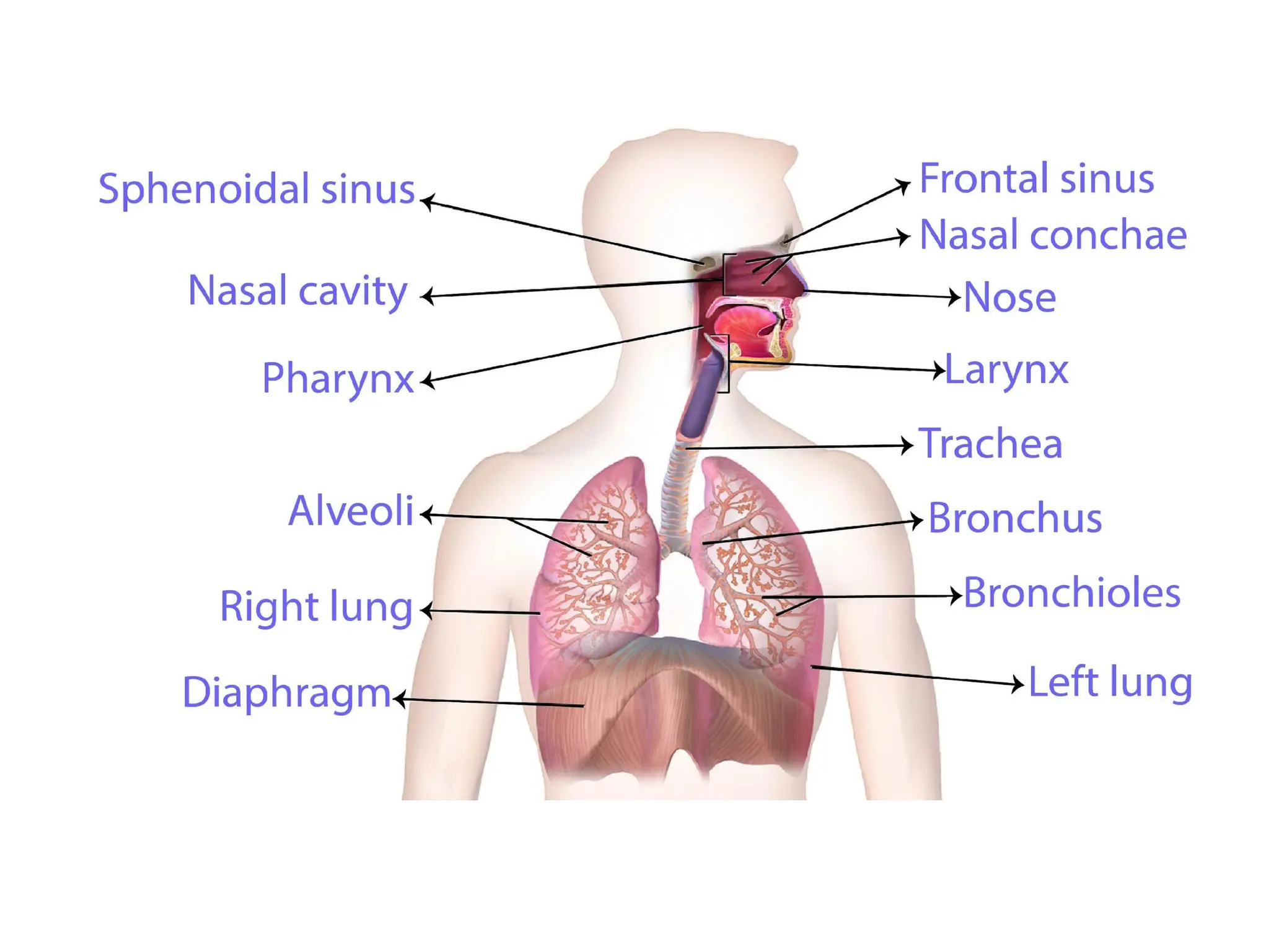
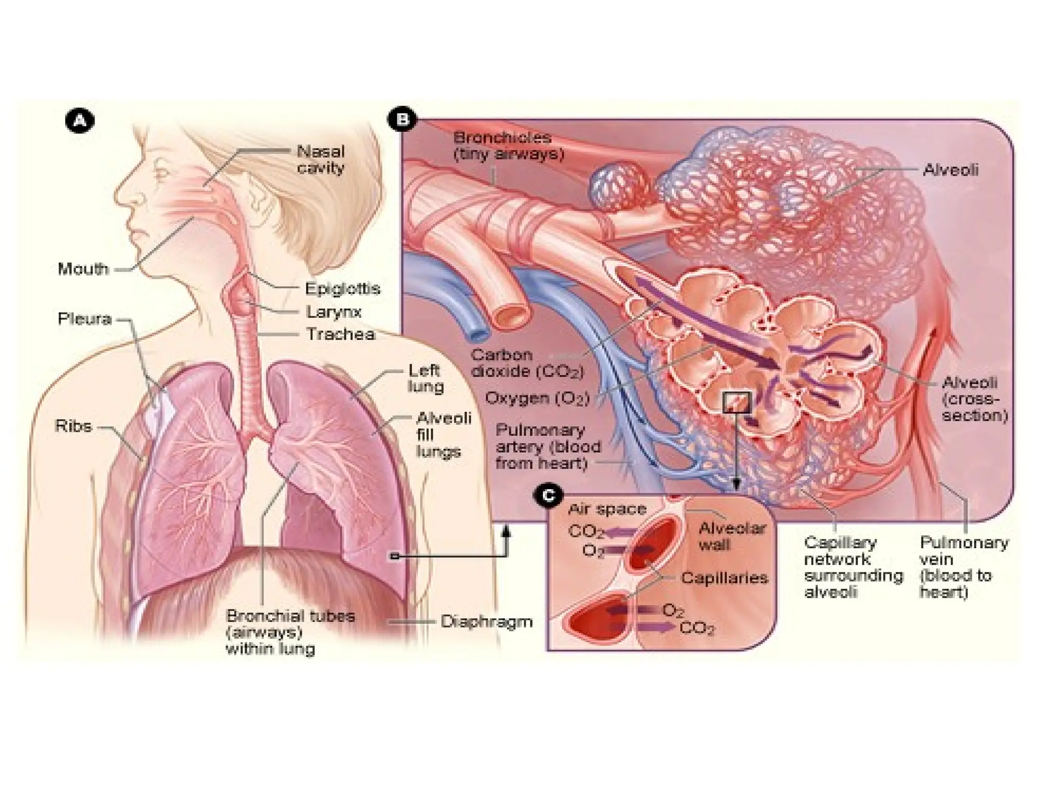
ARCHITECTURE OF LUNGSAS PURIFICATION SYSTEM
The lung is divided into several parts
▪Trachea: The trachea is the main airway that leads from the larynx (voice box) to
the lungs. It is lined with cilia and mucus-secreting glands that help to filter out
harmful substances and trap them in the mucus
▪Bronchi: The trachea branches into two main bronchi, one for each lung. The
bronchi are larger airways that continue to branch into smaller airways called
bronchioles.
▪Bronchioles: The bronchioles are smaller airways that eventually lead to the
alveoli. They are surrounded by tiny air sacs called alveoli, which are the sites of
gas exchange.
▪Alveoli: The alveoli are tiny air sacs that are lined with a network of capillaries.
This close proximity of the alveoli and capillaries allows for efficient diffusion of
oxygen and carbon dioxide between the air in the alveoli and the bloodstream.
Overall, the architecture of the lung is designed to provide a large surface area for
gas exchange, while filtering out harmful substances and humidifying the air. The
close proximity of the alveoli and capillaries, along with the moist lining of the
respiratory tract, ensures that the air is properly purified and the bloodstream is
supplied with fresh, oxygen-rich air.
81.
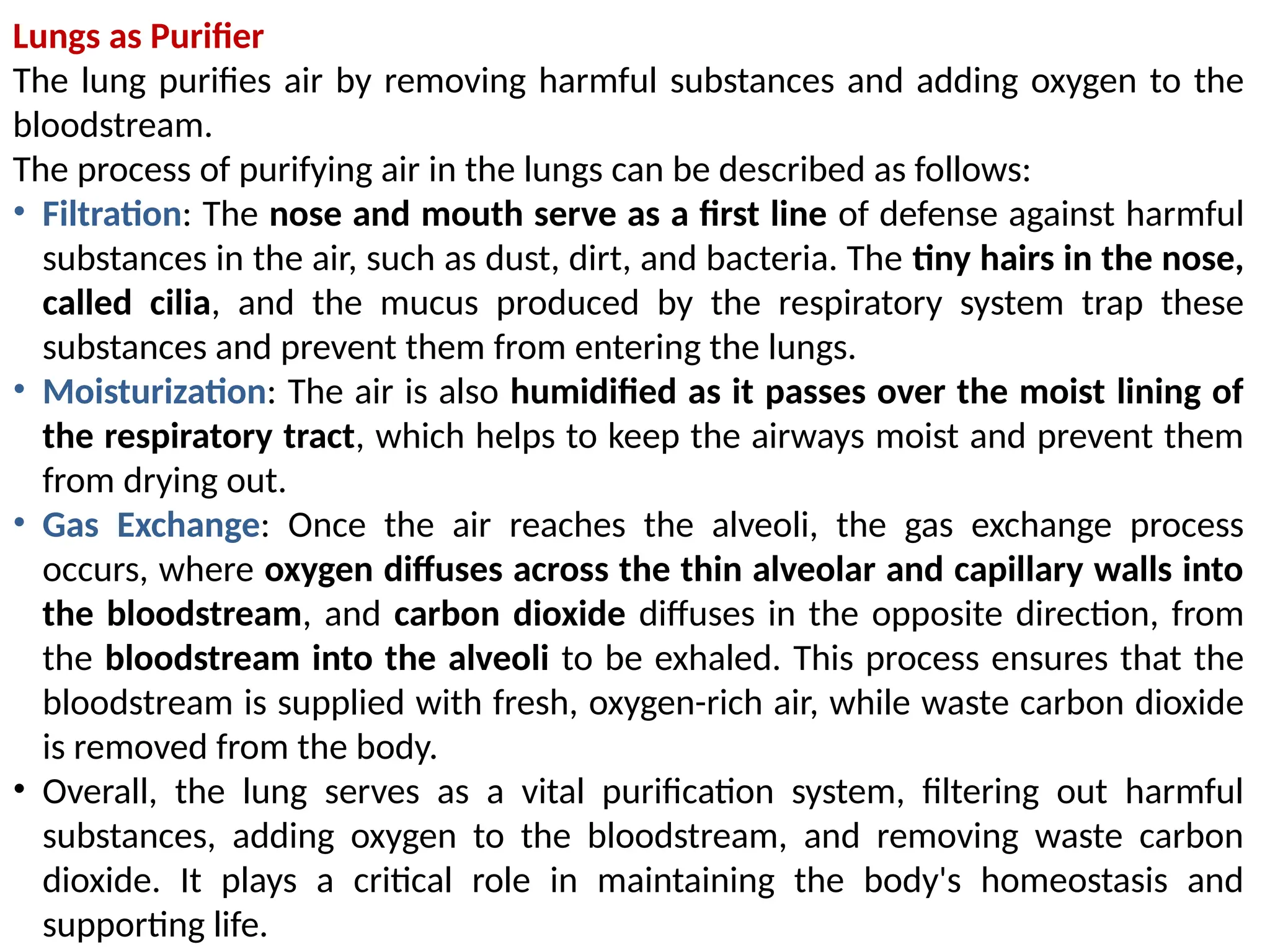
Lungs as Purifier
Thelung purifies air by removing harmful substances and adding oxygen to the
bloodstream.
The process of purifying air in the lungs can be described as follows:
• Filtration: The nose and mouth serve as a first line of defense against harmful
substances in the air, such as dust, dirt, and bacteria. The tiny hairs in the nose,
called cilia, and the mucus produced by the respiratory system trap these
substances and prevent them from entering the lungs.
• Moisturization: The air is also humidified as it passes over the moist lining of
the respiratory tract, which helps to keep the airways moist and prevent them
from drying out.
• Gas Exchange: Once the air reaches the alveoli, the gas exchange process
occurs, where oxygen diffuses across the thin alveolar and capillary walls into
the bloodstream, and carbon dioxide diffuses in the opposite direction, from
the bloodstream into the alveoli to be exhaled. This process ensures that the
bloodstream is supplied with fresh, oxygen-rich air, while waste carbon dioxide
is removed from the body.
• Overall, the lung serves as a vital purification system, filtering out harmful
substances, adding oxygen to the bloodstream, and removing waste carbon
dioxide. It plays a critical role in maintaining the body's homeostasis and
supporting life.
82.
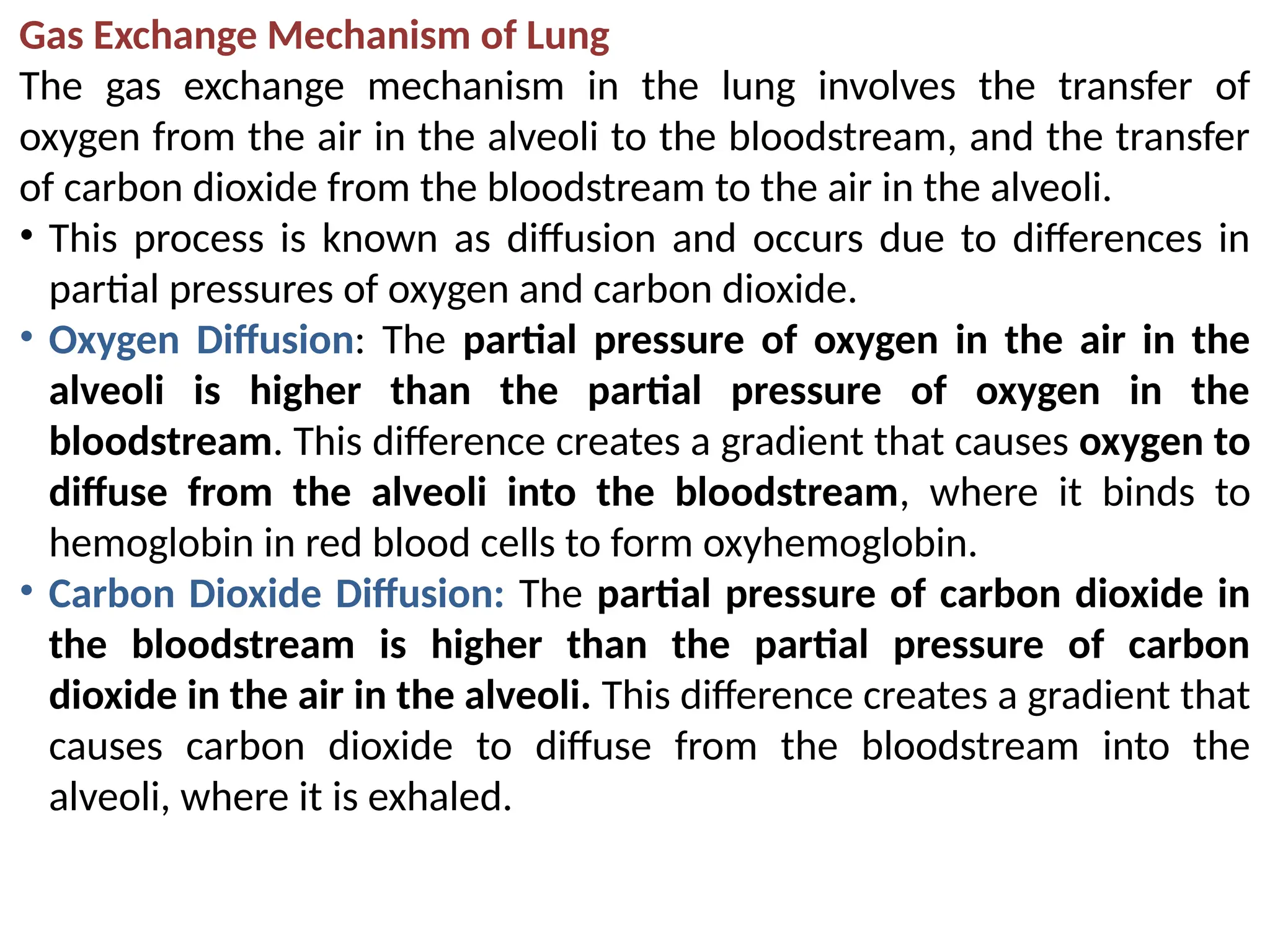
Gas Exchange Mechanismof Lung
The gas exchange mechanism in the lung involves the transfer of
oxygen from the air in the alveoli to the bloodstream, and the transfer
of carbon dioxide from the bloodstream to the air in the alveoli.
• This process is known as diffusion and occurs due to differences in
partial pressures of oxygen and carbon dioxide.
• Oxygen Diffusion: The partial pressure of oxygen in the air in the
alveoli is higher than the partial pressure of oxygen in the
bloodstream. This difference creates a gradient that causes oxygen to
diffuse from the alveoli into the bloodstream, where it binds to
hemoglobin in red blood cells to form oxyhemoglobin.
• Carbon Dioxide Diffusion: The partial pressure of carbon dioxide in
the bloodstream is higher than the partial pressure of carbon
dioxide in the air in the alveoli. This difference creates a gradient that
causes carbon dioxide to diffuse from the bloodstream into the
alveoli, where it is exhaled.
84.
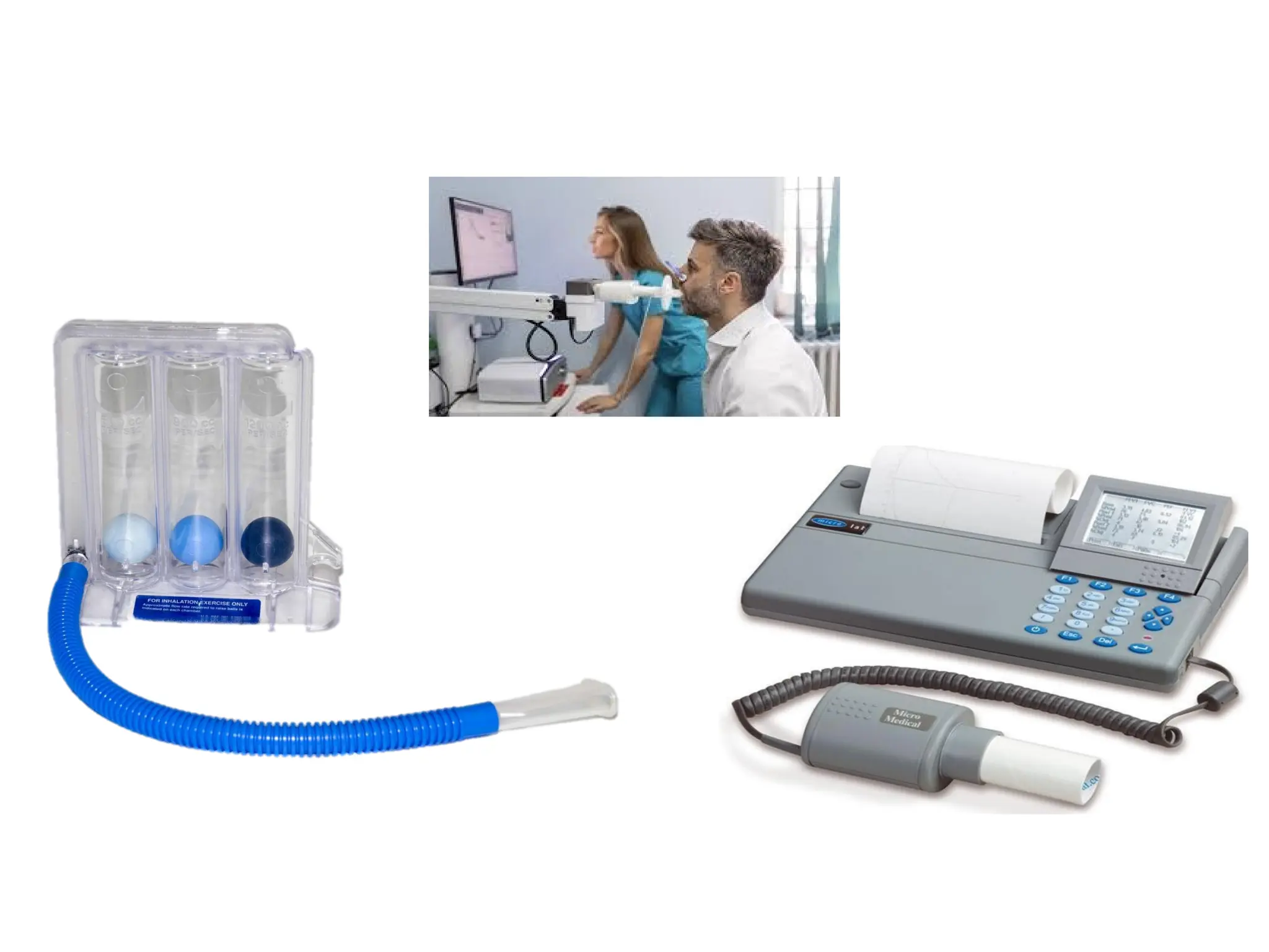
SPIROMETRY
Spirometry is adiagnostic test that measures the function of the lungs by
measuring the amount and flow rate of air that can be exhaled.
• The test is commonly used to diagnose lung conditions such as asthma, chronic
obstructive pulmonary disease (COPD), and interstitial lung disease.
Principle: The principle behind spirometry is to measure the volume of air that
can be exhaled from the lungs in a given time period. By measuring the volume of
air exhaled, spirometry can provide information about the functioning of the
lungs and the ability of the lungs to move air in and out.
Working: Spirometry is performed using a spirometer, a device that consists of a
mouthpiece, a flow sensor, and a volume sensor.
• The patient is asked to exhale as much air as possible into the spirometer, and
the spirometer measures the volume and flow rate of the exhaled air.
• The volume of air exhaled is displayed on a graph called a flow-volume loop,
which provides information about the lung function.
Interpretation of Results
The results of spirometry can be used to determine if the lungs are functioning
normally and to diagnose lung conditions.
For example, a decrease in the volume of air exhaled or a decrease in the flow
rate of the exhaled air can indicate a restriction in the airways, which can be a
sign of a lung condition.
86.
Abnormal Lung Physiology– COPD
Abnormal lung physiology refers to any deviation from the normal functioning of
the respiratory system.
This can be caused by a variety of factors, including diseases, injuries, or genetic
conditions.
Some common examples of abnormal lung physiology include:
• Asthma: the airways to narrow, making it difficult to breathe.
• Chronic obstructive pulmonary disease (COPD): A progressive lung disease that
makes it hard to breathe and can include conditions such as emphysema and
chronic bronchitis.
• Pulmonary fibrosis: A disease in which scar tissue builds up in the lungs,
making it difficult to breathe and reducing lung function.
• Pneumonia: inflammation and fluid buildup in the air sacs.
• Pulmonary embolism: A blockage in one of the pulmonary arteries, usually by
a blood clot, which can cause lung damage and reduce oxygen flow to the body.
• Lung cancer: A type of cancer that originates in the lung and can impair lung
function by interfering with normal air flow and oxygen exchange.
Treatment for abnormal lung physiology depends on the underlying cause and
may include medications, lifestyle changes, or surgery.
88.
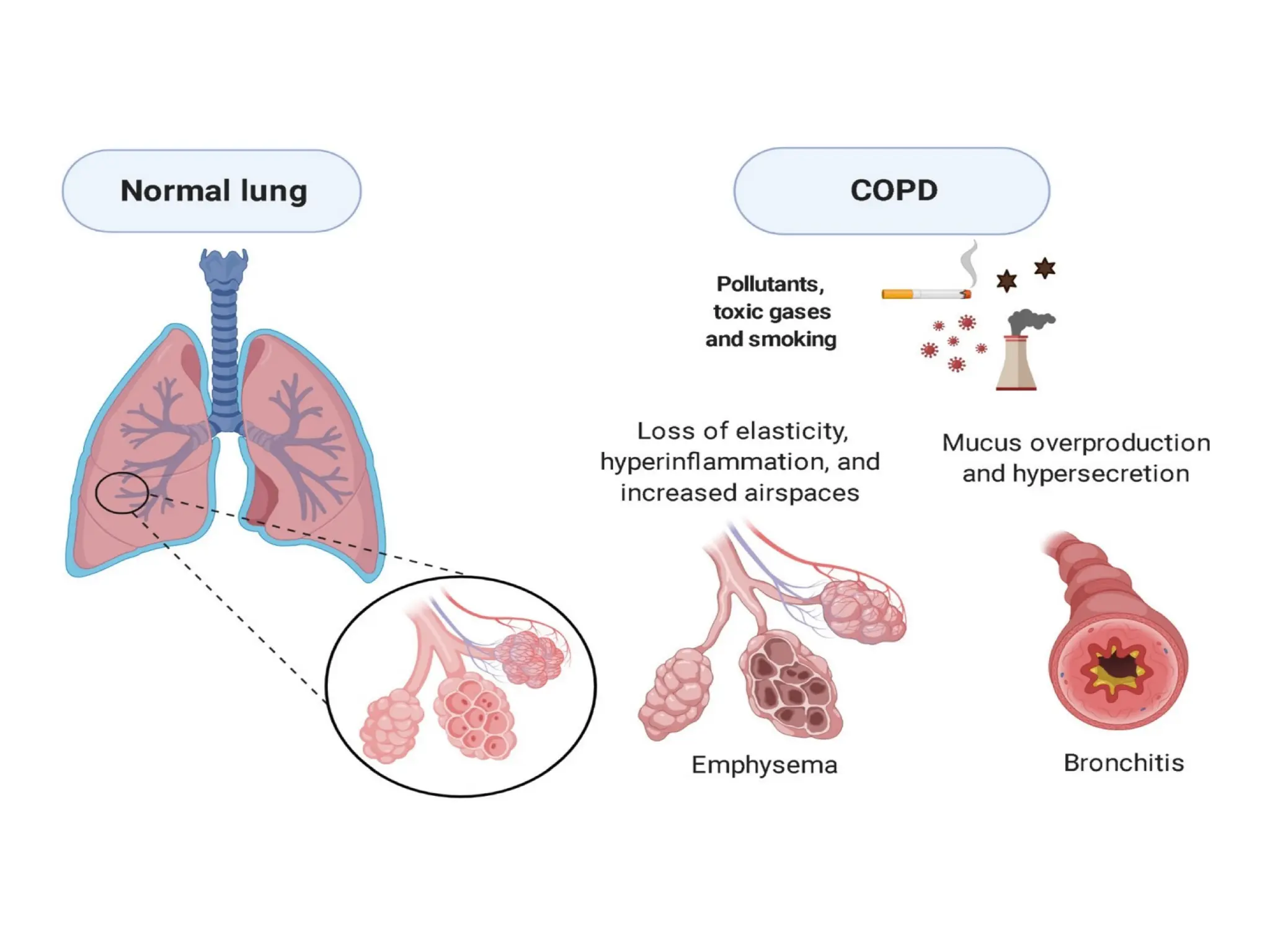
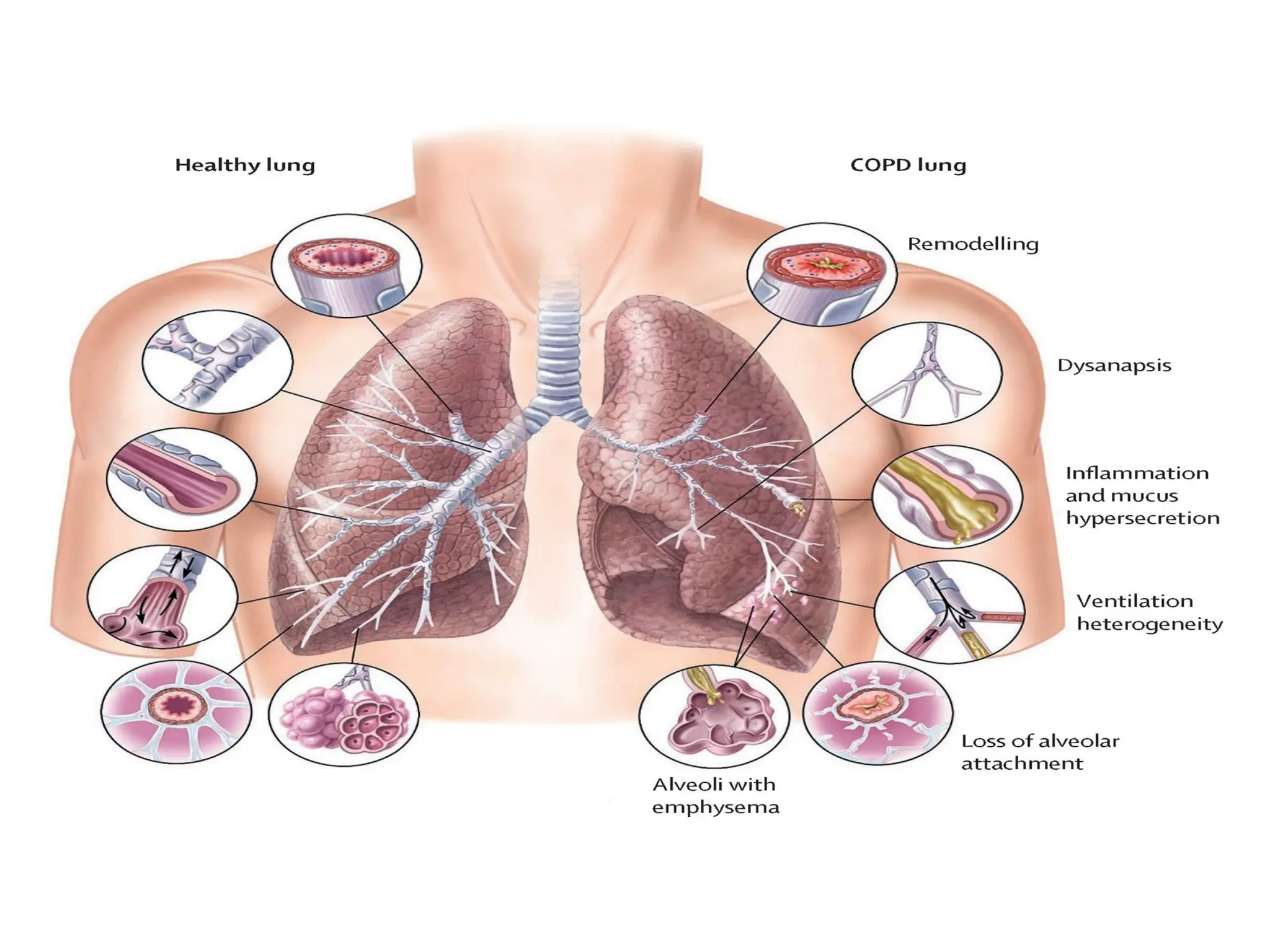
Chronic Obstructive PulmonaryDisease (COPD) is a group of progressive lung
diseases that cause breathing difficulties.
• It's characterized by persistent airflow limitation that is not fully reversible.
• The two main forms of COPD are chronic bronchitis and emphysema.
• In COPD, the airways and small air sacs (alveoli) in the lungs become
damaged or blocked, leading to difficulty in exhaling air.
• This results in a decrease in lung function, leading to shortness of breath,
wheezing, and coughing.
• Over time, these symptoms can get worse and limit a person's ability to
perform everyday activities.
• The primary cause of COPD is long-term exposure to irritants such as
tobacco smoke, air pollution, and dust, a history of frequent lung infections,
a family history of lung disease, and exposure to second-hand smoke.
• There is no cure for COPD
• Treatment options include medication, such as bronchodilators and steroids,
oxygen therapy, and lung rehabilitation. In severe cases, surgery may also be
an option. In addition, quitting smoking and avoiding exposure to irritants is
crucial in managing COPD.
90.
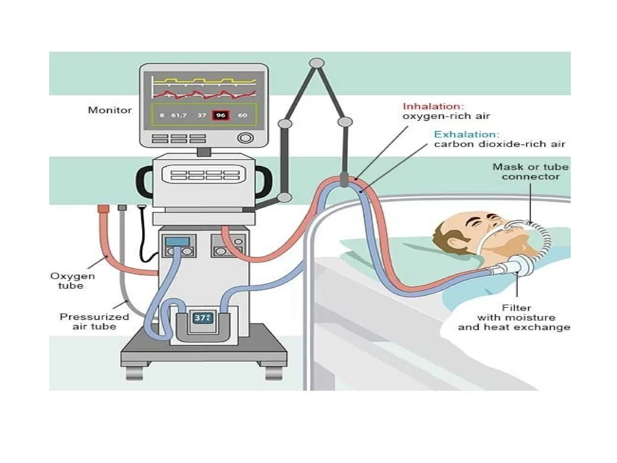
Ventilators to assistor control breathing
• They are commonly used in the treatment of acute respiratory failure,
which can occur as a result of a variety of conditions such as
pneumonia, severe asthma, and chronic obstructive pulmonary disease
(COPD)
Types of ventilators, including volume-controlled ventilators, pressure-
controlled ventilators, and bilevel positive airway pressure (BiPAP)
devices.
• Ventilators work by delivering pressurized air or oxygen into the lungs
through a breathing tube or mask.
• The pressure can be adjusted to match the patient's needs and to help
maintain adequate oxygen levels in the blood.
• While ventilators can be lifesaving for individuals with acute respiratory
failure, they also come with potential risks and complications.
• For example, prolonged use of a ventilator can increase the risk of
ventilator-associated pneumonia, and patients may experience
discomfort or pain from the breathing tube.
The use of ventilators is carefully monitored and managed by healthcare
92.
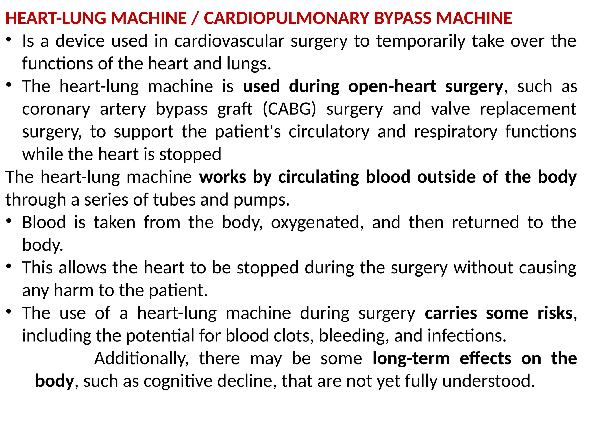
HEART-LUNG MACHINE /CARDIOPULMONARY BYPASS MACHINE
• Is a device used in cardiovascular surgery to temporarily take over the
functions of the heart and lungs.
• The heart-lung machine is used during open-heart surgery, such as
coronary artery bypass graft (CABG) surgery and valve replacement
surgery, to support the patient's circulatory and respiratory functions
while the heart is stopped
The heart-lung machine works by circulating blood outside of the body
through a series of tubes and pumps.
• Blood is taken from the body, oxygenated, and then returned to the
body.
• This allows the heart to be stopped during the surgery without causing
any harm to the patient.
• The use of a heart-lung machine during surgery carries some risks,
including the potential for blood clots, bleeding, and infections.
Additionally, there may be some long-term effects on the
body, such as cognitive decline, that are not yet fully understood.
94.
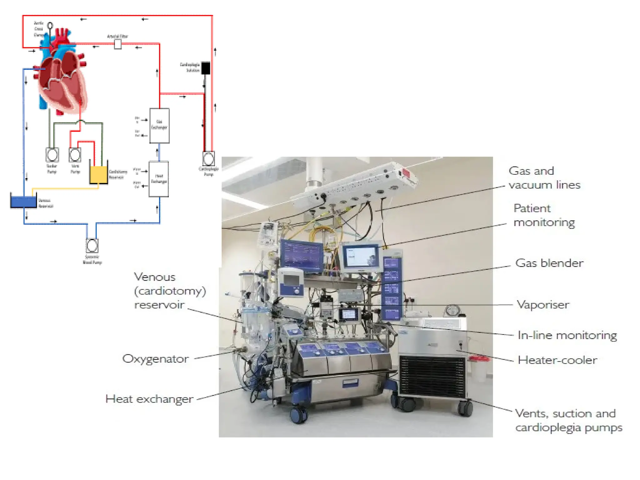
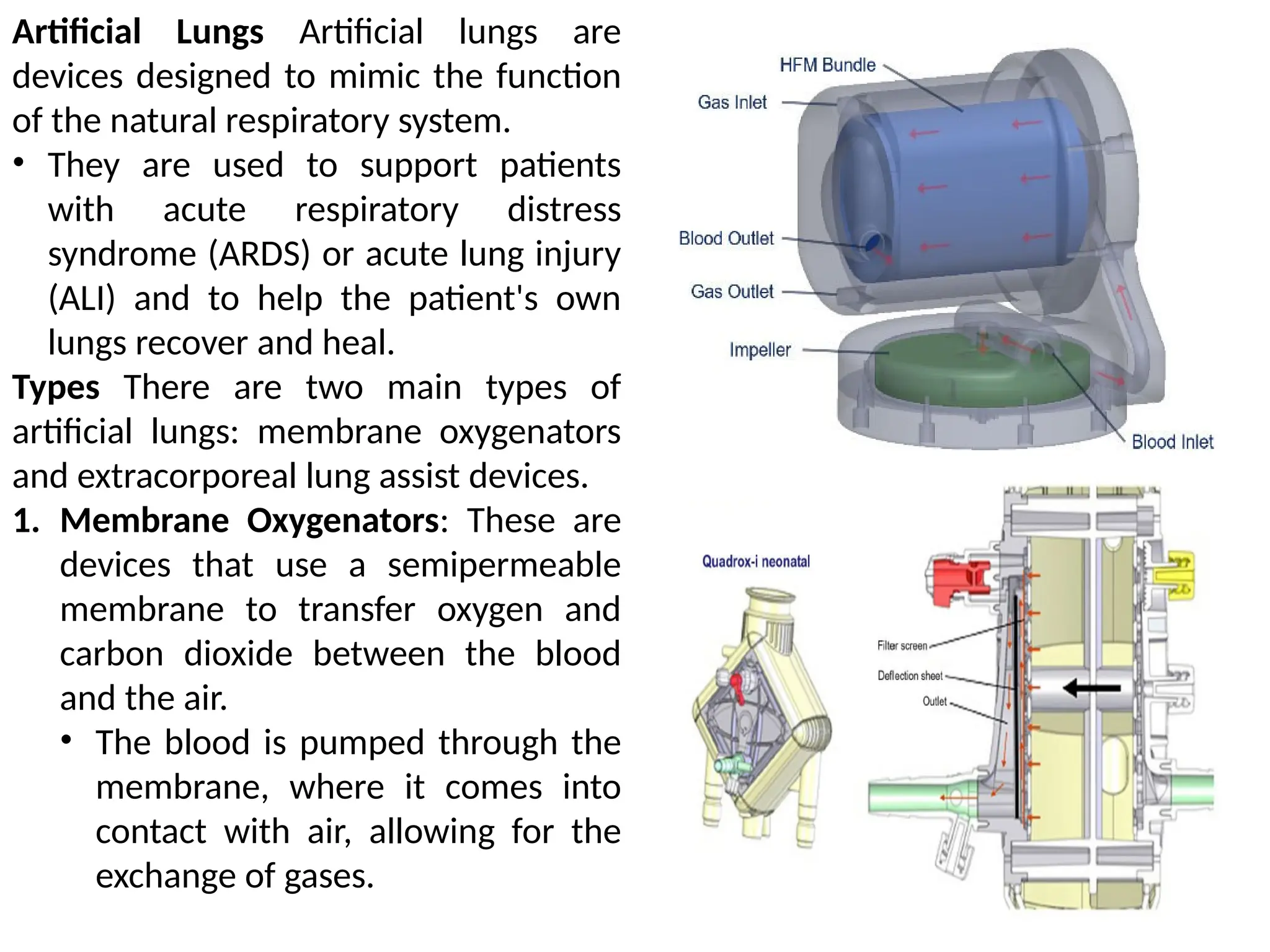
Artificial Lungs Artificiallungs are
devices designed to mimic the function
of the natural respiratory system.
• They are used to support patients
with acute respiratory distress
syndrome (ARDS) or acute lung injury
(ALI) and to help the patient's own
lungs recover and heal.
Types There are two main types of
artificial lungs: membrane oxygenators
and extracorporeal lung assist devices.
1. Membrane Oxygenators: These are
devices that use a semipermeable
membrane to transfer oxygen and
carbon dioxide between the blood
and the air.
• The blood is pumped through the
membrane, where it comes into
contact with air, allowing for the
exchange of gases.
95.
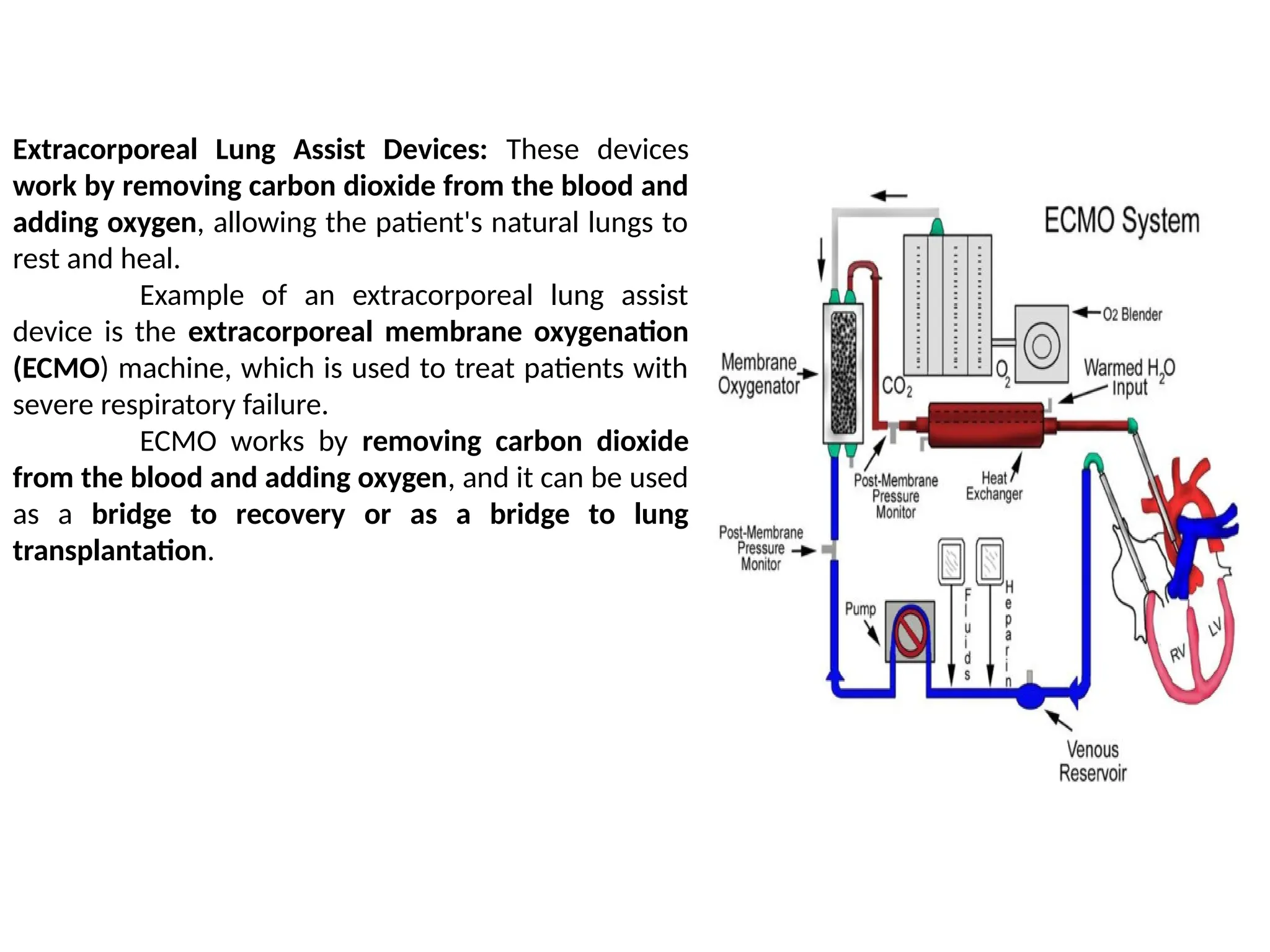
Extracorporeal Lung AssistDevices: These devices
work by removing carbon dioxide from the blood and
adding oxygen, allowing the patient's natural lungs to
rest and heal.
Example of an extracorporeal lung assist
device is the extracorporeal membrane oxygenation
(ECMO) machine, which is used to treat patients with
severe respiratory failure.
ECMO works by removing carbon dioxide
from the blood and adding oxygen, and it can be used
as a bridge to recovery or as a bridge to lung
transplantation.
▪ The kidneyis a complex organ that acts as a filtration system for the body.
▪ It removes waste and excess fluid from the bloodstream and maintains a
delicate balance of electrolytes, hormones, and other substances that are
critical for the body's normal functioning.
▪ The kidney also plays an important role in regulating blood pressure by
secreting the hormone renin, which helps control the balance of fluid and
electrolytes in the body.
▪ It also regulates red blood cell production and the levels of various minerals
in the blood, such as calcium and phosphorus.
▪ Without the kidney, waste and excess fluid would accumulate in the body,
leading to serious health problems
99.
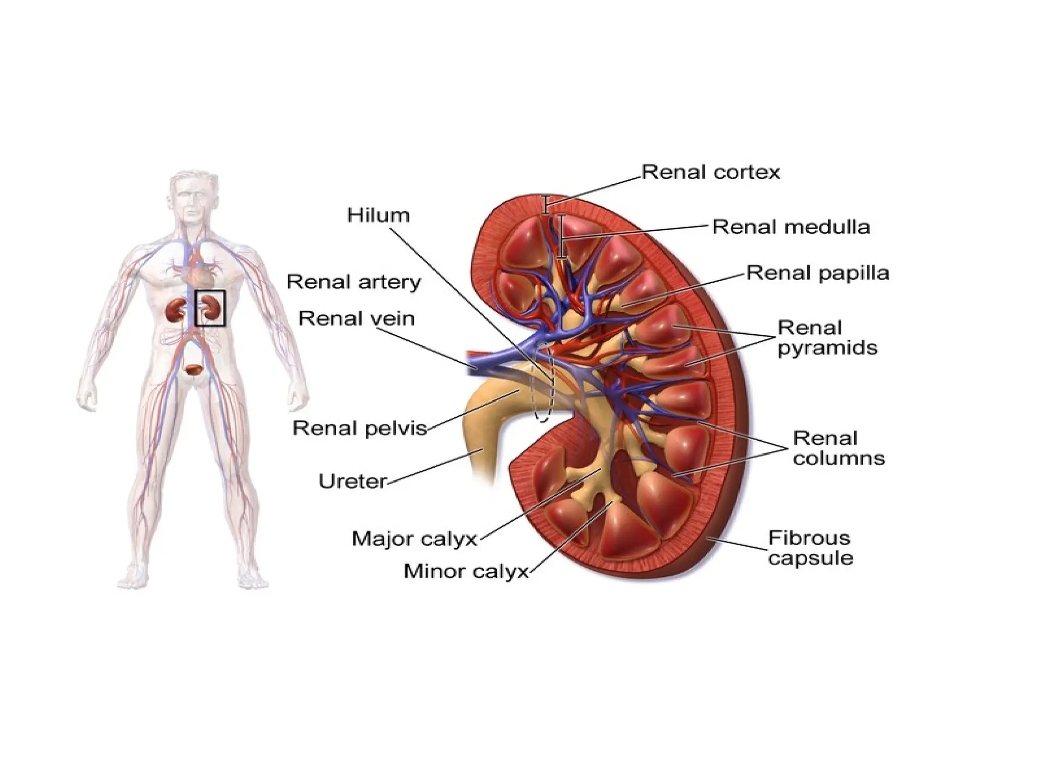
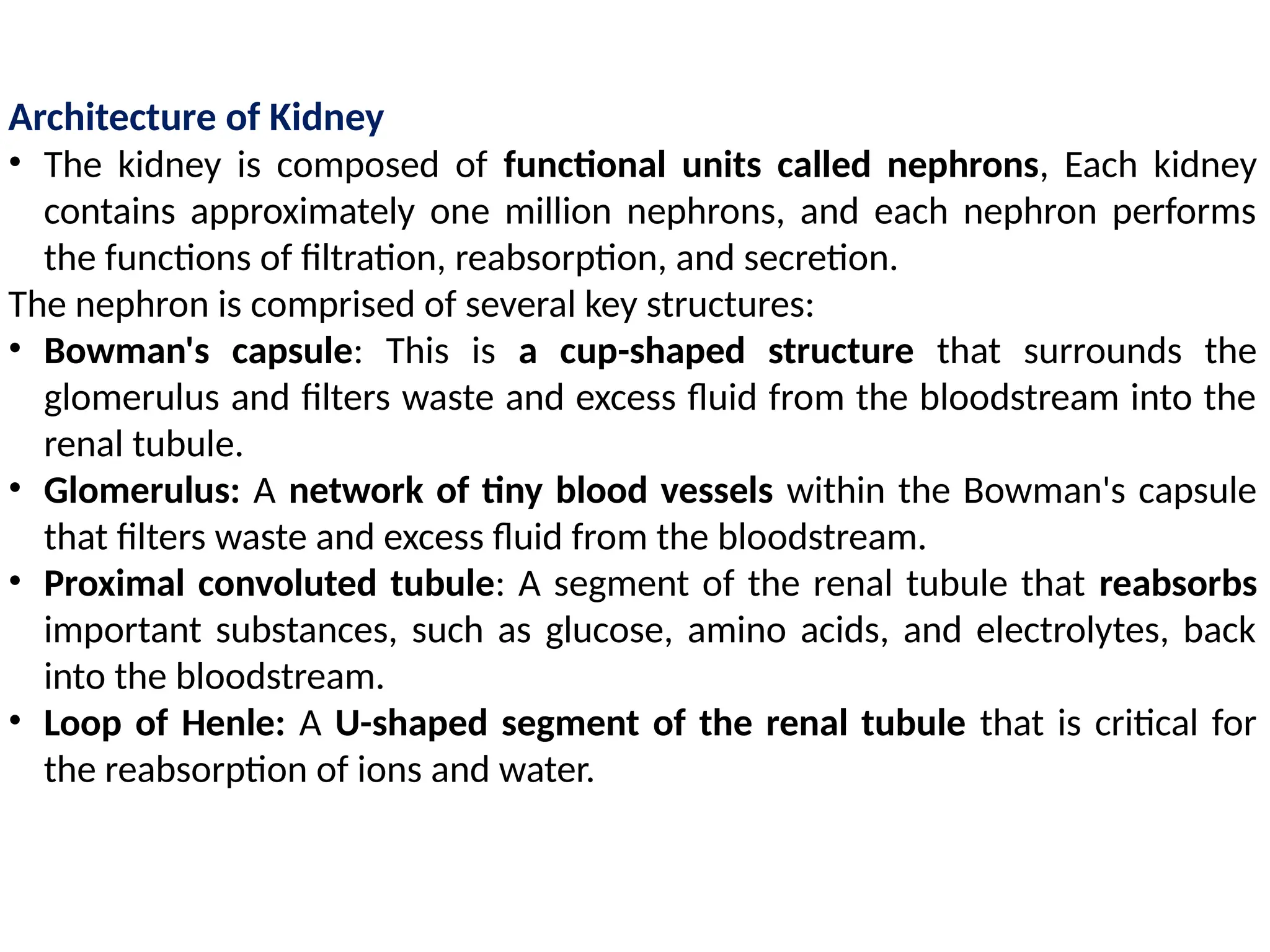
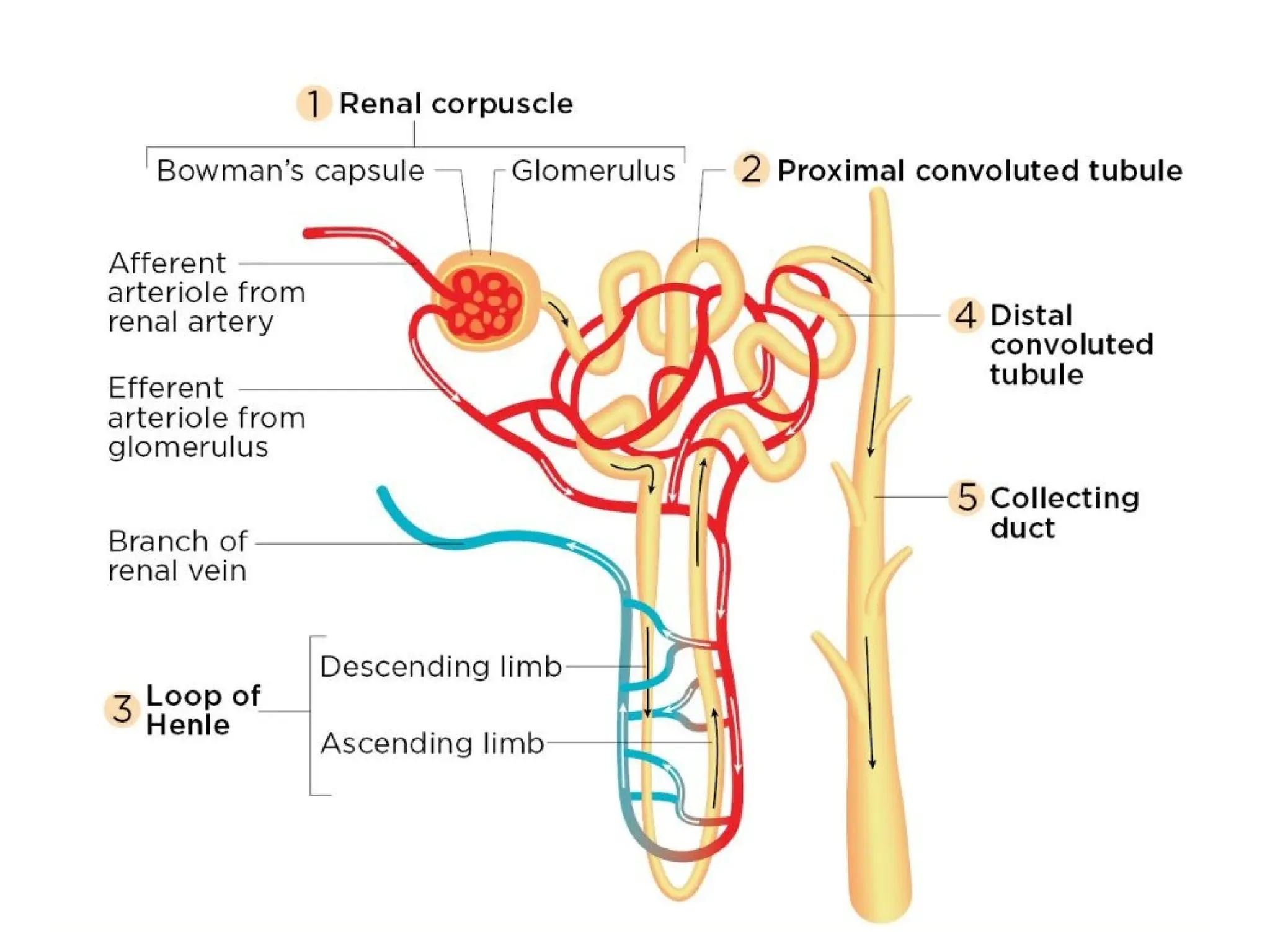
Architecture of Kidney
•The kidney is composed of functional units called nephrons, Each kidney
contains approximately one million nephrons, and each nephron performs
the functions of filtration, reabsorption, and secretion.
The nephron is comprised of several key structures:
• Bowman's capsule: This is a cup-shaped structure that surrounds the
glomerulus and filters waste and excess fluid from the bloodstream into the
renal tubule.
• Glomerulus: A network of tiny blood vessels within the Bowman's capsule
that filters waste and excess fluid from the bloodstream.
• Proximal convoluted tubule: A segment of the renal tubule that reabsorbs
important substances, such as glucose, amino acids, and electrolytes, back
into the bloodstream.
• Loop of Henle: A U-shaped segment of the renal tubule that is critical for
the reabsorption of ions and water.
100.
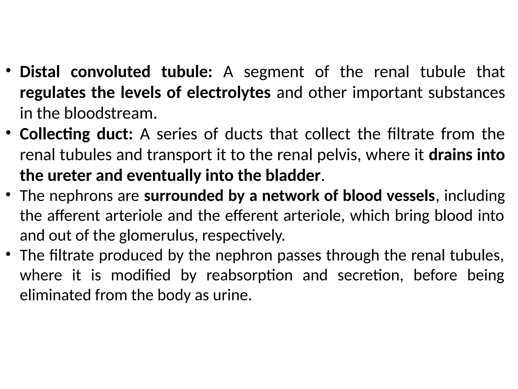
• Distal convolutedtubule: A segment of the renal tubule that
regulates the levels of electrolytes and other important substances
in the bloodstream.
• Collecting duct: A series of ducts that collect the filtrate from the
renal tubules and transport it to the renal pelvis, where it drains into
the ureter and eventually into the bladder.
• The nephrons are surrounded by a network of blood vessels, including
the afferent arteriole and the efferent arteriole, which bring blood into
and out of the glomerulus, respectively.
• The filtrate produced by the nephron passes through the renal tubules,
where it is modified by reabsorption and secretion, before being
eliminated from the body as urine.
102.
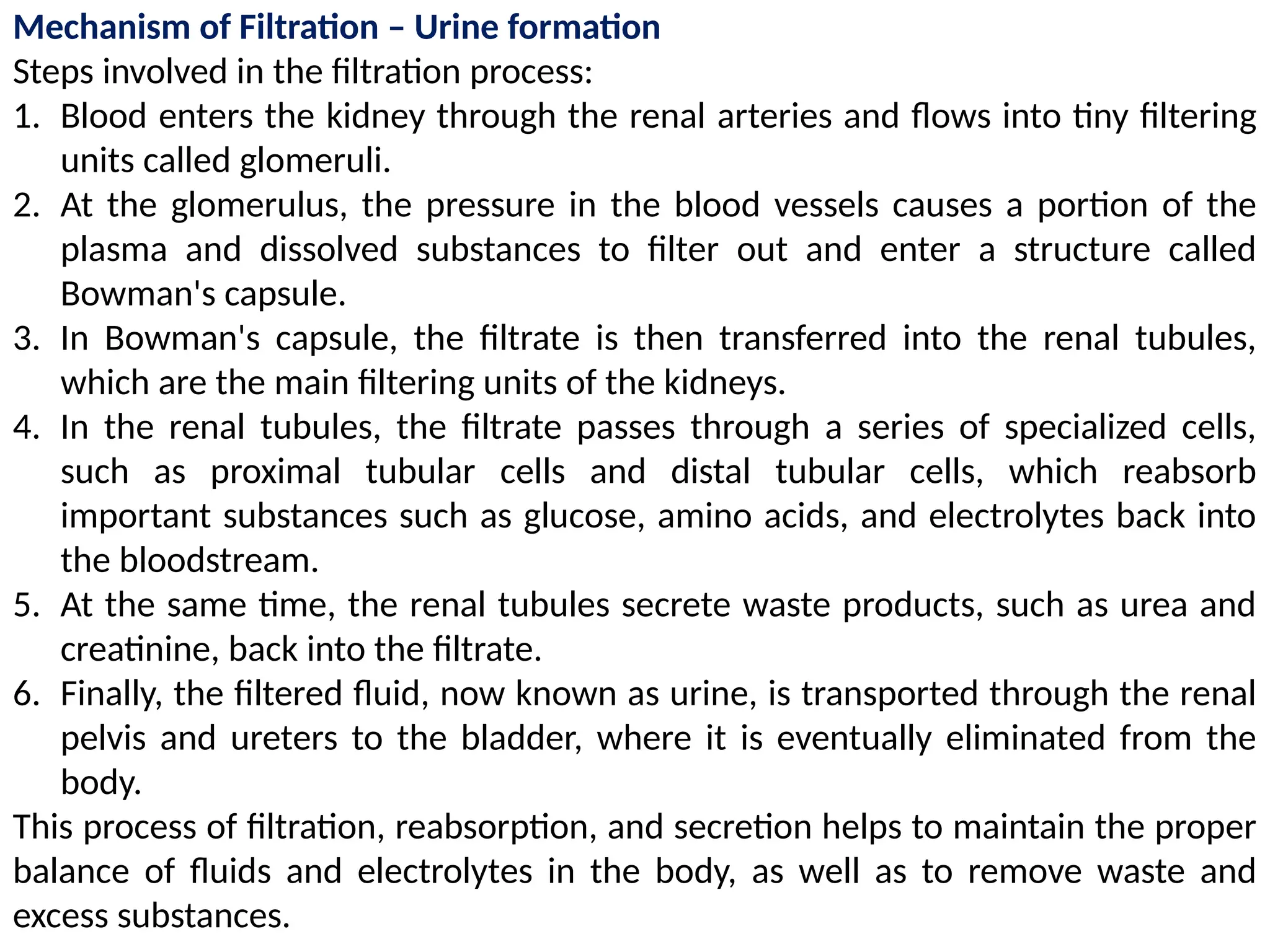
Mechanism of Filtration– Urine formation
Steps involved in the filtration process:
1. Blood enters the kidney through the renal arteries and flows into tiny filtering
units called glomeruli.
2. At the glomerulus, the pressure in the blood vessels causes a portion of the
plasma and dissolved substances to filter out and enter a structure called
Bowman's capsule.
3. In Bowman's capsule, the filtrate is then transferred into the renal tubules,
which are the main filtering units of the kidneys.
4. In the renal tubules, the filtrate passes through a series of specialized cells,
such as proximal tubular cells and distal tubular cells, which reabsorb
important substances such as glucose, amino acids, and electrolytes back into
the bloodstream.
5. At the same time, the renal tubules secrete waste products, such as urea and
creatinine, back into the filtrate.
6. Finally, the filtered fluid, now known as urine, is transported through the renal
pelvis and ureters to the bladder, where it is eventually eliminated from the
body.
This process of filtration, reabsorption, and secretion helps to maintain the proper
balance of fluids and electrolytes in the body, as well as to remove waste and
excess substances.
103.
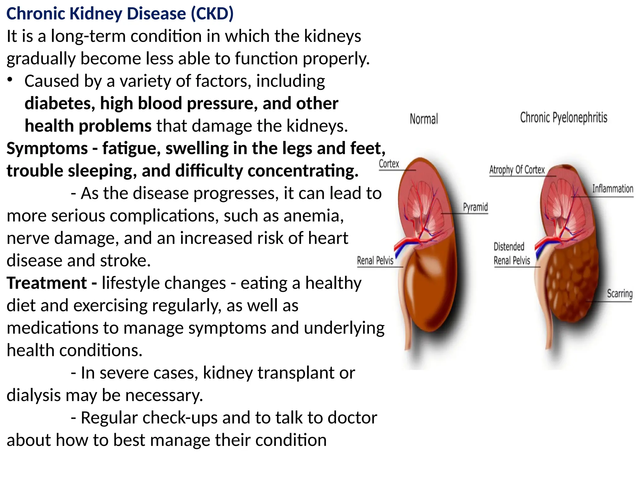
Chronic Kidney Disease(CKD)
It is a long-term condition in which the kidneys
gradually become less able to function properly.
• Caused by a variety of factors, including
diabetes, high blood pressure, and other
health problems that damage the kidneys.
Symptoms - fatigue, swelling in the legs and feet,
trouble sleeping, and difficulty concentrating.
- As the disease progresses, it can lead to
more serious complications, such as anemia,
nerve damage, and an increased risk of heart
disease and stroke.
Treatment - lifestyle changes - eating a healthy
diet and exercising regularly, as well as
medications to manage symptoms and underlying
health conditions.
- In severe cases, kidney transplant or
dialysis may be necessary.
- Regular check-ups and to talk to doctor
about how to best manage their condition
104.
Dialysis Systems
• Dialysisis a medical treatment that helps to filter waste and excess fluids
from the blood when the kidneys are unable to function properly.
There are two main types of dialysis systems:
1. Hemodialysis is a procedure that uses a machine to clean the blood.
• During hemodialysis, blood is removed from the body, passed through a
dialysis machine that filters out waste and excess fluids, and then returned to
the body.
• Hemodialysis typically takes place in a hospital or dialysis center, and is
typically performed three times a week for three to four hours at a time.
2. Peritoneal dialysis is a type of dialysis that uses the lining of the abdomen,
called the peritoneum, to filter waste and excess fluids from the blood.
• A sterile solution is introduced into the abdomen, where it absorbs waste
and excess fluids, and is then drained and replaced with fresh solution.
• Peritoneal dialysis can be performed at home and allows for more flexibility
in scheduling.
Both hemodialysis and peritoneal dialysis can effectively treat the symptoms of
kidney failure, but each has its own advantages and disadvantages.
The choice of dialysis system depends on various factors such as the
individual's overall health, lifestyle, and personal preferences.
105.
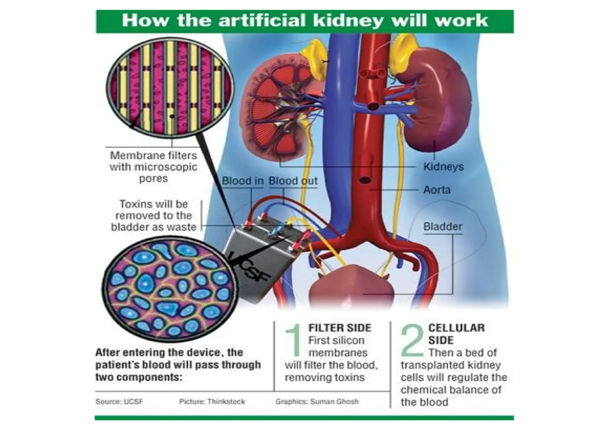
Artificial Kidney
• Itis still in the experimental stage and is not yet widely available.
• Further research and development is needed to improve the efficiency and
safety of artificial kidney devices, and to ensure that they can be widely
adopted as a treatment for chronic kidney disease
An artificial kidney is a device that is being developed to mimic the functions
of the human kidney.
• The goal of an artificial kidney is to provide a more effective and efficient
means of treating patients with chronic kidney disease, who currently rely
on dialysis or kidney transplantation.
There are currently two main approaches to developing an artificial kidney:
1. The biological approach involves using living cells, such as kidney cells or
stem cells, to create a functional, implantable artificial kidney.
2. The technological approach involves using synthetic materials, such as
silicon or polymer, to create a dialysis device that can filter the blood and
remove waste and excess fluids.
It's important to note that while the development of an artificial kidney holds
great promise, it is not a cure for chronic kidney disease and patients with
kidney failure will still need dialysis or kidney transplantation in the
meantime.