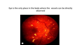
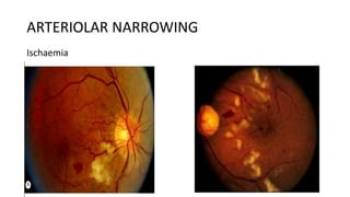
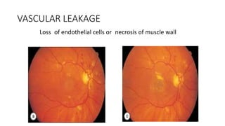
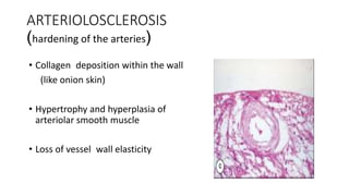
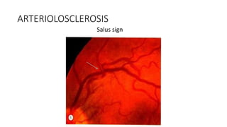
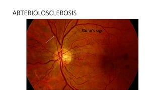
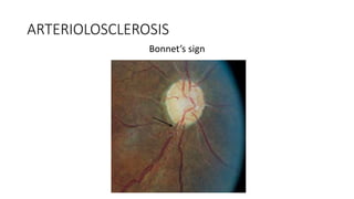
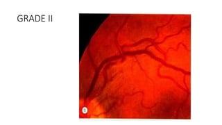
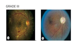
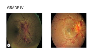
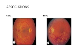
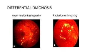
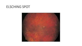
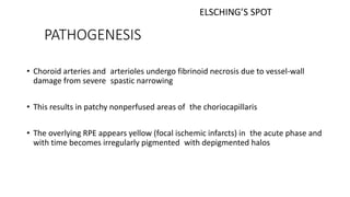
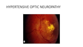
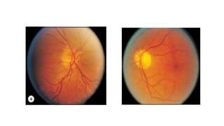
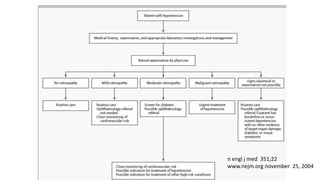
This document discusses hypertensive retinopathy, which refers to retinal vascular changes caused by high blood pressure. It notes that the prevalence of hypertensive retinopathy is approximately 15% among hypertensive patients without other vascular diseases. The key signs include arteriolar narrowing, arteriovenous nicking, and cotton wool spots. The pathogenesis involves initial vasoconstriction, then exudation and fluid leakage due to damaged blood vessels, and finally arteriolar sclerosis. Hypertensive choroidopathy and optic neuropathy can also occur in severe untreated cases and are characterized by lesions such as Elsching spots. Strict blood pressure control and regular eye screening are important for treatment and prevention of further vision loss
Hypertensive Retinopathy (HTN-R) for undergraduate MBBS Students.
Covers the basics of Aetiology, pathophysiology, clinical features, Classification and management of HTN-R.
Also encompasses salient points for PGMEE
Hypertensive retinopathy is a very important topic for PG examinations of all types. Especially, the fundal changes are important; Keith and Wegner Grading is also a repeated topic in PG. This slide represents all information in a compressed fashion. Have fun!
Retinal vein occlusion (RVO) is an obstruction of the retinal venous system by thrombus formation and may involve the central, hemi-central or branch retinal vein.
The most common aetiological factor is compression by adjacent atherosclerotic retinal arteries.
Other possible causes are external compression or disease of the vein wall e.g. vasculitis.
This lecture is based on medical students those are preparing for postgraduate degree namely FCPS/MS/MD/ any any subject coz hypertension is a systemic disease and by seeing the ocular fundus we can asses the general condition of blood vessels in major organ.
Most retinal surgeons are trained to create formal retinal drawings of the fundus.
Retinal drawings are useful to document pathology, although more and more people now prefer fundus photographs.
Can be used for serial follow up of patients to document changes in the pathology.
Ischemic condition affecting the eye.
The ischemia can occur secondary to systemically problem [or] particulary the eye.
Many retinal vascular disorders {like CRAO,CRVO,Diabetic retinopathy,Hypertensive Retinopathy} shows ischemic signs.
Cerebrovascular Vasospasm - Etiopathogenesis and ManagementDr. Rahul Jain
Cerebrovascular vasospasm is a consequence of subarachnoid hemorrhage following aneurysmal rupture. its types, causes, etiology, incidence, diagnois and treatment protocols should be understood for better identification and management of this condition.
microvascular complications of DM 09-12-2023.pptxmanjujanhavi
etiopathogenesis of microvascular complications , pathophysiology of each type of retino, nephropathy ,neuropathy & diabetic foot , prevention , early detection ,patient education
These simplified slides by Dr. Sidra Arshad present an overview of the non-respiratory functions of the respiratory tract.
Learning objectives:
1. Enlist the non-respiratory functions of the respiratory tract
2. Briefly explain how these functions are carried out
3. Discuss the significance of dead space
4. Differentiate between minute ventilation and alveolar ventilation
5. Describe the cough and sneeze reflexes
Study Resources:
1. Chapter 39, Guyton and Hall Textbook of Medical Physiology, 14th edition
2. Chapter 34, Ganong’s Review of Medical Physiology, 26th edition
3. Chapter 17, Human Physiology by Lauralee Sherwood, 9th edition
4. Non-respiratory functions of the lungs https://academic.oup.com/bjaed/article/13/3/98/278874
Ozempic: Preoperative Management of Patients on GLP-1 Receptor Agonists Saeid Safari
Preoperative Management of Patients on GLP-1 Receptor Agonists like Ozempic and Semiglutide
ASA GUIDELINE
NYSORA Guideline
2 Case Reports of Gastric Ultrasound
Basavarajeeyam is an important text for ayurvedic physician belonging to andhra pradehs. It is a popular compendium in various parts of our country as well as in andhra pradesh. The content of the text was presented in sanskrit and telugu language (Bilingual). One of the most famous book in ayurvedic pharmaceutics and therapeutics. This book contains 25 chapters called as prakaranas. Many rasaoushadis were explained, pioneer of dhatu druti, nadi pareeksha, mutra pareeksha etc. Belongs to the period of 15-16 century. New diseases like upadamsha, phiranga rogas are explained.
These lecture slides, by Dr Sidra Arshad, offer a quick overview of the physiological basis of a normal electrocardiogram.
Learning objectives:
1. Define an electrocardiogram (ECG) and electrocardiography
2. Describe how dipoles generated by the heart produce the waveforms of the ECG
3. Describe the components of a normal electrocardiogram of a typical bipolar lead (limb II)
4. Differentiate between intervals and segments
5. Enlist some common indications for obtaining an ECG
6. Describe the flow of current around the heart during the cardiac cycle
7. Discuss the placement and polarity of the leads of electrocardiograph
8. Describe the normal electrocardiograms recorded from the limb leads and explain the physiological basis of the different records that are obtained
9. Define mean electrical vector (axis) of the heart and give the normal range
10. Define the mean QRS vector
11. Describe the axes of leads (hexagonal reference system)
12. Comprehend the vectorial analysis of the normal ECG
13. Determine the mean electrical axis of the ventricular QRS and appreciate the mean axis deviation
14. Explain the concepts of current of injury, J point, and their significance
Study Resources:
1. Chapter 11, Guyton and Hall Textbook of Medical Physiology, 14th edition
2. Chapter 9, Human Physiology - From Cells to Systems, Lauralee Sherwood, 9th edition
3. Chapter 29, Ganong’s Review of Medical Physiology, 26th edition
4. Electrocardiogram, StatPearls - https://www.ncbi.nlm.nih.gov/books/NBK549803/
5. ECG in Medical Practice by ABM Abdullah, 4th edition
6. Chapter 3, Cardiology Explained, https://www.ncbi.nlm.nih.gov/books/NBK2214/
7. ECG Basics, http://www.nataliescasebook.com/tag/e-c-g-basics
Title: Sense of Smell
Presenter: Dr. Faiza, Assistant Professor of Physiology
Qualifications:
MBBS (Best Graduate, AIMC Lahore)
FCPS Physiology
ICMT, CHPE, DHPE (STMU)
MPH (GC University, Faisalabad)
MBA (Virtual University of Pakistan)
Learning Objectives:
Describe the primary categories of smells and the concept of odor blindness.
Explain the structure and location of the olfactory membrane and mucosa, including the types and roles of cells involved in olfaction.
Describe the pathway and mechanisms of olfactory signal transmission from the olfactory receptors to the brain.
Illustrate the biochemical cascade triggered by odorant binding to olfactory receptors, including the role of G-proteins and second messengers in generating an action potential.
Identify different types of olfactory disorders such as anosmia, hyposmia, hyperosmia, and dysosmia, including their potential causes.
Key Topics:
Olfactory Genes:
3% of the human genome accounts for olfactory genes.
400 genes for odorant receptors.
Olfactory Membrane:
Located in the superior part of the nasal cavity.
Medially: Folds downward along the superior septum.
Laterally: Folds over the superior turbinate and upper surface of the middle turbinate.
Total surface area: 5-10 square centimeters.
Olfactory Mucosa:
Olfactory Cells: Bipolar nerve cells derived from the CNS (100 million), with 4-25 olfactory cilia per cell.
Sustentacular Cells: Produce mucus and maintain ionic and molecular environment.
Basal Cells: Replace worn-out olfactory cells with an average lifespan of 1-2 months.
Bowman’s Gland: Secretes mucus.
Stimulation of Olfactory Cells:
Odorant dissolves in mucus and attaches to receptors on olfactory cilia.
Involves a cascade effect through G-proteins and second messengers, leading to depolarization and action potential generation in the olfactory nerve.
Quality of a Good Odorant:
Small (3-20 Carbon atoms), volatile, water-soluble, and lipid-soluble.
Facilitated by odorant-binding proteins in mucus.
Membrane Potential and Action Potential:
Resting membrane potential: -55mV.
Action potential frequency in the olfactory nerve increases with odorant strength.
Adaptation Towards the Sense of Smell:
Rapid adaptation within the first second, with further slow adaptation.
Psychological adaptation greater than receptor adaptation, involving feedback inhibition from the central nervous system.
Primary Sensations of Smell:
Camphoraceous, Musky, Floral, Pepperminty, Ethereal, Pungent, Putrid.
Odor Detection Threshold:
Examples: Hydrogen sulfide (0.0005 ppm), Methyl-mercaptan (0.002 ppm).
Some toxic substances are odorless at lethal concentrations.
Characteristics of Smell:
Odor blindness for single substances due to lack of appropriate receptor protein.
Behavioral and emotional influences of smell.
Transmission of Olfactory Signals:
From olfactory cells to glomeruli in the olfactory bulb, involving lateral inhibition.
Primitive, less old, and new olfactory systems with different path
Rasamanikya is a excellent preparation in the field of Rasashastra, it is used in various Kushtha Roga, Shwasa, Vicharchika, Bhagandara, Vatarakta, and Phiranga Roga. In this article Preparation& Comparative analytical profile for both Formulationon i.e Rasamanikya prepared by Kushmanda swarasa & Churnodhaka Shodita Haratala. The study aims to provide insights into the comparative efficacy and analytical aspects of these formulations for enhanced therapeutic outcomes.
2. INTRODUCTION
The overall prevalence of hypertension was 20.5 % and pre-
hypertension was 46.6%.
Prevalence and Associated Factors of Hypertension among Adults in Rural Nepal: A Community Based Study.
Chataut j et al,
Kathmandu univ med J, 2015
3. Prevalence of Hypertensive retinopathy
• The incidence of hypertensive retinal changes is variable and often
masked by the presence of other retinal vascular disease such as
diabetes.
• In the Beaver Dam Eye Study, which evaluated hypertensive patients
without coexisting vascular diseases, the incidence of hypertensive
retinopathy was about 15%; specifically, 8% showed retinopathy, 13%
showed arteriolar narrowing, and 2% showed arteriovenous nicking.
5. • First described in 1859 as ‘albuminuric retinitis’ by Liebreich
• Angiospastic retinopathy, Hypertensive neuroretinopathy and
Hypertensive retinopathy
6. ETIOLOGY
• Race:
Blacks > Whites
Afro-Caribbeans > Europeans
but prevalence of retinopathy
Afro-Caribbeans < Europeans
8. ETIOLOGY
• Genetic factors
The D (deletion) allele of the angio-tensin converting enzyme (ACE)
gene is an independent risk factor for the development of end-organ
damage
2.4 fold higher chance of retinopathy
9. ETIOLOGY
• Renal status:
persistent microalbuminuria - early end-organ damage including
retinopathy
• Cardiac status:
more common in concentric hypertrophy
10. ETIOLOGY
• Secondary hypertension:
Renal hypertension secondary to focal segmental sclerosis and
membranoproliferative glomerulonephritis - severe retinopathy
Pheochromocytoma - grade III or IV retinopathy
12. VASO-CONSTRICTIVE PHASE
• Reversible stimulation of the vascular tone of muscular retinal
arteries as an autoregulatory mechanism
• Prolonged blood pressure elevation, definitive narrowing
(angiotensin II, vasopressin) of the vascular lumen at the
precapillary level occurs
13. VASO-CONSTRICTIVE PHASE
• Cotton wool spots (feature of inner retinal ischemia)
• Interruption of axonal orthograde and retrograde energy dependent
organelle transport in the ganglion cell axons, swollen interrupted
nerve
14. EXUDATIVE PHASE
Vascular endothelial necrosis or disruption (Disruption of the
blood–retinal barrier )
Transudation of plasma into the vessel wall, around pericytes and
arteriolar muscle cells
Retinal HGE of various shapes and locations
Edema and exudates (the macular region)
15. SCLEROTIC PHASE
• Fibrinoid necrosis or hyaline degeneration
• Hyperplasia of the vascular tunica media
• Arteriosclerosis
• Narrowing of arteries
• A-V crossing abnormalities
17. RETINOPATHY
• Hypertensive retinopathy consists of spectrum of retinal vascular
changes that are pathologically related to microvascular damage
from elevated blood pressure
21. ARTERIOLOSCLEROSIS
(hardening of the arteries)
• Collagen deposition within the wall
(like onion skin)
• Hypertrophy and hyperplasia of
arteriolar smooth muscle
• Loss of vessel wall elasticity
39. INTRODUCTION
• Seen in Malignant hypertension in which the blood pressure is
200/140 mm Hg associated with ocular, cardiac, renal and cerebral
involvement
• Visual disturbances : scotoma, diplopia, dimunition of vision,
photopsia and headache
40. INTRODUCTION
• Seen typically in young patients with pliable vessels that are not yet
sclerotic from long-term hypertension
• Toxemia of pregnancy, renal disease, pheochromocytoma, essential
hypertension, and connective tissue diseases
41. PATHOGENESIS
• Greater effect on the choroidal circulation than on retinal circulation
• The retinal vessels - autoregulatory mechanisms transiently maintain
the vascular tone in response to sudden rise in BP
42. PATHOGENESIS
• The sympathetic nervous system - choroidal vasculature - constrict
in response to rise in BP
• Due to shorter distance and fewer branchings of the choroidal
arteries, systemic hypertension is transmitted more effectively
45. PATHOGENESIS
• Choroid arteries and arterioles undergo fibrinoid necrosis due to vessel-wall
damage from severe spastic narrowing
• This results in patchy nonperfused areas of the choriocapillaris
• The overlying RPE appears yellow (focal ischemic infarcts) in the acute phase and
with time becomes irregularly pigmented with depigmented halos
ELSCHING’S SPOT
50. HYPERTENSIVE OPTIC NEUROPATHY
• Some propose that it occurs secondary to encephalopathy
• Others believe that it occurs in the absence of the raised ICP and is
secondary to the ischemic changes of the optic disc
51. HYPERTENSIVE OPTIC NEUROPATHY
• Optic nerve head is susceptible to ischemia by virtue of its tightly
arranged nerve fibers within a nonexpandable intrascleral canal
• Vasoconstriction of the posterior ciliary arteries: results from the
release of angiotensin II and other vasoconstricting agents
52. PATHOGENESIS
Ischaemia of optic nerve head
delay in the axoplasmic transport and a subsequent accumulation
of axonal components in the lamina scleralis region
plasma leakage and disruption of nerve fibers leading to
subsequent gliosis
53.
54. COURSE
• It rarely results in significant loss of vision
• Ongoing end-organ damage is more important than actual blood
pressure
55. TREATMENT
• Treatment of the underlying systemic condition can halt the
progression but arteriolar narrowing and AV nicking usually are
permanent
• Hypertensive emergencies: sodium nitroprusside, nitroglycerin,
calcium channel blockers, beta blockers, and angiotensin-converting
enzyme inhibitors
• Blood pressure should be lowered in a controlled fashion
57. n engl j med 351;22
www.nejm.org november 25, 2004
58. SUMMARY
• Retina is the only place where hypertensive vascular changes can be
directly observed
• Pathogenesis: Vasoconstriction, exudation, arteriolosclerosis
• Manifestations: Retinopathy, choroidopathy, neuropathy
• Hallmark: AV changes and cotton wool spots
• Classification:
• Strict control of Blood pressure and periodic screening
59. References
• Retina Ryan 5th edition
• American Academy of Ophthalmology. 2013-14; Section 12- Vitreous and Retina
• Kanski JJ. Clinical Ophthalmology-A Systemic Approach.
• Yanoff M, Duker JD. Ophthalmology. 4th edition
• 6th ed. ELSEVIER; 2008
DD of cotton wool spots: acquired immunodeficiency syndrome, collagen diseases, blood dyscrasias and malignancy, central retinal vein occlusion, diabetic retinopathy
Early phase before the onset of sclerosis
(cotton wool spots)
(flame shaped hge and hard exudates in henle layer around fovea leading to macular star)
arteriolovenous ‘nicking.
Sclerosis may shorten or elongate retinal arterioles, with the branches coming off at right angles. This change in length deflects the veins at the common sheath and changes the course of the vein (Salus sign)
In arteriovenous nicking (the Gunn sign), impeded circulation results in a dilated or swollen vein peripheral to the crossing, causing hourglass constrictions on both sides of the crossing and aneurysmal-like swellings. Ikui noted that arteriole and venous basement membranes are adherent with shared collagen fibers at the crossing points. Thickening of the basement membrane and the media of the arteriole in hypertension impinge on the vein and cause the crossing phenomenon
Banking of veins distal to AV crossing
Thrombosis or accumulation of fibrin or other plasma products beneath the endothelium in the vessel wall
RVO- >50yrs- 64% have htn
<50yrs- 25% have htn
Thrombosis or accumulation of fibrin or other plasma products beneath the endothelium in the vessel wall
Exudates form a ring or circinate pattern in diabetes
Av crossing changes are not present
Vessels are dilated and torturous in altitudnal
h/o radiation exposure and the exudates are discretely present
(innervation and the anatomy of the choroidal vessels may explain the underlying pathophysiologic process )
Black spots surrounded by yellow halos
Due to clumping and atrophy of infarcted pigment epithelium.
Typically in the midperiphery and in the vicinity of the optic disc
FFA, choroidal vasculopathy is demonstrated by irregular filling patterns, delays in filling time, and areas of diffuse leakage (new) or window defects (old), depending on the stage of the Elschnig’s spot formation
Fibrinoid necrosis-(replacement of smooth muscle fibers by fibrin-platelet and other plasma protein materials)
Are areas of hyperpigmentation (RPE hyperplasia and hypertrophy) overlying sclerotic choroidal arteries
Flakes arranged linearly around the choroidal vessels and are indicative of fibrinoid necrosis
Poor visual prognosis
Optic nerve head swelling in hypertension
Optic nerve head is supplied by multiple blood vessels, including the central retinal artery, short posterior ciliary arteries (through the circle of Zinn), and pial vessels