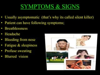
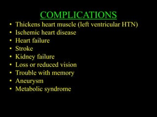
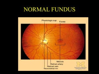
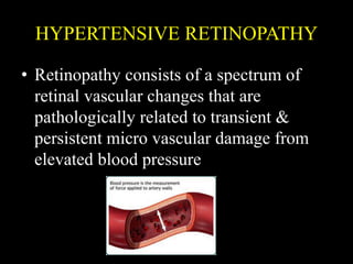
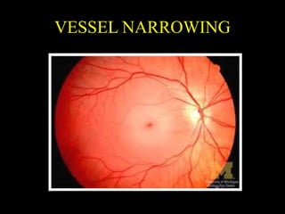
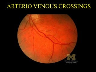
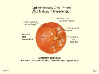
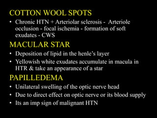
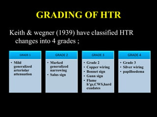
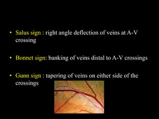
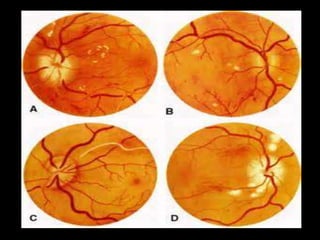
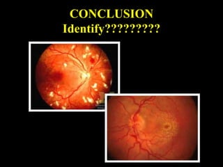
HYPERTENSION
LINTA K P
LIMSAR
LF Hospital
INTRODUCTION
Hypertension is one of the most common worldwide
diseases affecting humans
• Normal BP : 140/80 mm of hg
• Hypertension is the high blood pressure
• BP is the force of blood against the artery wall as it
flows through them
• A patient is said to be hypertensive when his
SBP≥140 mm of hg & DBP≥90mm of hg that the
patient is not on antihypertensive drugs
CLASSIFICATION
Etiological classification
Essential HTN (1* HTN)
• Idiopathic
• BP is regulated by the renal , hormonal , vascular &
neurologic systems
Secondary HTN
• In this class , etiology of the high BP can be identified
• Its due to Renal , Endocrine , Neurogenic ,
Mechanical , Exogenous , & miscellaneous
Based on recommendations of the 7th report of
the joint national committee of Prevention ,
detection , Evaluation & Tx of high BP the
classification of BP for adults aged 18 years or
older is as follows;
• Normal : SBP <120 , DBP <80 mm of hg
• Prehypertension : SBP 120-139 , DBP 80-89
• Stage 1 : SBP 140-159 , DBP 90-99
• Stage 2: SBP ≥ 160 , DBP ≥ 100
According to severity
Mild ( SBP : 140-160 & DBP : 90-100)
Moderate (SBP :160-200 & DBP : 100-120)
Severe (SBP : Above 200 & DBP : Above 120)
Another classification
Border line
Labile.
Sustained
Malignant
Accelerated
OTHER CAUSES
• Kidney disease
• Adrenal gland tumors
• Increased salt intake
• Tobacco use
• Alcohol abuse
• Stress
• Certain medications etc
SYMPTOMS & SIGNS
• Usually asymptomatic (that’s why its called silent killer)
• Patient can have following symptoms;
• Breathlessness
• Headache
• Bleeding from nose
• Fatigue & sleepiness
• Profuse sweating
• Blurred vision
COMPLICATIONS
• Thickens heart muscle (left ventricular HTN)
• Ischemic heart disease
• Heart failure
• Stroke
• Kidney failure
• Loss or reduced vision
• Trouble with memory
• Aneurysm
• Metabolic syndrome
TIPS TO CONTROL HTN
Eat less salt
Exercise
Stop smoking
& alcohol
Healthy diet
Reduce
weight
Hypertensive retinopathy
Hypertensive choroidopathy
Hypertensive optic neuropathy
NORMAL FUNDUS
HYPERTENSIVE RETINOPATHY
• Retinopathy consists of a spectrum of
retinal vascular changes that are
pathologically related to transient &
persistent micro vascular damage from
elevated blood pressure
FUNDUS CHANGES
Prolonged systemic hypertension results in
retinal vascular effects ;
 Vessel narrowing
Arterio venous crossings
Microaneurysms
Hemorrhages
Cotton-wool spots
Macular star
Papilledema
VESSEL NARROWING
• Chronic HTN with significant elevation in diastolic
pressure directly related to narrowing of caliber of
vessel
• Arteriole narrowing is best judged by comparing the
caliber of artery with that of adjacent retinal venule
• Atherosclerosis & ↑sed DBP – Vessel wall
hyperplasia - Fibrosis – Luminal narrowing
ARTERIO VENOUS CROSSINGS
• The retinal arteriole & venule share a common
adventitial sheath in the area where they cross each
other & artery compress vein (A-V nicking / nipping)
• Compression of vein → venule deviation , vein
humping
• BANKING – Distal : dilation of vein
- Proximal : Narrowing of vein
MICROANEURYSMS
• Small round dark red dots on retinal surface
• Best detected on FFA
• Beginning as dilations in areas in capillary wall
where pericytes are absent
• Initially they are thin walled & Later endothelial
cells proliferate & lay down layers of basement
membrane material around themselves
• ↑se in number as the degree of retinal
involvement
• ↑se in no. – capillary occlusion – retinal ischemia
HEMMORRHAGES
• H’ges occurs superficially in the Nerve fibre
layer
• Streak appearance - FLAME shaped h’ge –
macular edema & subsequent vision loss
• It’s a result of chronic hypertensive damage to
the capillary wall endothelium – extravasation
of plasma from lumen into the extracapillary
space
COTTON WOOL SPOTS
• Chronic HTN + Arteriolar sclerosis - Arteriole
occlusion - focal ischemia - formation of soft
exudates - CWS
MACULAR STAR
• Deposition of lipid in the henle’s layer
• Yellowish white exudates accumulate in macula in
HTR & take an appearance of a star
PAPILLEDEMA
• Unilateral swelling of the optic nerve head
• Due to direct effect on optic nerve or its blood supply
• Its an imp sign of malignant HTN
GRADING OF HTR
Keith & wegner (1939) have classified HTR
changes into 4 grades ;
GRADE 1
• Mild
generalized
arteriolar
attenuation
GRADE 2
• Marked
generalized
narrowing
• Salus sign
GRADE 3
• Grade 2
• Copper wiring
• Bonnet sign
• Gunn sign
• Flame
h’ge,CWS,hard
exudates
GRADE 4
• Grade 3
• Silver wiring
• papilloedema
• Salus sign : right angle deflection of veins at A-V
crossing
• Bonnet sign: banking of veins distal to A-V crossings
• Gunn sign : tapering of veins on either side of the
crossings
HYPERTENSIVE
CHOROIDOPATHY
• Its a rare but may occur as the result of accelerated
HTN in young adults
• Choroidal vascular bed shows impaired circulation &
extensive occlusive & ischemic changes
ELSCHING SPOTS
-small black spots surrounded by yellow halos
-represents focal choroidal infarcts
SIEGRIST STREAKS
-Linear hyperpigmented streaks oor flecks arranged
over choroidal arteries
EXUDATIVE RETINAL DETACHMENT
-sometimes bilateral , may occur in severe acute
HTN
HYPERTENSIVE OPTIC
NEUROPATHY
• Optic disc edema has been described as an essential
manifestation of malignant HTN
• Optic disc edema is the initial manifestation of
HTON
• Our studies indicated that HTON represents a form of
AION
CONCLUSION
Identify?????????
REFERENCE
• The eye in systemic disease : Daniel H Hold
• Clinical ophthalmology : jack j kanski & brad
bowling
• BOS014
• www.ophthobook.com
• www.mayoclinic.com
THANK YOU

Hypertension & HYPERTENSIVE RETINOPATHY

  • 1.
  • 2.
    INTRODUCTION Hypertension is oneof the most common worldwide diseases affecting humans • Normal BP : 140/80 mm of hg • Hypertension is the high blood pressure • BP is the force of blood against the artery wall as it flows through them • A patient is said to be hypertensive when his SBP≥140 mm of hg & DBP≥90mm of hg that the patient is not on antihypertensive drugs
  • 3.
    CLASSIFICATION Etiological classification Essential HTN(1* HTN) • Idiopathic • BP is regulated by the renal , hormonal , vascular & neurologic systems Secondary HTN • In this class , etiology of the high BP can be identified • Its due to Renal , Endocrine , Neurogenic , Mechanical , Exogenous , & miscellaneous
  • 4.
    Based on recommendationsof the 7th report of the joint national committee of Prevention , detection , Evaluation & Tx of high BP the classification of BP for adults aged 18 years or older is as follows; • Normal : SBP <120 , DBP <80 mm of hg • Prehypertension : SBP 120-139 , DBP 80-89 • Stage 1 : SBP 140-159 , DBP 90-99 • Stage 2: SBP ≥ 160 , DBP ≥ 100
  • 5.
    According to severity Mild( SBP : 140-160 & DBP : 90-100) Moderate (SBP :160-200 & DBP : 100-120) Severe (SBP : Above 200 & DBP : Above 120) Another classification Border line Labile. Sustained Malignant Accelerated
  • 15.
    OTHER CAUSES • Kidneydisease • Adrenal gland tumors • Increased salt intake • Tobacco use • Alcohol abuse • Stress • Certain medications etc
  • 16.
    SYMPTOMS & SIGNS •Usually asymptomatic (that’s why its called silent killer) • Patient can have following symptoms; • Breathlessness • Headache • Bleeding from nose • Fatigue & sleepiness • Profuse sweating • Blurred vision
  • 17.
    COMPLICATIONS • Thickens heartmuscle (left ventricular HTN) • Ischemic heart disease • Heart failure • Stroke • Kidney failure • Loss or reduced vision • Trouble with memory • Aneurysm • Metabolic syndrome
  • 18.
    TIPS TO CONTROLHTN Eat less salt Exercise Stop smoking & alcohol Healthy diet Reduce weight
  • 20.
  • 21.
  • 22.
    HYPERTENSIVE RETINOPATHY • Retinopathyconsists of a spectrum of retinal vascular changes that are pathologically related to transient & persistent micro vascular damage from elevated blood pressure
  • 23.
    FUNDUS CHANGES Prolonged systemichypertension results in retinal vascular effects ;  Vessel narrowing Arterio venous crossings Microaneurysms Hemorrhages Cotton-wool spots Macular star Papilledema
  • 24.
  • 25.
    • Chronic HTNwith significant elevation in diastolic pressure directly related to narrowing of caliber of vessel • Arteriole narrowing is best judged by comparing the caliber of artery with that of adjacent retinal venule • Atherosclerosis & ↑sed DBP – Vessel wall hyperplasia - Fibrosis – Luminal narrowing
  • 26.
  • 27.
    • The retinalarteriole & venule share a common adventitial sheath in the area where they cross each other & artery compress vein (A-V nicking / nipping) • Compression of vein → venule deviation , vein humping • BANKING – Distal : dilation of vein - Proximal : Narrowing of vein
  • 28.
  • 29.
    • Small rounddark red dots on retinal surface • Best detected on FFA • Beginning as dilations in areas in capillary wall where pericytes are absent • Initially they are thin walled & Later endothelial cells proliferate & lay down layers of basement membrane material around themselves • ↑se in number as the degree of retinal involvement • ↑se in no. – capillary occlusion – retinal ischemia
  • 30.
  • 31.
    • H’ges occurssuperficially in the Nerve fibre layer • Streak appearance - FLAME shaped h’ge – macular edema & subsequent vision loss • It’s a result of chronic hypertensive damage to the capillary wall endothelium – extravasation of plasma from lumen into the extracapillary space
  • 33.
    COTTON WOOL SPOTS •Chronic HTN + Arteriolar sclerosis - Arteriole occlusion - focal ischemia - formation of soft exudates - CWS MACULAR STAR • Deposition of lipid in the henle’s layer • Yellowish white exudates accumulate in macula in HTR & take an appearance of a star PAPILLEDEMA • Unilateral swelling of the optic nerve head • Due to direct effect on optic nerve or its blood supply • Its an imp sign of malignant HTN
  • 34.
    GRADING OF HTR Keith& wegner (1939) have classified HTR changes into 4 grades ; GRADE 1 • Mild generalized arteriolar attenuation GRADE 2 • Marked generalized narrowing • Salus sign GRADE 3 • Grade 2 • Copper wiring • Bonnet sign • Gunn sign • Flame h’ge,CWS,hard exudates GRADE 4 • Grade 3 • Silver wiring • papilloedema
  • 35.
    • Salus sign: right angle deflection of veins at A-V crossing • Bonnet sign: banking of veins distal to A-V crossings • Gunn sign : tapering of veins on either side of the crossings
  • 37.
    HYPERTENSIVE CHOROIDOPATHY • Its arare but may occur as the result of accelerated HTN in young adults • Choroidal vascular bed shows impaired circulation & extensive occlusive & ischemic changes
  • 38.
    ELSCHING SPOTS -small blackspots surrounded by yellow halos -represents focal choroidal infarcts SIEGRIST STREAKS -Linear hyperpigmented streaks oor flecks arranged over choroidal arteries EXUDATIVE RETINAL DETACHMENT -sometimes bilateral , may occur in severe acute HTN
  • 39.
    HYPERTENSIVE OPTIC NEUROPATHY • Opticdisc edema has been described as an essential manifestation of malignant HTN • Optic disc edema is the initial manifestation of HTON • Our studies indicated that HTON represents a form of AION
  • 40.
  • 41.
    REFERENCE • The eyein systemic disease : Daniel H Hold • Clinical ophthalmology : jack j kanski & brad bowling • BOS014 • www.ophthobook.com • www.mayoclinic.com
  • 42.