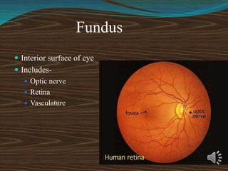
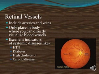
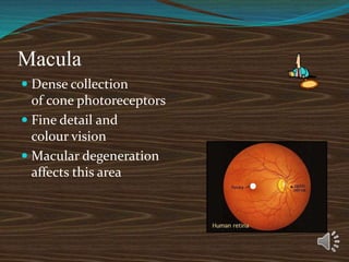
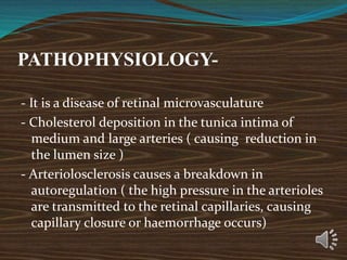
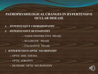
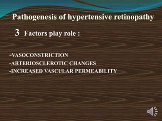
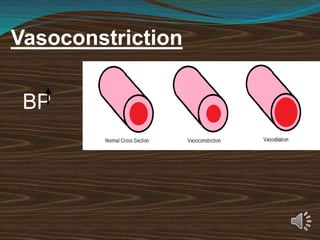
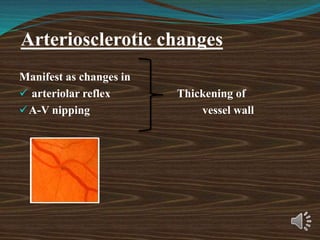
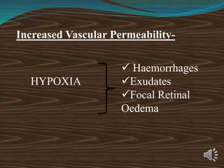
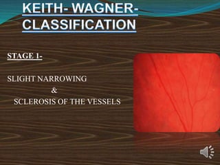
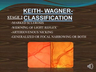
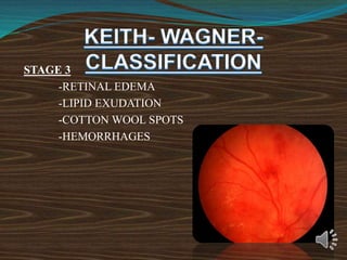
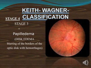
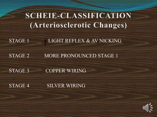
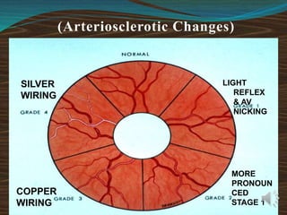
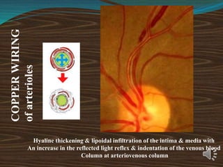
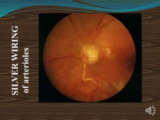
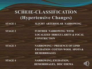
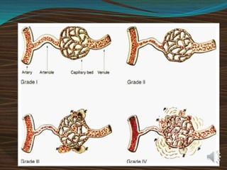
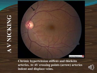
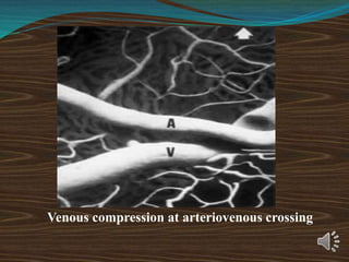
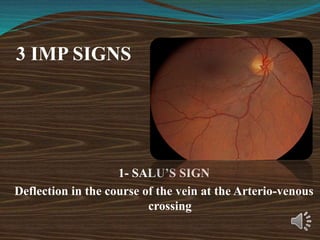
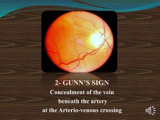
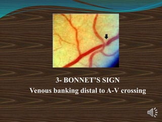
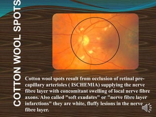
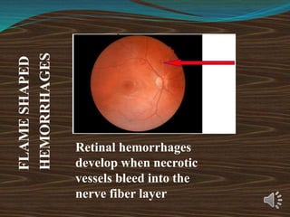
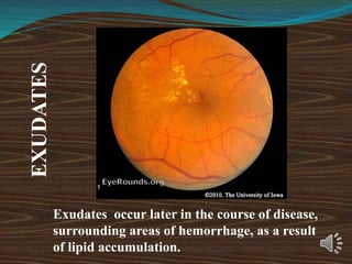
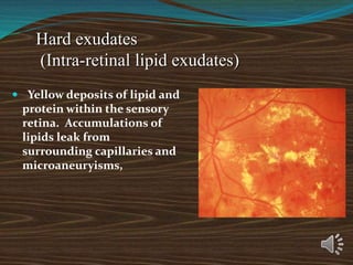
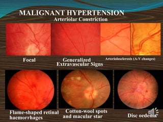
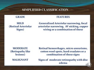
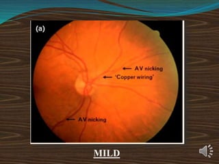
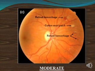
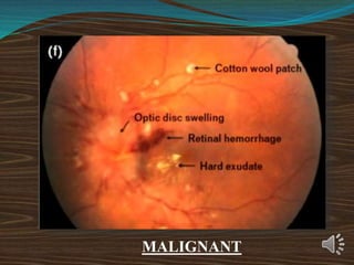
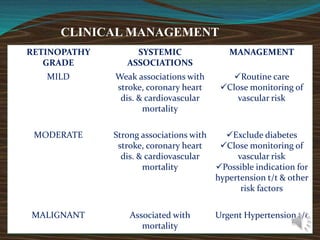
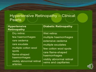
HYPERTENSIVE
RETINOPATHY
PRESENTED BY- DR. SHIKHA
ASSISTANT PROFESSOR
SHALAKYA TANTRA
HAMC&H DEHRADUN
9454908322
Fundus
 Interior surface of eye
 Includes-
 Optic nerve
 Retina
 Vasculature
Optic Nerve Head
 Collection of nerve
fibers and blood vessels
from retina
 Transfers info to brain’s
visual cortex
 Slightly yellow-pink
when healthy
 White ‘full moon’
appearance can mean
trouble!
Optic Nerve Head
 Cup is natural
depression in center
of nerve
 Cup size varies
between people
 Very large cup, or
change in appearance
over time, can
indicate glaucoma
Physiologic Cup
Optic Disc
Retinal Vessels
 Include arteries and veins
 Only place in body
where you can directly
visualize blood vessels
 Excellent indicators
of systemic diseases like-
 HTN
 Diabetes
 High cholesterol
 Carotid disease
Macula
 Dense collection
of cone photoreceptors
 Fine detail and
colour vision
 Macular degeneration
affects this area
INTRODUCTION Hypertensive
Retinopathy -
-Bilateral
-Symmetrical
-Small blood vessel disease
-Caused by systemic hypertention -
- acute or chronic
- systolic or diastolic-
PREVALENCE
-The second most common retinal vascular
disease
- Systemic hypertention ( > 160/90 mmHg)
-Malignent hypertention ( 240/140 mmHg)
- Hypertensive retinopathy 4- 10 %
Risk Factors-
Age
Family history
Obesity
Smoking
Stress
Alcohol consumption
Lack of exercises
Diagnostic techniques
- Ophthalmoscopy
- Sphygmomanometry
PATHOPHYSIOLOGY-
- It is a disease of retinal microvasculature
- Cholesterol deposition in the tunica intima of
medium and large arteries ( causing reduction in
the lumen size )
- Arteriolosclerosis causes a breakdown in
autoregulation ( the high pressure in the arterioles
are transmitted to the retinal capillaries, causing
capillary closure or haemorrhage occurs)
PATHOPHYSIOLOGICAL CHANGES IN HYPERTENSIVE
OCULAR DISEASE
1. HYPERTENSIVE CHOROIDOPATHY
2. HYPERTENSIVE RETINOPATHY
-- VASOCONSTRICTIVE PHASE
-- SCLEROTIC PHASE
-- EXUDATIVE PHASE
3. HYPERTENSIVE OPTIC NEUROPATHY
-- OPTIC DISC EDEMA
-- OPTIC ATROPHY
-- ISCHEMIC OPTIC NEUROPATHY
3 Factors play role :
-VASOCONSTRICTION
-ARTERIOSCLEROTIC CHANGES
-INCREASED VASCULAR PERMEABILITY
Vasoconstriction
BP
Manifest as changes in
 arteriolar reflex Thickening of
A-V nipping vessel wall
Arteriosclerotic changes
HYPOXIA
Increased Vascular Permeability-
 Haemorrhages
Exudates
Focal Retinal
Oedema
STAGE 1-
SLIGHT NARROWING
&
SCLEROSIS OF THE VESSELS
STAGE 2
-MARKED SCLEROSIS
-WIDENING OF LIGHT REFLEX
-ARTERIOVENOUS NICKING
-GENERALIZED OR FOCAL NARROWING OR BOTH
STAGE 3
-RETINAL EDEMA
-LIPID EXUDATION
-COTTON WOOL SPOTS
-HEMORRHAGES
STAGE 4
STAGE 3
+
Papilledema
(DISK EDEMA
blurring of the borders of the
optic disk with hemorrhages)
-Poor reliability and reproducibility
-Grades do not correlate with severity of
hypertension
-Not sequential in nature
-Grades do not correlate with prognosis,
cardiovascular events, and mortality.
STAGE 1 LIGHT REFLEX & AV NICKING
STAGE 2 MORE PRONOUNCED STAGE 1
STAGE 3 COPPER WIRING
STAGE 4 SILVER WIRING
(Arteriosclerotic Changes)
LIGHT
REFLEX
& AV
NICKING
MORE
PRONOUN
CED
STAGE 1
COPPER
WIRING
SILVER
WIRING
COPPER
WIRING
of
arterioles
Hyaline thickening & lipoidal infiltration of the intima & media with
An increase in the reflected light reflex & indentation of the venous blood
Column at arteriovenous column
SILVER
WIRING
of
arterioles
STAGE 1 SLIGHT ARTERIOLAR NARROWING
STAGE 2 FURTHER NARROWING WITH
LOCALIZED IRREGULARITY & FOCAL
CONSTRICTION
STAGE 3 NARROWING + PRESENCE OF LIPID
EXUDATION COTTON WOOL SPOTS &
HEMORRHAGES
STAGE 4 NARROWING, EXUDATION,
HEMORRHAGES, DISC EDEMA
Chronic hypertension stiffens and thickens
arteries. At AV crossing points (arrow) arteries
indent and displace veins.
A
V
NICKING
Venous compression at arteriovenous crossing
3 IMP SIGNS
1- SALU’S SIGN
Deflection in the course of the vein at the Arterio-venous
crossing
2- GUNN’S SIGN
Concealment of the vein
beneath the artery
at the Arterio-venous crossing
3- BONNET’S SIGN
Venous banking distal to A-V crossing
COTTON
WOOL
SPOT
Cotton wool spots result from occlusion of retinal pre-
capillary arterioles ( ISCHEMIA) supplying the nerve
fibre layer with concomitant swelling of local nerve fibre
axons. Also called "soft exudates" or "nerve fibre layer
infarctions" they are white, fluffy lesions in the nerve
fibre layer.
FLAME
SHAPED
HEMORRHAGES
Retinal hemorrhages
develop when necrotic
vessels bleed into the
nerve fiber layer
EXUDATES
Exudates occur later in the course of disease,
surrounding areas of hemorrhage, as a result
of lipid accumulation.
Hard exudates
(Intra-retinal lipid exudates)
 Yellow deposits of lipid and
protein within the sensory
retina. Accumulations of
lipids leak from
surrounding capillaries and
microaneuryisms,
MALIGNANT HYPERTENSION
Cotton-wool spots
and macular star Disc oedema
Focal Generalized
Arteriolar Constriction
Extravascular Signs
Flame-shaped retinal
haemorrhages
Arteriolosclerosis (A-V changes)
SIMPLIFIED CLASSIFICATION
GRADE FEATURES
MILD
(Retinal Arteriolar
Signs)
Generalized Arteriolar narrowing, focal
arteriolar narrowing, AV nicking, copper
wiring or a combination of these
MODERATE
(Retiopathy like
lesions)
Retinal hemorrhages, micro aneurisms,
cotton wool spots, hard exudates or a
combination of these signs
MALIGNANT Signs of moderate retinopathy with disc
edema
MILD
MODERATE
MALIGNANT
CLINICAL MANAGEMENT
RETINOPATHY
GRADE
SYSTEMIC
ASSOCIATIONS
MANAGEMENT
MILD Weak associations with
stroke, coronary heart
dis. & cardiovascular
mortality
Routine care
Close monitoring of
vascular risk
MODERATE Strong associations with
stroke, coronary heart
dis. & cardiovascular
mortality
Exclude diabetes
Close monitoring of
vascular risk
Possible indication for
hypertension t/t & other
risk factors
MALIGNANT Associated with
mortality
Urgent Hypertension t/t
THANK YOU

Hypertensive Retinopaty.ppt