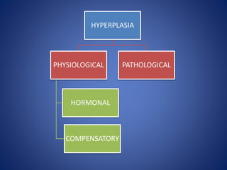
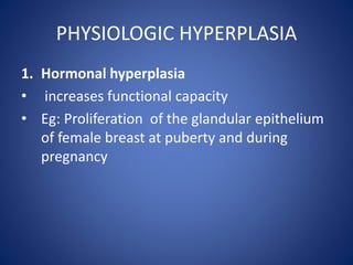
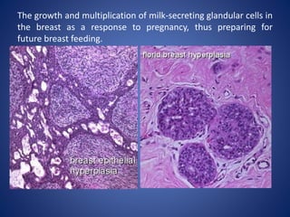
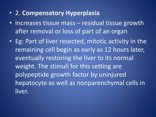
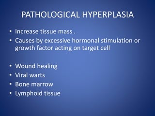
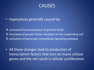
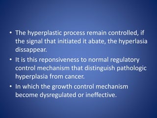
Enlargement of an organ associated
with increased number of cells
HYPERPLASIA
PHYSIOLOGICAL
COMPENSATORY
HORMONAL
PATHOLOGICAL
MECHANISM
• For both situation cellular proliferation is
stimulated by growth factors that are produce
by a variety of cell types.
PHYSIOLOGIC HYPERPLASIA
1. Hormonal hyperplasia
• increases functional capacity
• Eg: Proliferation of the glandular epithelium
of female breast at puberty and during
pregnancy
The growth and multiplication of milk-secreting glandular cells in
the breast as a response to pregnancy, thus preparing for
future breast feeding.
• 2. Compensatory Hyperplasia
• increases tissue mass – residual tissue growth
after removal or loss of part of an organ
• Eg: Part of liver resected, mitotic activity in the
remaining cell begin as early as 12 hours later,
eventually restoring the liver to its normal
weight. The stimuli for this setting are
polypeptide growth factor by uninjured
hepatocyte as well as nonparenchymal cells in
liver.
PATHOLOGICAL HYPERPLASIA
• Increase tissue mass .
• Causes by excessive hormonal stimulation or
growth factor acting on target cell
• Wound healing
• Viral warts
• Bone marrow
• Lymphoid tissue
ENDOMETRIUM HYPERPLASIA
PROSTATIC HYPERPLASIA
Example
• After a normal menstrual period tehre is a burst of uterine epithelial
proliferation that is normally tightly regulated by stimulation through
pituitary hormones and ovarian estrogen and by inhibition through
progesterone. However, a disturbed balance between estrogen and
progesterone causes endometrial hyperplasia, bleeding. Hyperplasia also
is an important response of connective tissue cells in wound healing, in
which prolifearting fibroblast and blood vessel aid in repair. In this process
growth factor are produced by leokocytes responding to injury and by cells
in wound healing
CAUSES
• Hyperplasia generally caused by
 increased local production of growth factor
 increased of growth factor receptor on the responding cell
 activation of particular intracellular signalling pathway
• All these changes lead to production of
transcription factors that turn on many cellular
genes and the net result is cellular proliferation
• In hormonal hyperlasia, the hormones act as
growth factor.
• The increase in tissue mass after some types
of cell loss is achieved not only by proliferation
of the remaining cell but also by the
development of new cells from stem cells
• The hyperplastic process remain controlled, if
the signal that initiated it abate, the hyperlasia
dissappear.
• It is this reponsiveness to normal regulatory
control mechanism that distinguish pathologic
hyperplasia from cancer.
• In which the growth control mechanism
become dysregulated or ineffective.

Hyperplasia

  • 1.
    Enlargement of anorgan associated with increased number of cells
  • 2.
  • 3.
    MECHANISM • For bothsituation cellular proliferation is stimulated by growth factors that are produce by a variety of cell types.
  • 4.
    PHYSIOLOGIC HYPERPLASIA 1. Hormonalhyperplasia • increases functional capacity • Eg: Proliferation of the glandular epithelium of female breast at puberty and during pregnancy
  • 5.
    The growth andmultiplication of milk-secreting glandular cells in the breast as a response to pregnancy, thus preparing for future breast feeding.
  • 6.
    • 2. CompensatoryHyperplasia • increases tissue mass – residual tissue growth after removal or loss of part of an organ • Eg: Part of liver resected, mitotic activity in the remaining cell begin as early as 12 hours later, eventually restoring the liver to its normal weight. The stimuli for this setting are polypeptide growth factor by uninjured hepatocyte as well as nonparenchymal cells in liver.
  • 7.
    PATHOLOGICAL HYPERPLASIA • Increasetissue mass . • Causes by excessive hormonal stimulation or growth factor acting on target cell • Wound healing • Viral warts • Bone marrow • Lymphoid tissue
  • 8.
  • 9.
  • 10.
    Example • After anormal menstrual period tehre is a burst of uterine epithelial proliferation that is normally tightly regulated by stimulation through pituitary hormones and ovarian estrogen and by inhibition through progesterone. However, a disturbed balance between estrogen and progesterone causes endometrial hyperplasia, bleeding. Hyperplasia also is an important response of connective tissue cells in wound healing, in which prolifearting fibroblast and blood vessel aid in repair. In this process growth factor are produced by leokocytes responding to injury and by cells in wound healing
  • 11.
    CAUSES • Hyperplasia generallycaused by  increased local production of growth factor  increased of growth factor receptor on the responding cell  activation of particular intracellular signalling pathway • All these changes lead to production of transcription factors that turn on many cellular genes and the net result is cellular proliferation
  • 12.
    • In hormonalhyperlasia, the hormones act as growth factor. • The increase in tissue mass after some types of cell loss is achieved not only by proliferation of the remaining cell but also by the development of new cells from stem cells
  • 13.
    • The hyperplasticprocess remain controlled, if the signal that initiated it abate, the hyperlasia dissappear. • It is this reponsiveness to normal regulatory control mechanism that distinguish pathologic hyperplasia from cancer. • In which the growth control mechanism become dysregulated or ineffective.