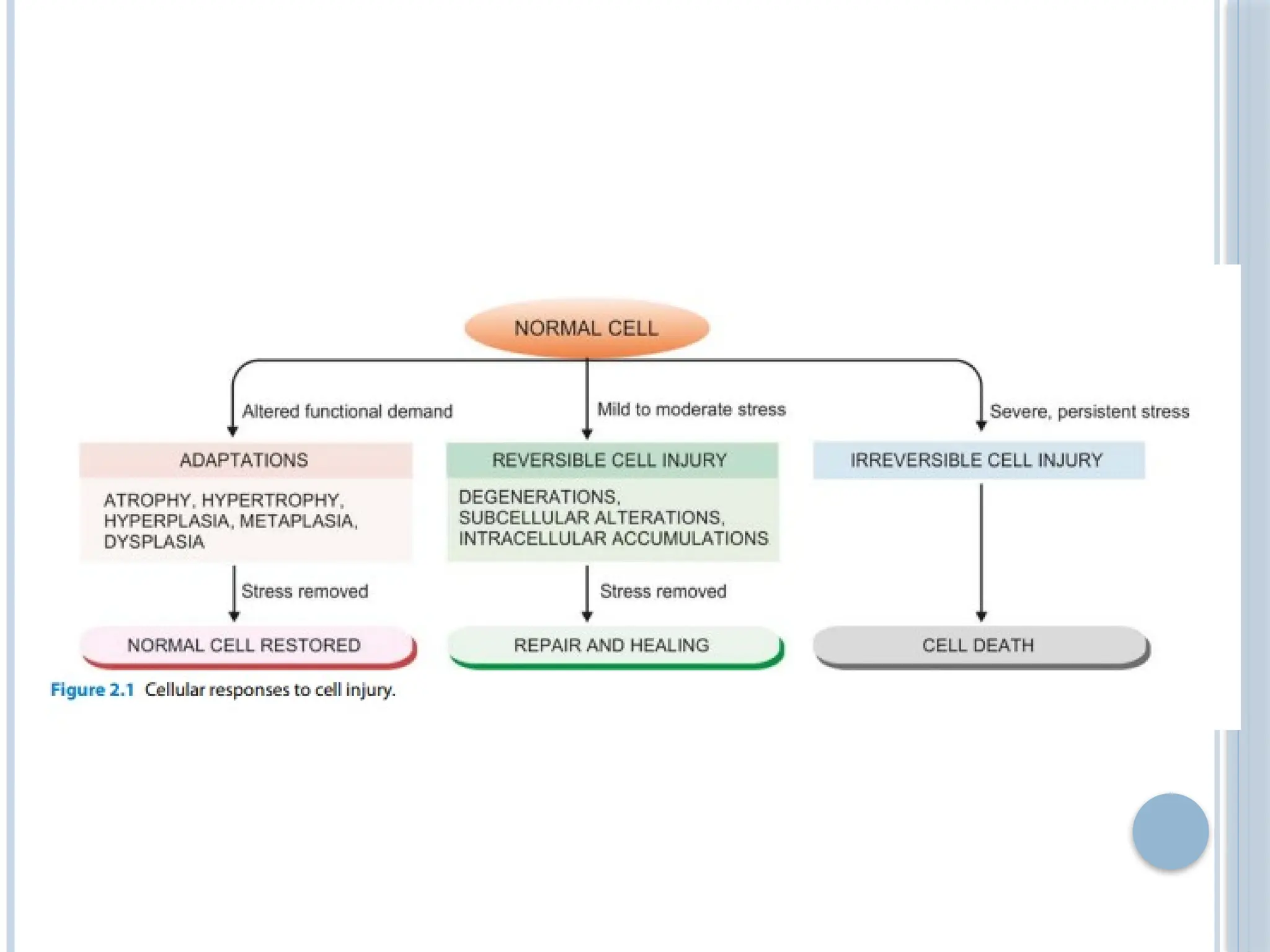
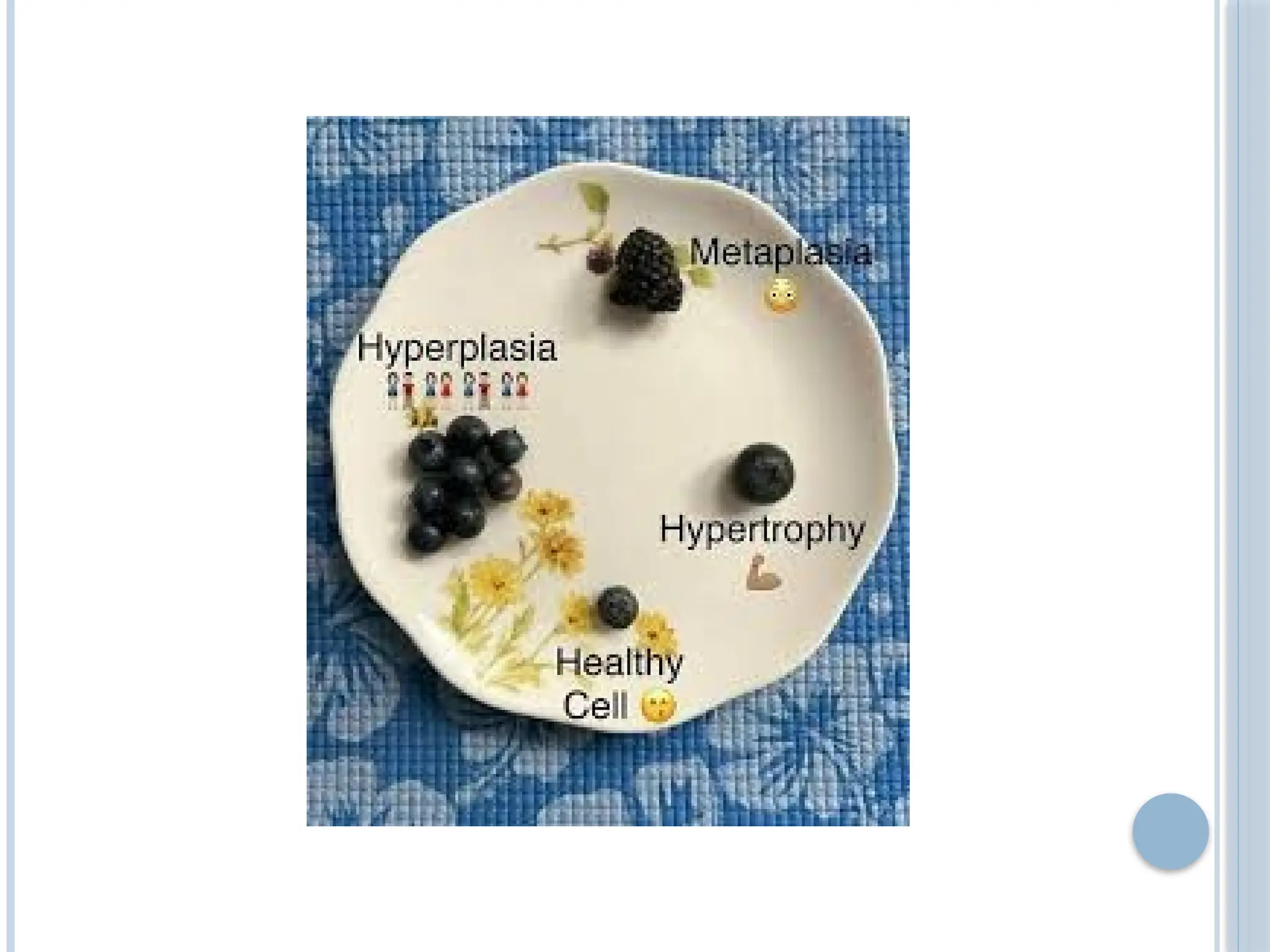
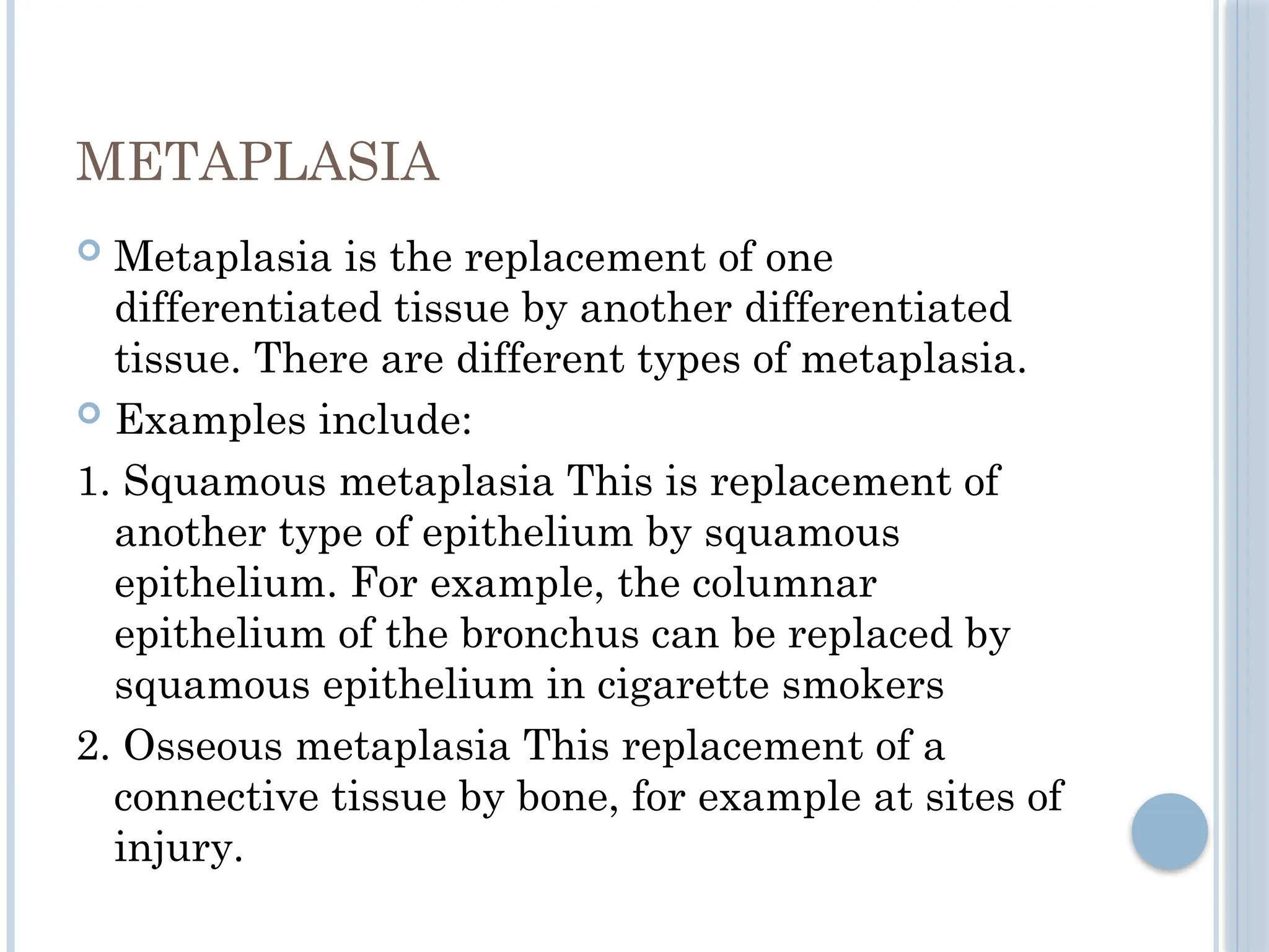
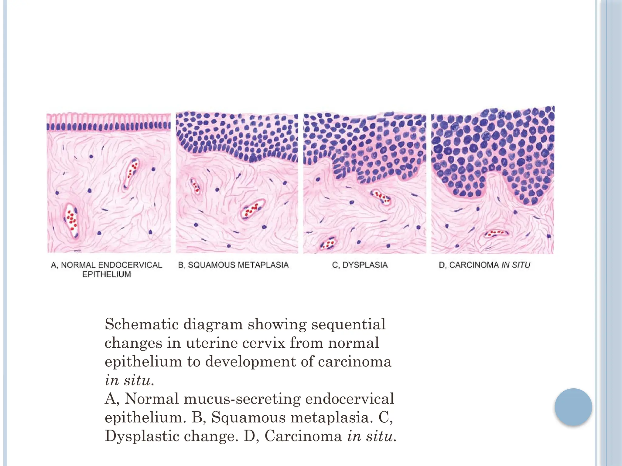
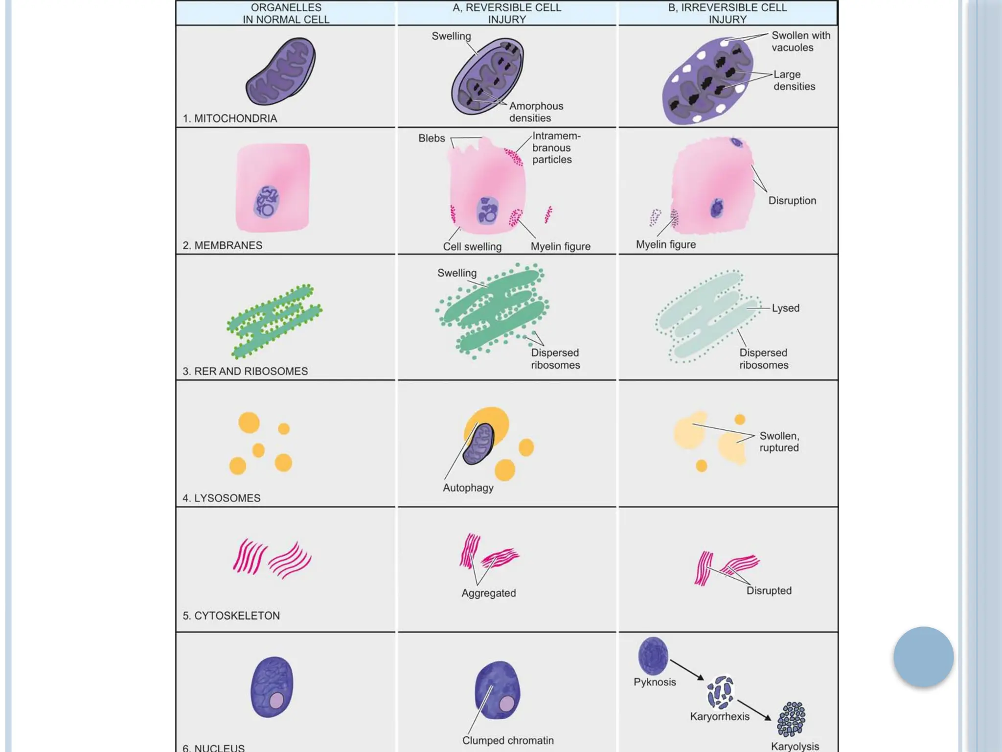
CELLULAR REACTIONS TO
INJURY
1. Define hyperplasia, hypertrophy, atrophy, &
Metaplasia & list some of their causes.
2. Reversible & irreversible forms of cell injury.
3. Necrosis .
4. Apoptosis.
5. Gangrene.
TYPES OF CELLULAR REACTIONS
TO INJURY
 CELLULAR ADATATIONS
 CELL INJURY
 Reversible cell injury : cellular, fatty change
 Irreversible cell injury : Necrosis , apotosis
 Intracellular accumulation
 Pathologic calcification
CELLULAR ADATATIONS
HYPERTROPHY
 Hypertrophy is increase in the size of cells.
 Increased workload leads to increased protein
synthesis & increased size & number of
intracellular organelles which, in turn, leads to
increased cell size.
 The increased cell size leads to increased size of
the organ.
 Examples: the enlargement of the left ventricle in
hypertensive heart disease & the increase in
skeletal muscle during sternous exercise.
PHYSIOLOGIC HYPERTROPHY
 Enlarged size of the uterus in pregnancy is an
example of physiologic hypertrophy as well as
hyperplasia.

 B. Pathologic hypertrophy Examples of certain
diseases associated with hypertrophy are as under:
1. Hypertrophy of cardiac muscle may occur in a
number of cardiovascular diseases.
A few conditions producing left ventricular
hypertrophy are as under: i) Systemic
hypertension ii) Aortic valve disease (stenosis
and insufficiency) iii) Mitral insufficiency
2. Hypertrophy of smooth muscle e.g. i) Cardiac
achalasia (in oesophagus) ii) Pyloric stenosis (in
stomach) iii) Intestinal strictures iv) Muscular
arteries in hypertension.
HYPERPLASIA
 Hyperplasia is an increase in the number of cells.
 It can lead to an increase in the size of the organ.
 It is usually caused by hormonal stimulation.
 It can be physiological as in enlargement of the
breast during pregnancy or it can pathological as
in endometrial hyperplasia.
PHYSIOLOGIC HYPERPLASIA
 The two most common types are hormonal and
compensatory:
1. Hormonal hyperplasia i.e. hyperplasia occurring
under the influence of hormonal stimulation e.g.
i) Hyperplasia of female breast at puberty, during
pregnancy and lactation.
ii) Hyperplasia of pregnant uterus.
iii) Proliferative activity of normal endometrium
after a normal menstrual cycle. iv) Prostatic
hyperplasia in old age
 Compensatory hyperplasia i.e. hyperplasia
occurring following removal of part of an organ or
in the contralateral organ in paired organ e.g.
i) Regeneration of the liver following partial
hepatectomy.
ii) Regeneration of epidermis after skin abrasion.
iii) Following nephrectomy on one side, there is
hyperplasia of nephrons of the other kidney.
PATHOLOGIC HYPERPLASIA
 Most examples of pathologic hyperplasia are due to
excessive stimulation of hormones or growth factors
e.g. i) Endometrial hyperplasia following oestrogen
excess.
ii) In wound healing, there is formation of granulation
tissue due to proliferation of fibroblasts and
endothelial cells.
iii) Formation of skin warts from hyperplasia of
epidermis due to human papilloma virus.
iv) Pseudocarcinomatous hyperplasia of the skin
occurring at the margin of a non-healing ulcer.
v) Intraductal epithelial hyperplasia in fi brocystic
change in the breast
ATROPHY
 Atrophy is a decrease in the size of a cell.
 This can lead to decreased size of the organ.
 The atrophic cell shows autophagic vacuoles
which contain cellular debris from degraded
organelles.
 Atrophy can be caused by:
1. Disuse
2. Under nutrition
3. Decreased endocrine stimulation
4. Denervation
5. Old age
PHYSIOLOGIC ATROPHY
 Atrophy is a normal process of ageing in some
tissues, which could be due to loss of endocrine
stimulation or arteriosclerosis.
For example: i) Atrophy of lymphoid tissue with
age.
ii) Atrophy of thymus in adult life.
iii) Atrophy of gonads after menopause.
iv) Atrophy of brain with ageing.
v) Osteoporosis with reduction in size of bony
trabeculae due to ageing
PATHOLOGIC ATROPHY
 The causes are as under:
1. Starvation atrophy In starvation, there is first
depletion of carbohydrate and fat stores followed
by protein catabolism. There is general weakness,
emaciation and anaemia referred to as cachexia
seen in cancer and severely ill patients.
2. Ischaemic atrophy Gradual diminution of blood
supply due to atherosclerosis may result in
shrinkage of the affected organ e.g.
i) Small atrophic kidney in atherosclerosis of renal
artery.
ii) ii) Atrophy of the brain in cerebral atherosclerosis.
3. Disuse atrophy Prolonged diminished
functional activity is associated with
disuseatrophy of the organ e.g.
i) Wasting of muscles of limb immobilised in cast.
ii) Atrophy of the pancreas in obstruction of
pancreatic duct.
4. Neuropathic atrophy Interruption in nerve
supply leads to wasting of muscles e.g.
i) Poliomyelitis
ii) Motor neuron disease
iii) Nerve section.
 Endocrine atrophy Loss of endocrine regulatory
mechanism results in reduced metabolic activity
of tissues and hence atrophy e.g.
i) Hypopituitarism may lead to atrophy of thyroid,
adrenal and gonads.
ii) Hypothyroidism may cause atrophy of the skin
and its adnexal structures
METAPLASIA
 Metaplasia is the replacement of one
differentiated tissue by another differentiated
tissue. There are different types of metaplasia.
 Examples include:
1. Squamous metaplasia This is replacement of
another type of epithelium by squamous
epithelium. For example, the columnar
epithelium of the bronchus can be replaced by
squamous epithelium in cigarette smokers
2. Osseous metaplasia This replacement of a
connective tissue by bone, for example at sites of
injury.
Schematic diagram showing sequential
changes in uterine cervix from normal
epithelium to development of carcinoma
in situ.
A, Normal mucus-secreting endocervical
epithelium. B, Squamous metaplasia. C,
Dysplastic change. D, Carcinoma in situ.
CELL INJURY
 Causes :
 Reduced oxygen supply (Hypoxia and ischaemia)
 Physical agents
 Chemical agents
 Infectious agents
 Abnormal immunologic reactions
 Nutritional imbalances
 Genetic factors
 idiopathic
TYPES OF CELL INJURY
 Reversible
 Irreversible cell injury
 If the stimulus is
acute and brief or
mild , the cell injury
produces changes in
the cells which are
reversible up to a
certain point
 If the cell cell is
exxposed to
continuous injurious
stimulus or if the
injury is severe , the
cells undergo cell
death.
 Two main types
 Necrosis
 Apoptosis
Reversible cell injury Irreversible cell injury
THANK YOU

cellular reactions to injury (PATHOLOGY)

  • 1.
  • 2.
    1. Define hyperplasia,hypertrophy, atrophy, & Metaplasia & list some of their causes. 2. Reversible & irreversible forms of cell injury. 3. Necrosis . 4. Apoptosis. 5. Gangrene.
  • 3.
    TYPES OF CELLULARREACTIONS TO INJURY  CELLULAR ADATATIONS  CELL INJURY  Reversible cell injury : cellular, fatty change  Irreversible cell injury : Necrosis , apotosis  Intracellular accumulation  Pathologic calcification
  • 5.
  • 6.
    HYPERTROPHY  Hypertrophy isincrease in the size of cells.  Increased workload leads to increased protein synthesis & increased size & number of intracellular organelles which, in turn, leads to increased cell size.  The increased cell size leads to increased size of the organ.  Examples: the enlargement of the left ventricle in hypertensive heart disease & the increase in skeletal muscle during sternous exercise.
  • 8.
    PHYSIOLOGIC HYPERTROPHY  Enlargedsize of the uterus in pregnancy is an example of physiologic hypertrophy as well as hyperplasia. 
  • 9.
     B. Pathologichypertrophy Examples of certain diseases associated with hypertrophy are as under: 1. Hypertrophy of cardiac muscle may occur in a number of cardiovascular diseases. A few conditions producing left ventricular hypertrophy are as under: i) Systemic hypertension ii) Aortic valve disease (stenosis and insufficiency) iii) Mitral insufficiency 2. Hypertrophy of smooth muscle e.g. i) Cardiac achalasia (in oesophagus) ii) Pyloric stenosis (in stomach) iii) Intestinal strictures iv) Muscular arteries in hypertension.
  • 11.
    HYPERPLASIA  Hyperplasia isan increase in the number of cells.  It can lead to an increase in the size of the organ.  It is usually caused by hormonal stimulation.  It can be physiological as in enlargement of the breast during pregnancy or it can pathological as in endometrial hyperplasia.
  • 12.
    PHYSIOLOGIC HYPERPLASIA  Thetwo most common types are hormonal and compensatory: 1. Hormonal hyperplasia i.e. hyperplasia occurring under the influence of hormonal stimulation e.g. i) Hyperplasia of female breast at puberty, during pregnancy and lactation. ii) Hyperplasia of pregnant uterus. iii) Proliferative activity of normal endometrium after a normal menstrual cycle. iv) Prostatic hyperplasia in old age
  • 13.
     Compensatory hyperplasiai.e. hyperplasia occurring following removal of part of an organ or in the contralateral organ in paired organ e.g. i) Regeneration of the liver following partial hepatectomy. ii) Regeneration of epidermis after skin abrasion. iii) Following nephrectomy on one side, there is hyperplasia of nephrons of the other kidney.
  • 14.
    PATHOLOGIC HYPERPLASIA  Mostexamples of pathologic hyperplasia are due to excessive stimulation of hormones or growth factors e.g. i) Endometrial hyperplasia following oestrogen excess. ii) In wound healing, there is formation of granulation tissue due to proliferation of fibroblasts and endothelial cells. iii) Formation of skin warts from hyperplasia of epidermis due to human papilloma virus. iv) Pseudocarcinomatous hyperplasia of the skin occurring at the margin of a non-healing ulcer. v) Intraductal epithelial hyperplasia in fi brocystic change in the breast
  • 17.
    ATROPHY  Atrophy isa decrease in the size of a cell.  This can lead to decreased size of the organ.  The atrophic cell shows autophagic vacuoles which contain cellular debris from degraded organelles.  Atrophy can be caused by: 1. Disuse 2. Under nutrition 3. Decreased endocrine stimulation 4. Denervation 5. Old age
  • 18.
    PHYSIOLOGIC ATROPHY  Atrophyis a normal process of ageing in some tissues, which could be due to loss of endocrine stimulation or arteriosclerosis. For example: i) Atrophy of lymphoid tissue with age. ii) Atrophy of thymus in adult life. iii) Atrophy of gonads after menopause. iv) Atrophy of brain with ageing. v) Osteoporosis with reduction in size of bony trabeculae due to ageing
  • 19.
    PATHOLOGIC ATROPHY  Thecauses are as under: 1. Starvation atrophy In starvation, there is first depletion of carbohydrate and fat stores followed by protein catabolism. There is general weakness, emaciation and anaemia referred to as cachexia seen in cancer and severely ill patients. 2. Ischaemic atrophy Gradual diminution of blood supply due to atherosclerosis may result in shrinkage of the affected organ e.g. i) Small atrophic kidney in atherosclerosis of renal artery. ii) ii) Atrophy of the brain in cerebral atherosclerosis.
  • 20.
    3. Disuse atrophyProlonged diminished functional activity is associated with disuseatrophy of the organ e.g. i) Wasting of muscles of limb immobilised in cast. ii) Atrophy of the pancreas in obstruction of pancreatic duct. 4. Neuropathic atrophy Interruption in nerve supply leads to wasting of muscles e.g. i) Poliomyelitis ii) Motor neuron disease iii) Nerve section.
  • 21.
     Endocrine atrophyLoss of endocrine regulatory mechanism results in reduced metabolic activity of tissues and hence atrophy e.g. i) Hypopituitarism may lead to atrophy of thyroid, adrenal and gonads. ii) Hypothyroidism may cause atrophy of the skin and its adnexal structures
  • 24.
    METAPLASIA  Metaplasia isthe replacement of one differentiated tissue by another differentiated tissue. There are different types of metaplasia.  Examples include: 1. Squamous metaplasia This is replacement of another type of epithelium by squamous epithelium. For example, the columnar epithelium of the bronchus can be replaced by squamous epithelium in cigarette smokers 2. Osseous metaplasia This replacement of a connective tissue by bone, for example at sites of injury.
  • 26.
    Schematic diagram showingsequential changes in uterine cervix from normal epithelium to development of carcinoma in situ. A, Normal mucus-secreting endocervical epithelium. B, Squamous metaplasia. C, Dysplastic change. D, Carcinoma in situ.
  • 27.
    CELL INJURY  Causes:  Reduced oxygen supply (Hypoxia and ischaemia)  Physical agents  Chemical agents  Infectious agents  Abnormal immunologic reactions  Nutritional imbalances  Genetic factors  idiopathic
  • 28.
    TYPES OF CELLINJURY  Reversible  Irreversible cell injury
  • 29.
     If thestimulus is acute and brief or mild , the cell injury produces changes in the cells which are reversible up to a certain point  If the cell cell is exxposed to continuous injurious stimulus or if the injury is severe , the cells undergo cell death.  Two main types  Necrosis  Apoptosis Reversible cell injury Irreversible cell injury
  • 31.