CELL INJURY ANDCELLULAR ADAPTATIONS
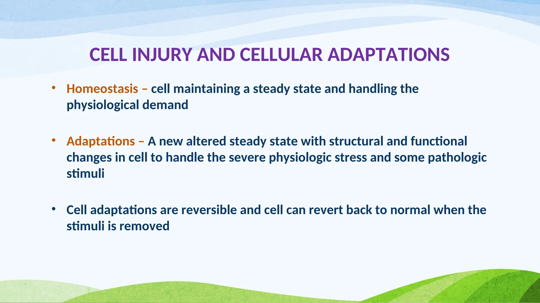
• Homeostasis – cell maintaining a steady state and handling the
physiological demand
• Adaptations – A new altered steady state with structural and functional
changes in cell to handle the severe physiologic stress and some pathologic
stimuli
• Cell adaptations are reversible and cell can revert back to normal when the
stimuli is removed
3.
CELL INJURY ANDCELLULAR ADAPTATIONS
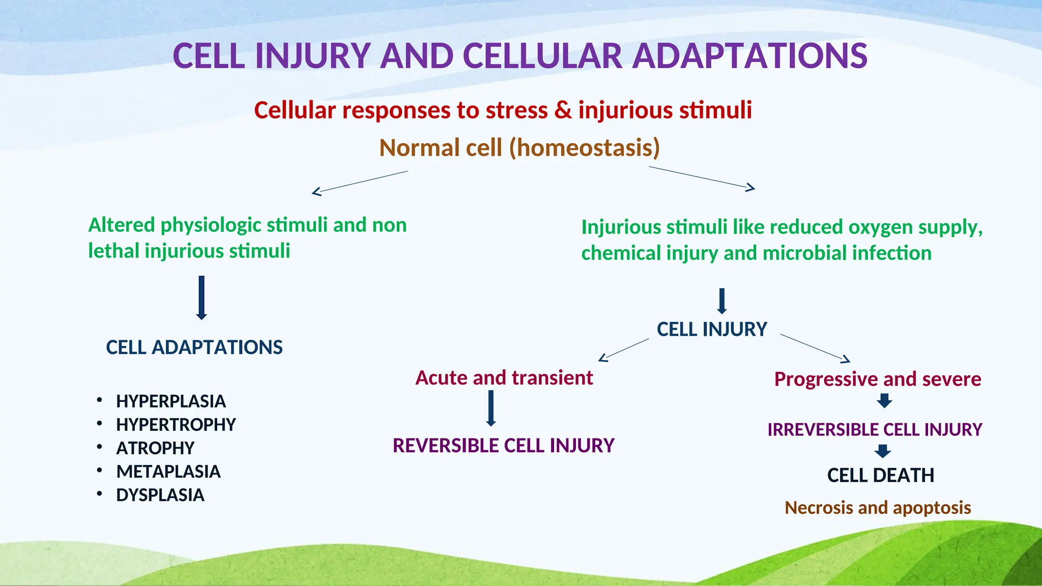
Cellular responses to stress & injurious stimuli
Normal cell (homeostasis)
Altered physiologic stimuli and non
lethal injurious stimuli
Injurious stimuli like reduced oxygen supply,
chemical injury and microbial infection
CELL ADAPTATIONS
CELL INJURY
Acute and transient Progressive and severe
REVERSIBLE CELL INJURY
IRREVERSIBLE CELL INJURY
CELL DEATH
• HYPERPLASIA
• HYPERTROPHY
• ATROPHY
• METAPLASIA
• DYSPLASIA
Necrosis and apoptosis
4.
CELL ADAPTATIONS
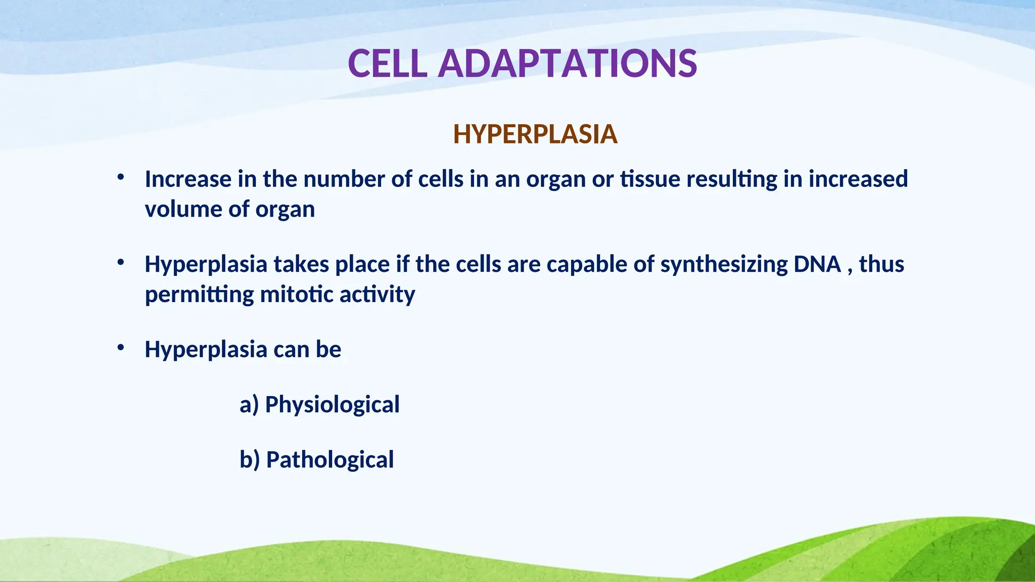
HYPERPLASIA
• Increasein the number of cells in an organ or tissue resulting in increased
volume of organ
• Hyperplasia takes place if the cells are capable of synthesizing DNA , thus
permitting mitotic activity
• Hyperplasia can be
a) Physiological
b) Pathological
5.
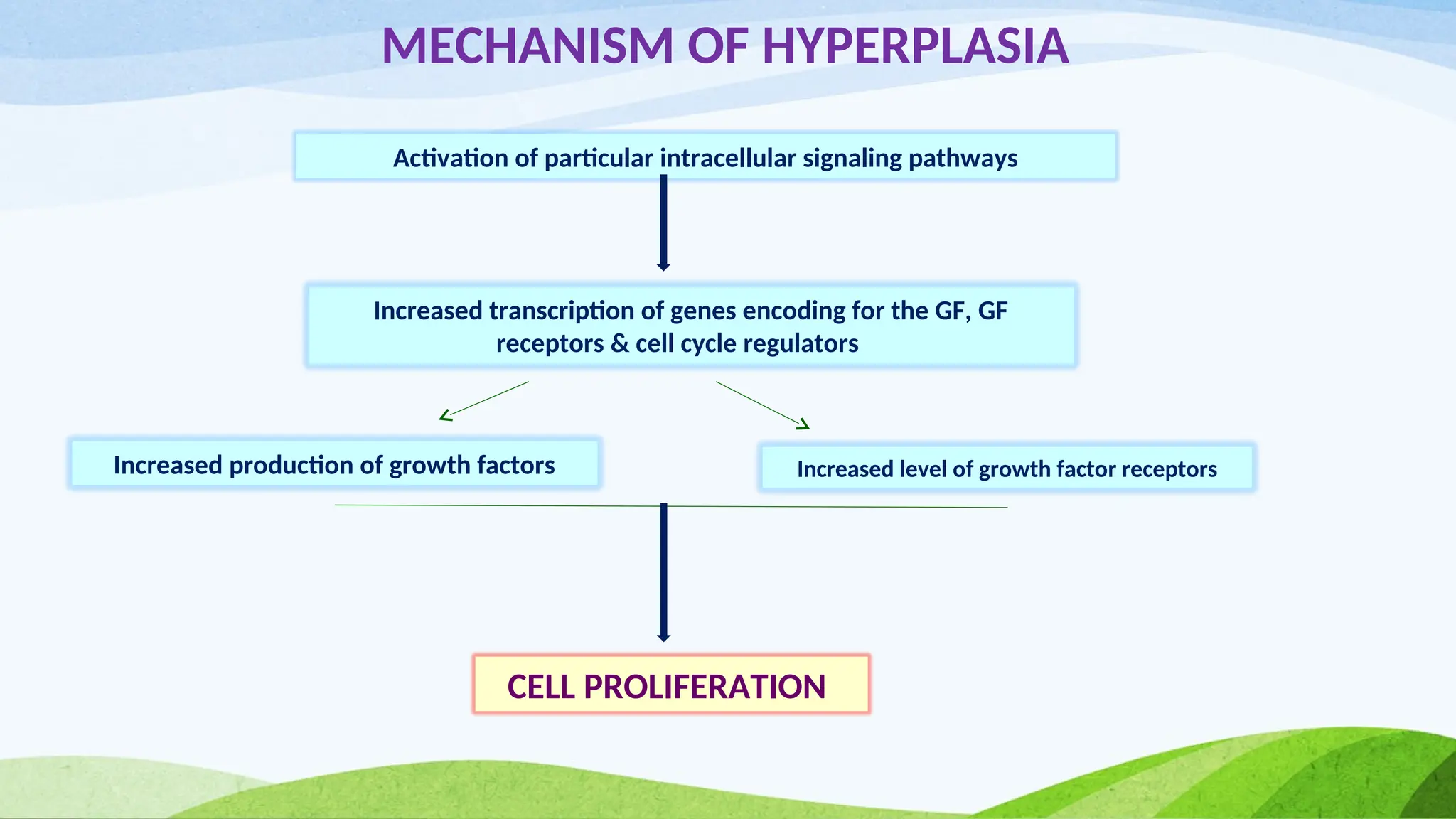
MECHANISM OF HYPERPLASIA
Activationof particular intracellular signaling pathways
Increased transcription of genes encoding for the GF, GF
receptors & cell cycle regulators
Increased production of growth factors Increased level of growth factor receptors
CELL PROLIFERATION
6.
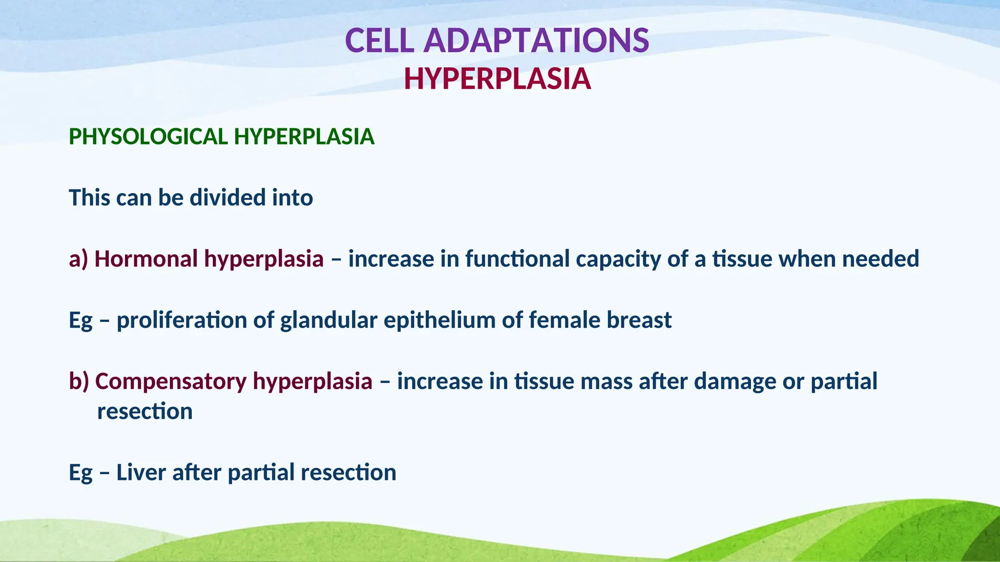
CELL ADAPTATIONS
HYPERPLASIA
PHYSOLOGICAL HYPERPLASIA
Thiscan be divided into
a) Hormonal hyperplasia – increase in functional capacity of a tissue when needed
Eg – proliferation of glandular epithelium of female breast
b) Compensatory hyperplasia – increase in tissue mass after damage or partial
resection
Eg – Liver after partial resection
7.
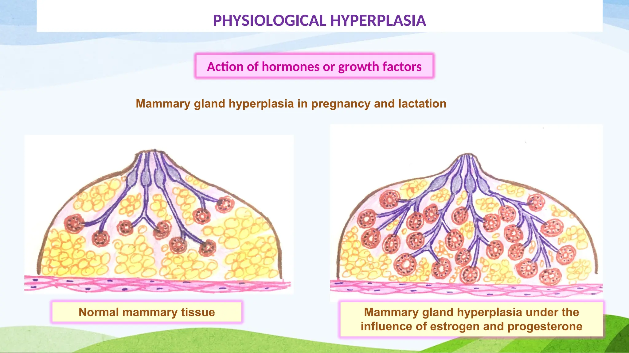
PHYSIOLOGICAL HYPERPLASIA
Action ofhormones or growth factors
Mammary gland hyperplasia in pregnancy and lactation
Normal mammary tissue Mammary gland hyperplasia under the
influence of estrogen and progesterone
8.
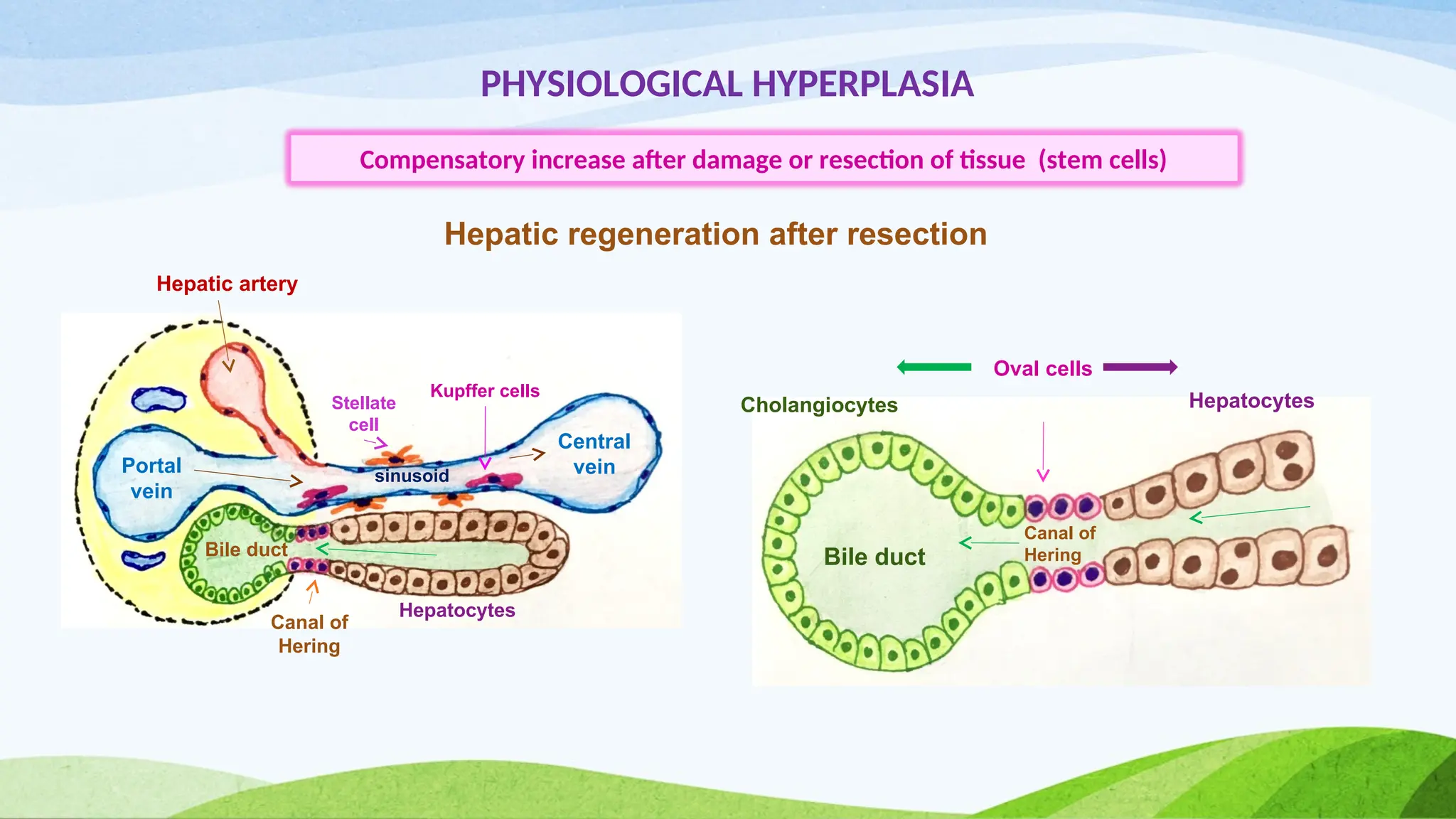
PHYSIOLOGICAL HYPERPLASIA
Compensatory increaseafter damage or resection of tissue (stem cells)
Portal
vein
Central
vein
Hepatic artery
Bile duct
Canal of
Hering
Stellate
cell
Kupffer cells
Hepatocytes
Oval cells
sinusoid
Cholangiocytes Hepatocytes
Bile duct
Canal of
Hering
Hepatic regeneration after resection
9.
CELL ADAPTATIONS
HYPERPLASIA
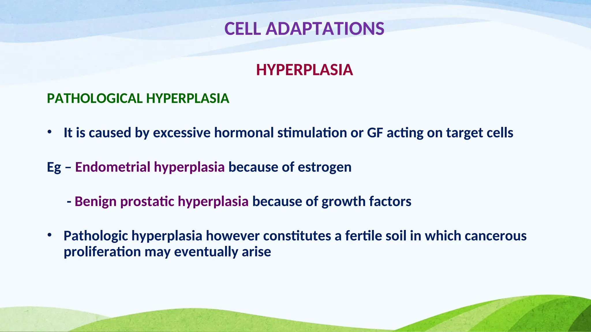
PATHOLOGICAL HYPERPLASIA
•It is caused by excessive hormonal stimulation or GF acting on target cells
Eg – Endometrial hyperplasia because of estrogen
- Benign prostatic hyperplasia because of growth factors
• Pathologic hyperplasia however constitutes a fertile soil in which cancerous
proliferation may eventually arise
10.
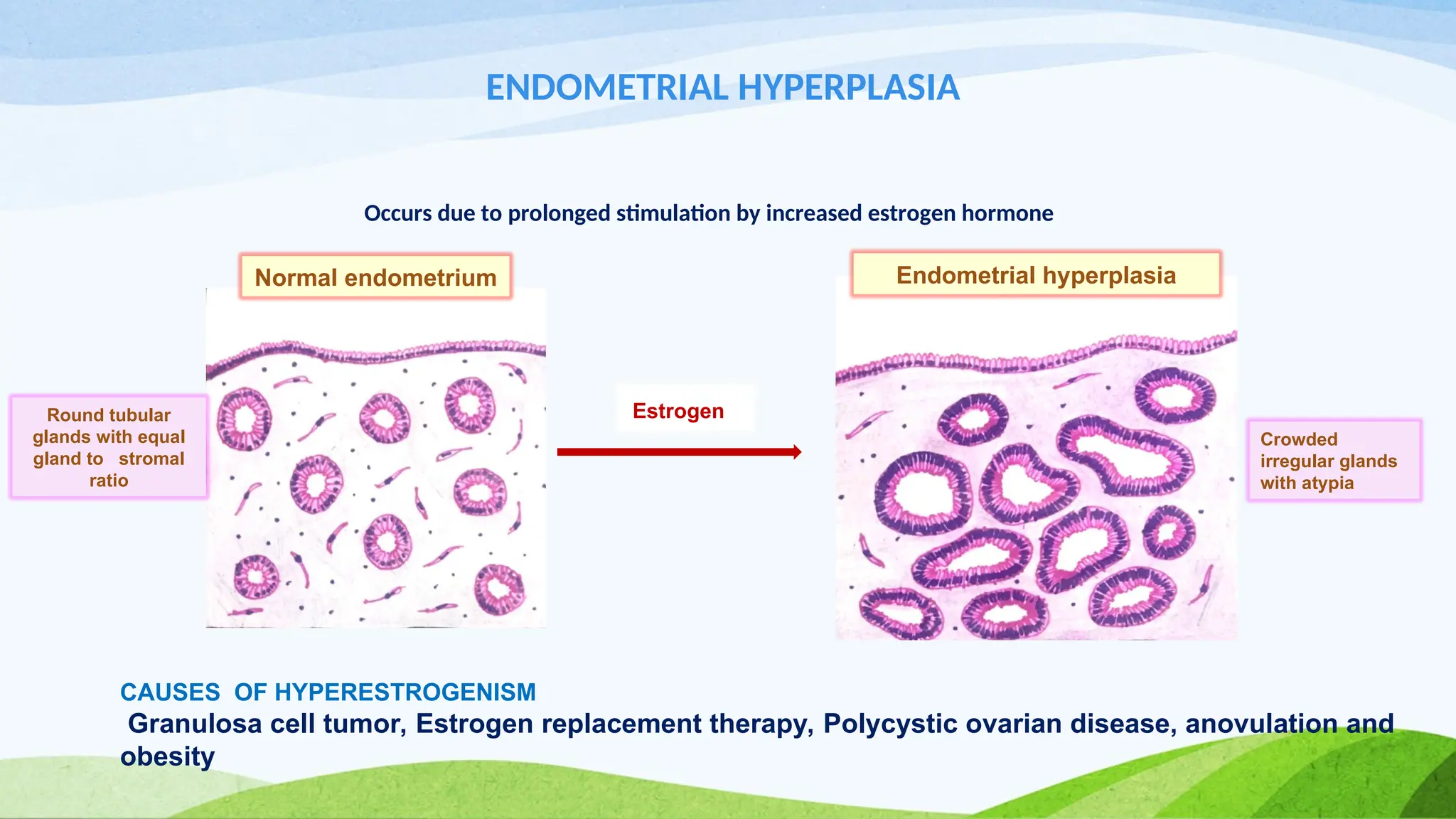
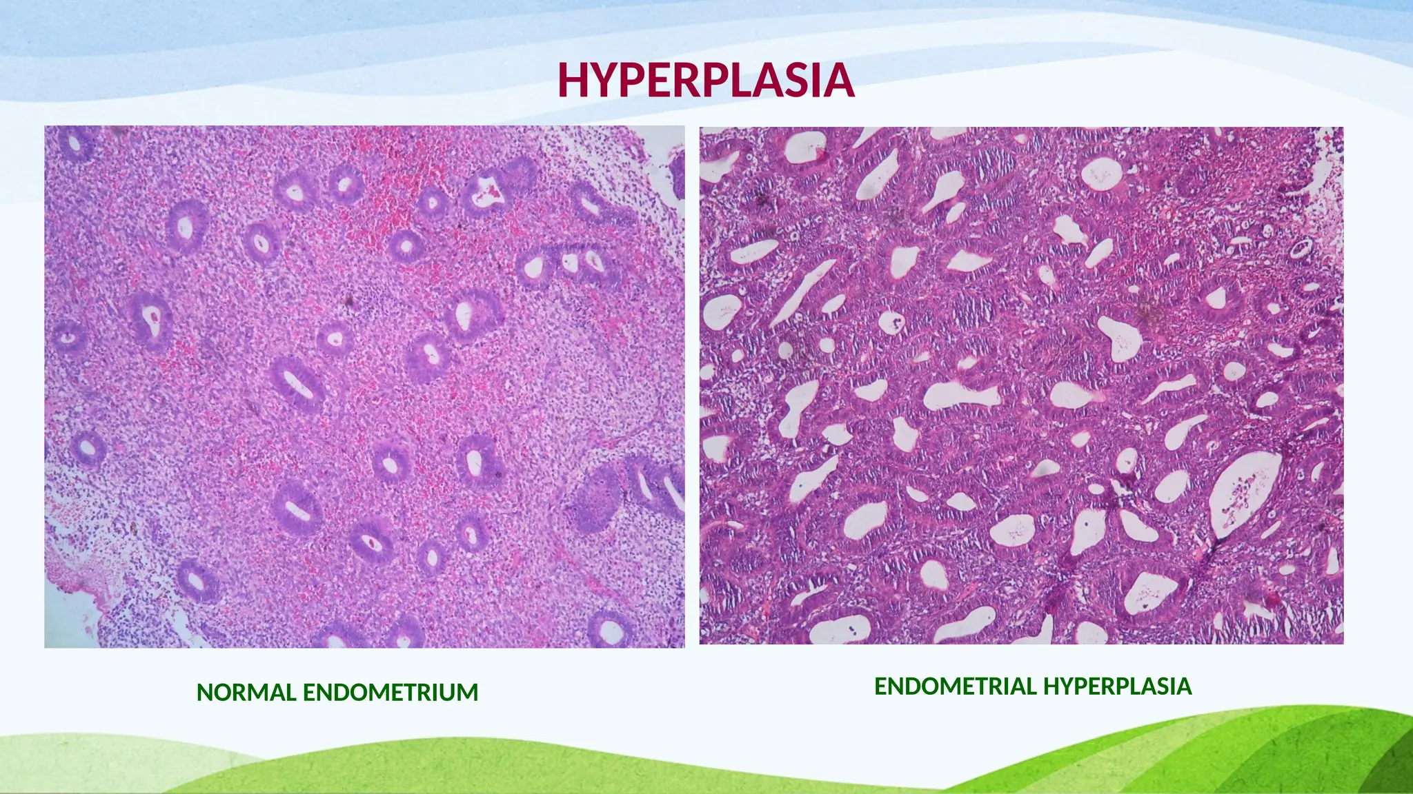
ENDOMETRIAL HYPERPLASIA
Occurs dueto prolonged stimulation by increased estrogen hormone
CAUSES OF HYPERESTROGENISM
Granulosa cell tumor, Estrogen replacement therapy, Polycystic ovarian disease, anovulation and
obesity
Estrogen
Crowded
irregular glands
with atypia
Round tubular
glands with equal
gland to stromal
ratio
Normal endometrium Endometrial hyperplasia
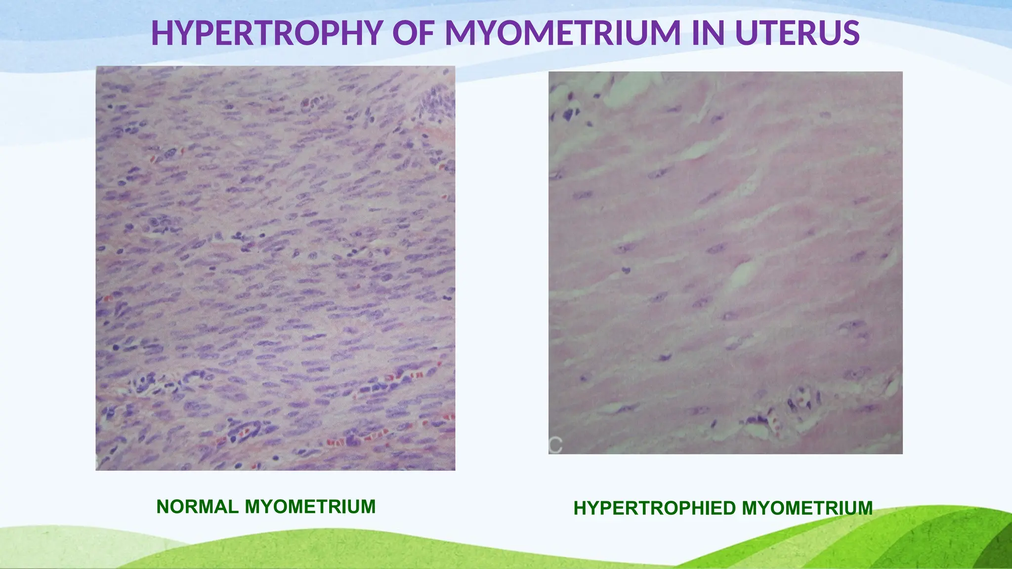
CELL ADAPTATIONS
HYPERTROPHY
• Hypertrophyrefers to an increase in the size of cells , resulting in an
increase in the size of the organ (no new cells but larger cells )
• This is more common in non-dividing cells
eg. Myocardial fibers
• It can be physiologic or pathologic & is caused by increased
functional demand or by specific hormonal stimulation
14.
CELL ADAPTATIONS
HYPERTROPHY
Some ofthe examples of hypertrophy are
a) Bulging of muscles in body builders
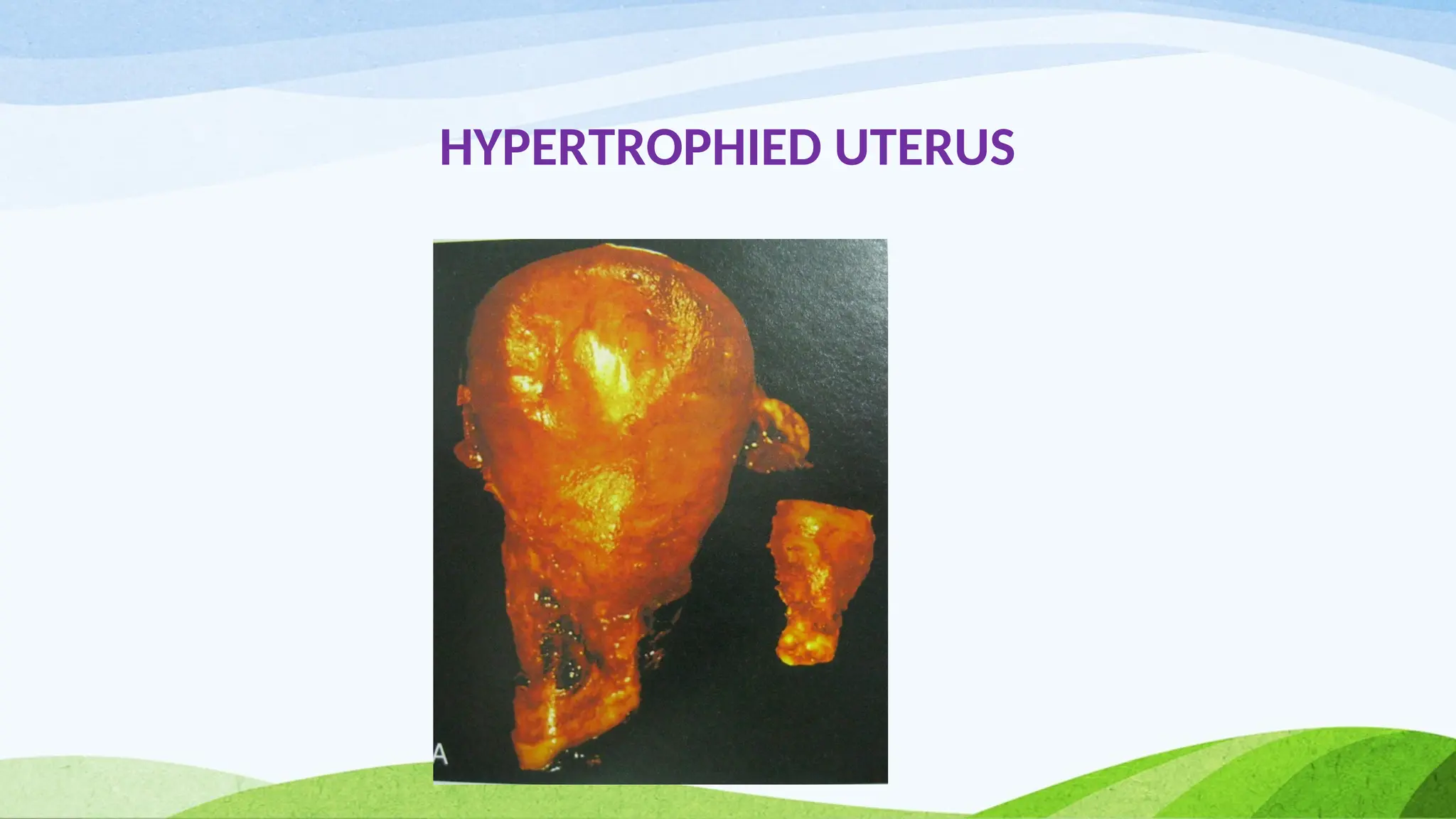
b) Massive physiological growth of the uterus during pregnancy – hormone
induced increase in size of an organ
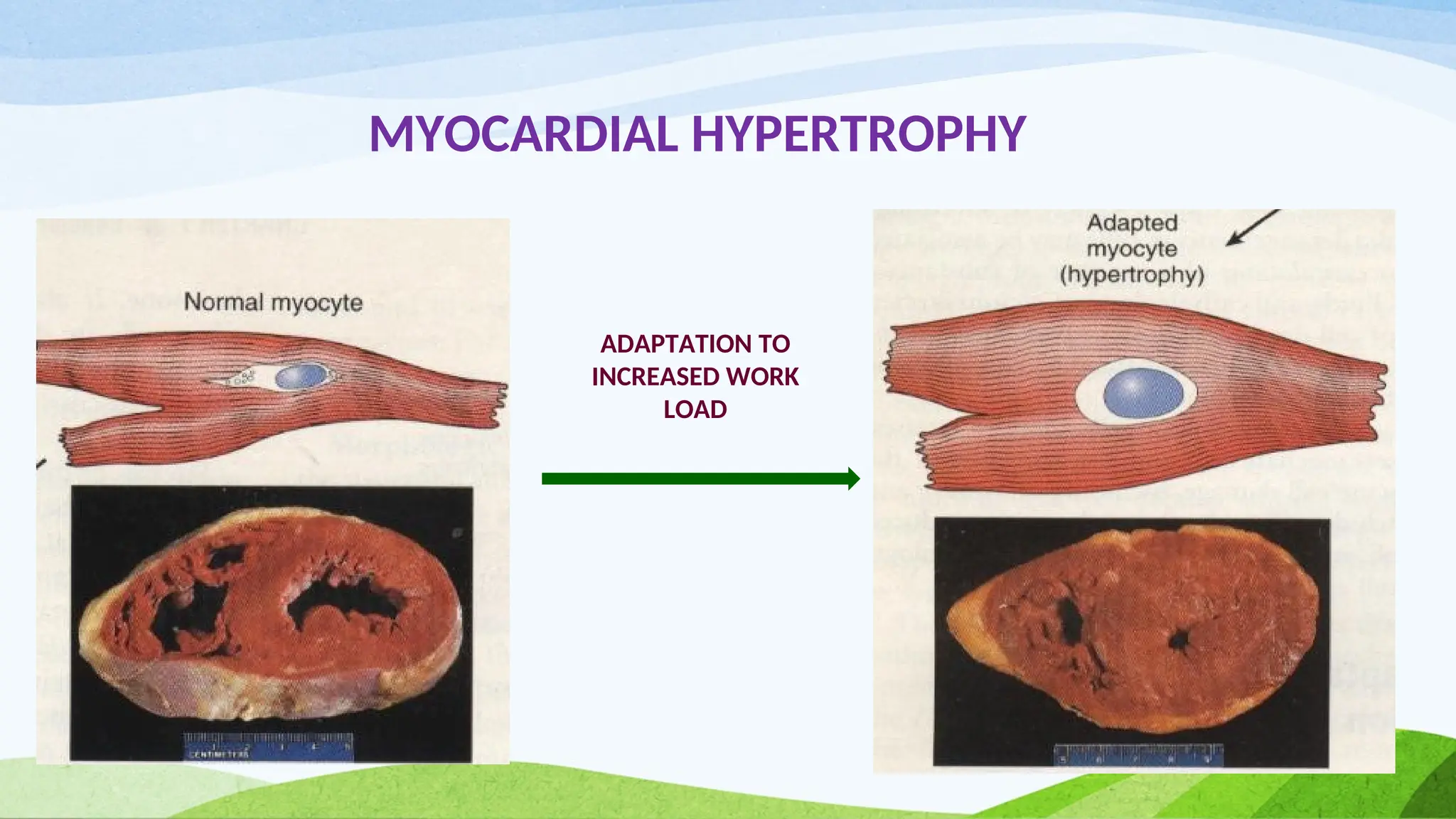
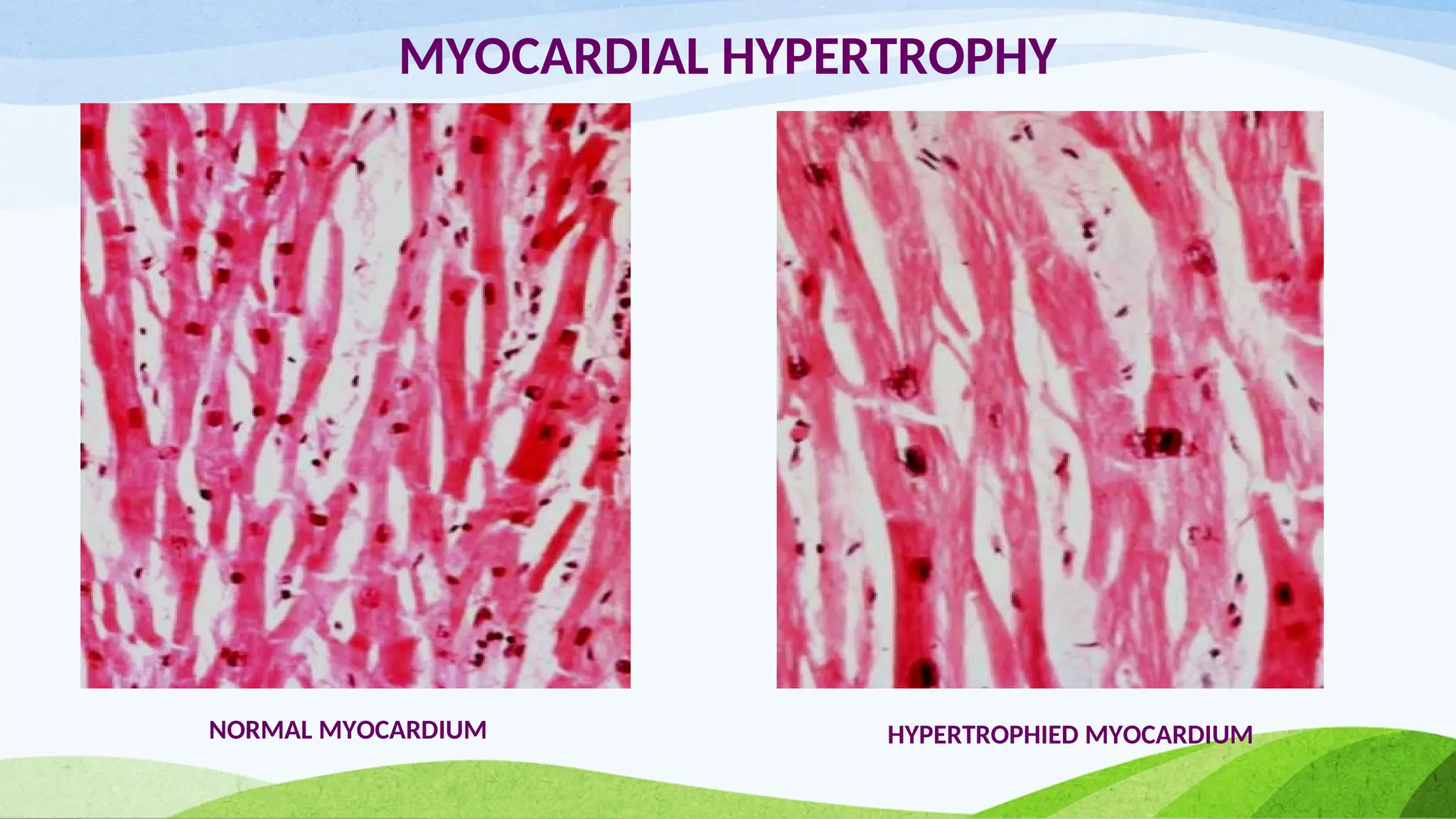
c) Hypertrophy of myocardium
15.
CELL ADAPTATIONS
HYPERTROPHY
• Hypertrophyresults from increased production of cellular proteins
• Mechanical stress and growth factors induce increased production of
proteins which causes hypertrophy
• Selective hypertrophy can occur at the subcellular organelle level also
eg – Individuals taking barbiturate drugs show hypertrophy of smooth
muscle ER in hepatocytes
CELL ADAPTATIONS
HYPERTROPHY
• Ifthe hypertrophied muscle mass cannot compensate for the increased work load
, degenerative changes occur
• The limiting factors for continued hypertrophy may be –
a) Limitations of vascular supply to the enlarged fibers
b) Diminished oxidative capabilities of mitochondria
c) Alterations in protein synthesis & degradation
d) Cytoskeletal alterations
21.
CELL ADAPTATIONS
ATROPHY
• Reductionin the size of the organ or tissue resulting from
decrease in cell size and number is called atrophy
• This is divided into -
a) Physiologic
b) Pathologic
CELL ADAPTATIONS
ATROPHY
PATHOLOGIC ATROPHY
Thisdepends on the underlying cause –
a) Decreased work load – immobilized broken limb in plaster cast
b) Denervation atrophy
c) Diminished blood supply – e.g. Brain in aged
d) Inadequate nutrition – Marasmus and cachexia in cancer pts
e) Loss of endocrine stimulation
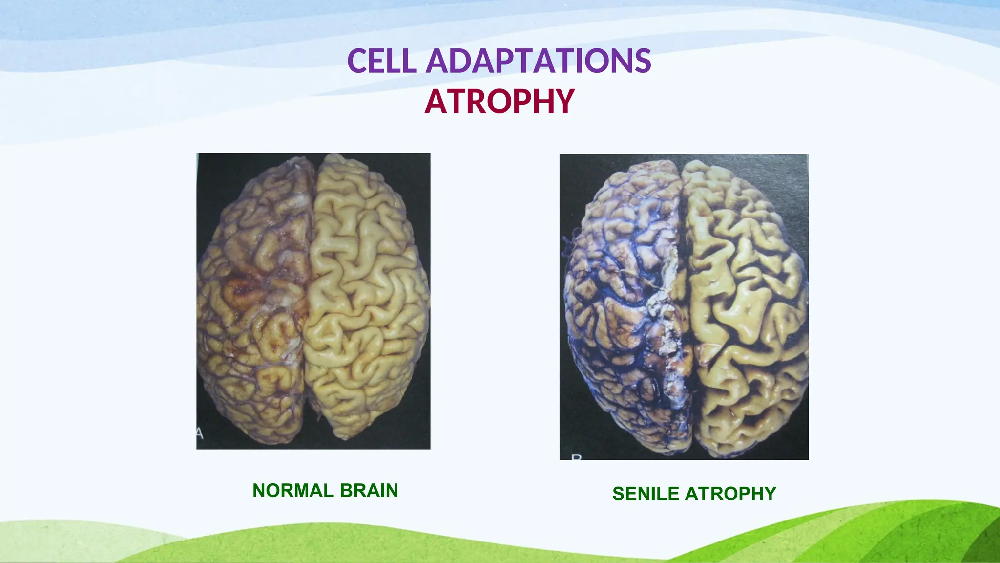
f) Ageing ( senile atrophy ) - in brain & heart
g) Pressure – tissue compression for long time by enlarging benign tumor
24.
CELL ADAPTATIONS
ATROPHY
MECHANISM OFATROPHY
• Results from decreased protein synthesis and increased protein
degradation by ubiquitin proteosome pathway
• Nutrient deficiency and disuse may activate ubiquitin ligases which
attach the small peptide ubiquitin to cellular proteins and target
these proteins for degradation in proteosomes
• In some cases atrophy is due to autophagy of cells own organelles
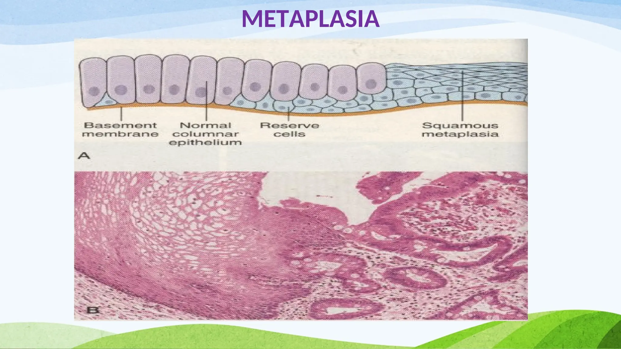
CELL ADAPTATIONS
METAPLASIA
Metaplasia isa reversible change in which one adult cell type
(epithelial or mesenchymal) is replaced by another adult cell
type.
E.g. a) Columnar to squamous – In habitual smokers , the normal
ciliated columnar epithelial cells of trachea & bronchi are
replaced by squamous epithelium
b) Squamous to columnar – Barrett’s oesophagus
27.
CELL ADAPTATIONS
METAPLASIA
Connective tissuemetaplasia is the formation of cartilage, bone or
adipose tissue in tissues that normally do not contain these
elements
E.g. Myositis ossificans
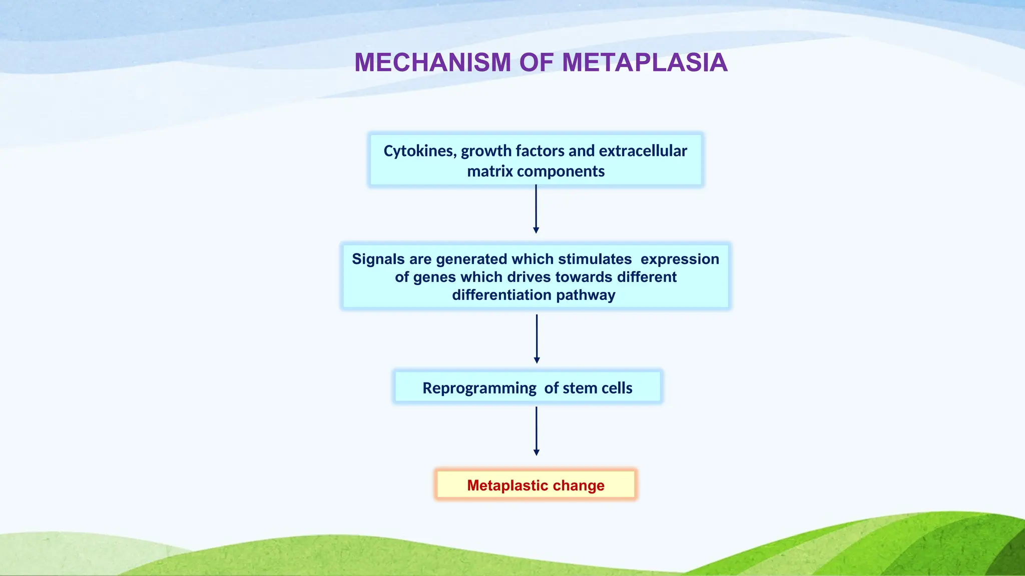
The influences that predispose to metaplasia if persistent , may
induce malignant transformation in metaplastic epithelium
Cytokines, growth factorsand extracellular
matrix components
Signals are generated which stimulates expression
of genes which drives towards different
differentiation pathway
Reprogramming of stem cells
Metaplastic change
MECHANISM OF METAPLASIA
30.
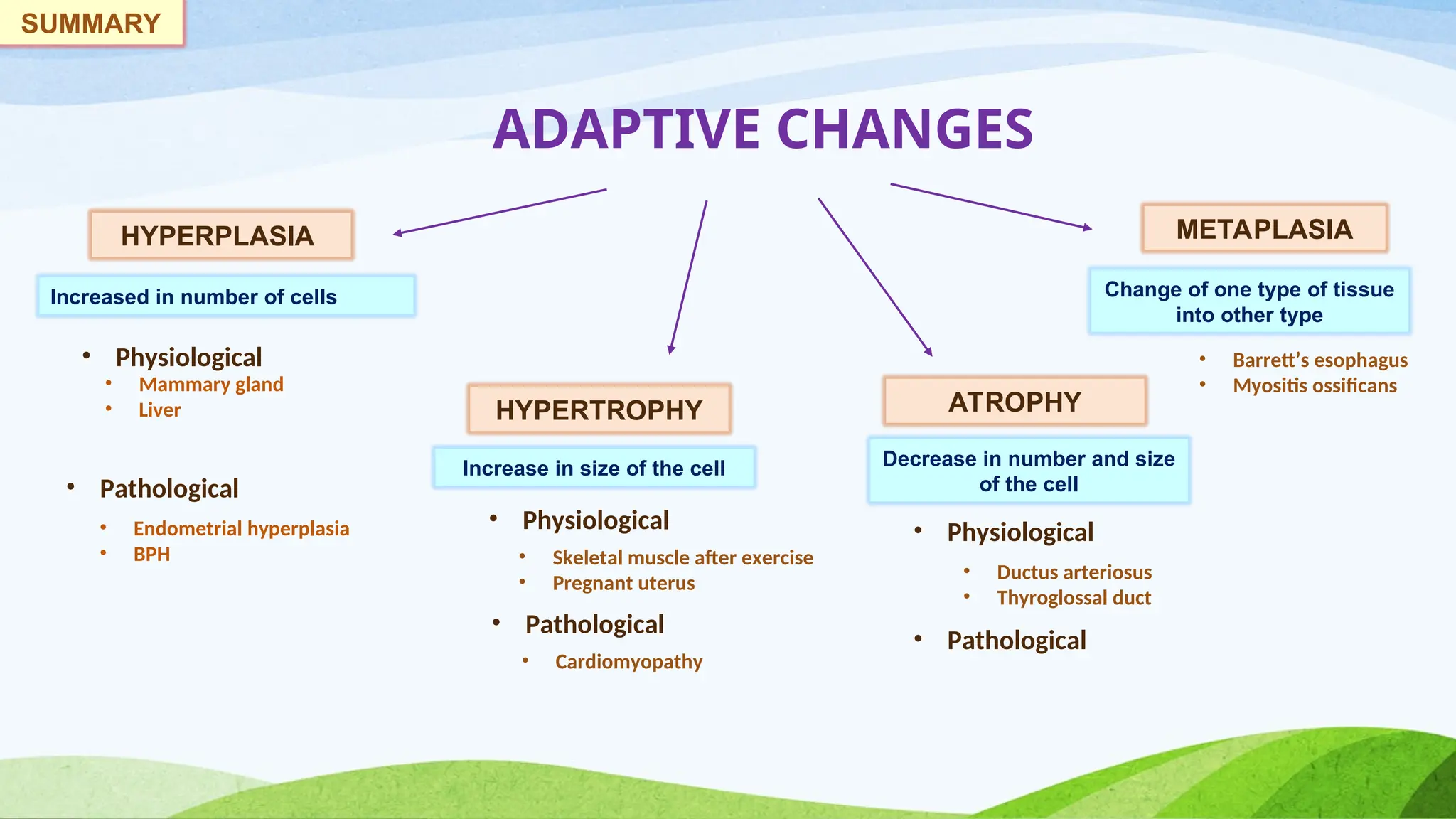
ADAPTIVE CHANGES
HYPERPLASIA
HYPERTROPHY ATROPHY
METAPLASIA
Increasedin number of cells
Increase in size of the cell Decrease in number and size
of the cell
Change of one type of tissue
into other type
• Physiological
• Pathological
• Mammary gland
• Liver
• Endometrial hyperplasia
• BPH
• Physiological
• Pathological
• Pathological
• Physiological
• Skeletal muscle after exercise
• Pregnant uterus
• Cardiomyopathy
• Ductus arteriosus
• Thyroglossal duct
• Barrett’s esophagus
• Myositis ossificans
SUMMARY
31.
THANK YOU
The realmeasure of
your wealth is how
much you would be
worth if you lost all your
money
Editor's Notes
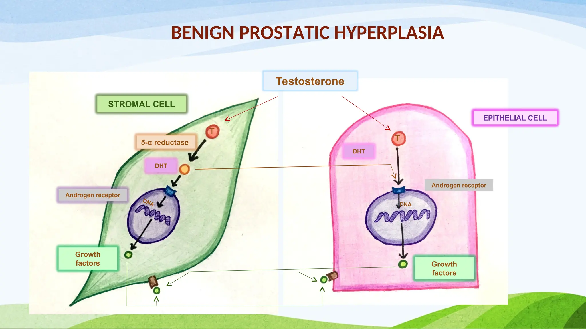
#12 This occurs due to androgenic (testosterone ) stimulation of prostate in elderly male patients
Testosterone in blood 5 α reductase in stromal cells of prostate Dihydrotestosterone (DHT) Acts on the stromal cells and epithelial cells of the prostate proliferation of the cells BPH
DHT is 10 times more potent than testosterone


![CELLULAR ADAPTATION AND ABERRANT CELL GROWTH [Autosaved].pptx](https://cdn.slidesharecdn.com/ss_thumbnails/cellularadaptationandaberrantcellgrowthautosaved-240328115316-03dc0e35-thumbnail.jpg?width=640&height=640&fit=bounds)


