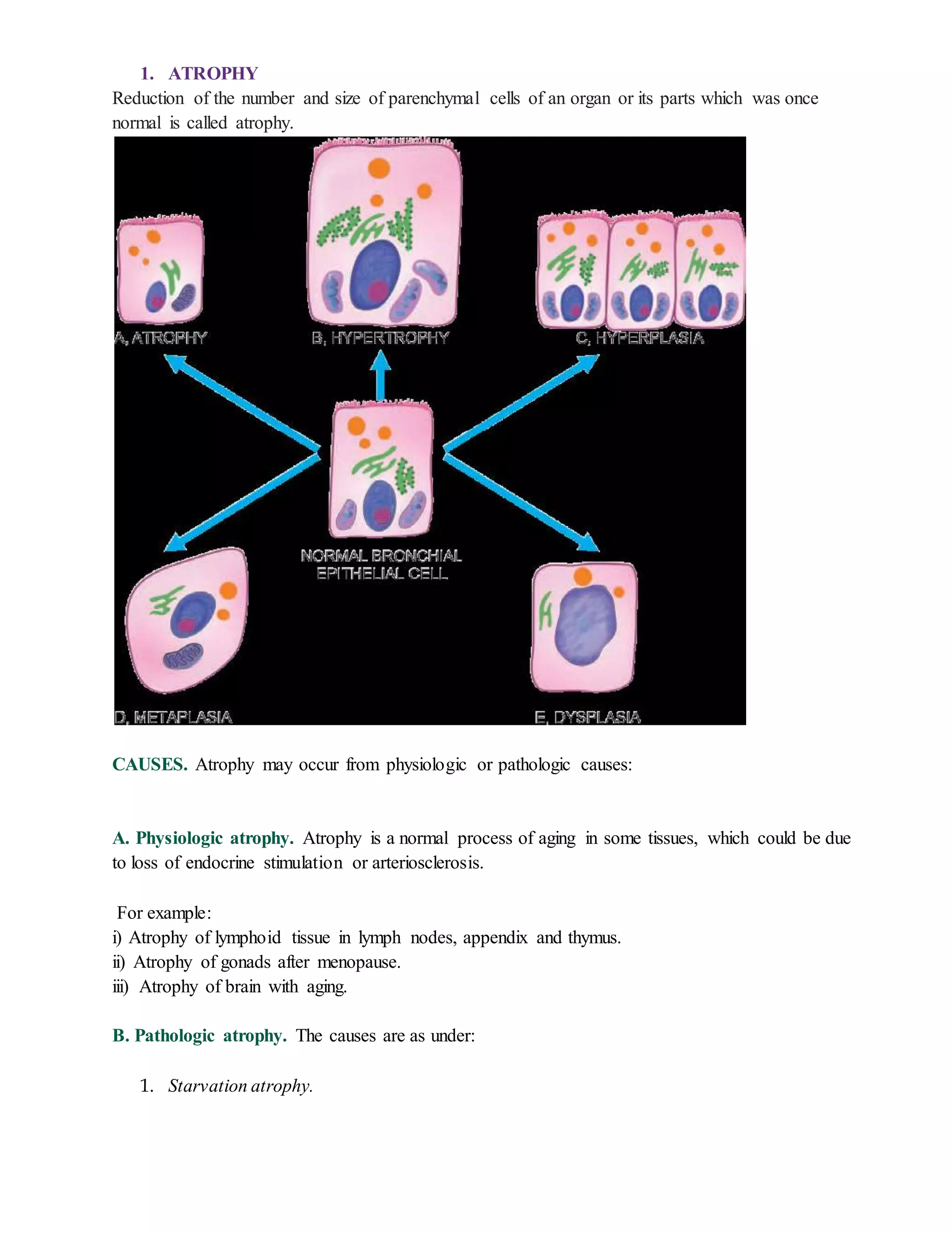
1. ATROPHY
Reduction of the number and size of parenchymal cells of an organ or its parts which was once
normal is called atrophy.
CAUSES. Atrophy may occur from physiologic or pathologic causes:
A. Physiologic atrophy. Atrophy is a normal process of aging in some tissues, which could be due
to loss of endocrine stimulation or arteriosclerosis.
For example:
i) Atrophy of lymphoid tissue in lymph nodes, appendix and thymus.
ii) Atrophy of gonads after menopause.
iii) Atrophy of brain with aging.
B. Pathologic atrophy. The causes are as under:
1. Starvation atrophy.
2. Ischaemic atrophy. Gradual diminution of blood supply due to atherosclerosis may result
in shrinkage of the affected organ.
3. Disuse atrophy. Prolonged diminished functional activity is associated with disuse
atrophy of the organ
4. Neuropathic atrophy. Interruption in nerve supply leads to wasting of muscles
5. Endocrine atrophy. Loss of endocrine regulatory mechanism results in reduced metabolic
activity of tissues and hence atrophy.
2. HYPERTROPHY
Hypertrophy is an increase in the size of parenchymal cells resulting in enlargement of
the organ or tissue, without any change in the number of cells.
CAUSES. Hypertrophy may be physiologic or pathologic. In both cases, it is caused either by
increased functional demand or by hormonal stimulation.
A. Physiologic hypertrophy. Enlarged size of the uterus in pregnancy is an excellent example of
physiologic
hypertrophy as well as hyperplasia.
B. Pathologic hypertrophy. Examples of certain diseases associated with hypertrophy are as
under:
1. Hypertrophy of cardiac muscle may occur in a number of cardiovascular diseases.
2. Hypertrophy of smooth muscle
3. HYPERPLASIA Hyperplasia is an increase in the number of parenchymal cells
resulting in enlargement of the organ or tissue. Quite often, both hyperplasia and
hypertrophy occur together.
CAUSES.
A. Physiologic hyperplasia. The two most common types are as follows:
1. Hormonal hyperplasia i.e. hyperplasia occurring under the influence of hormonal stimulation
e.g.
i) Hyperplasia of female breast at puberty, during pregnancy and lactation.
ii) Hyperplasia of pregnant uterus.
iii) Proliferative activity of normal endometrium after a normal menstrual cycle.
iv) Prostatic hyperplasia in old age.
2. Compensatory hyperplasia i.e. hyperplasia occurring following removal of part of an organ or
a contralateral organ in paired organ e.g.
i) Regeneration of the liver following partial hepatectomy
ii) Regeneration of epidermis after skin abrasion
B. Pathologic hyperplasia. Most examples of pathologic hyperplasia are due to excessive
stimulation of hormones or growth factors e.g.
i) Endometrial hyperplasia following oestrogen excess.
ii) In wound healing, there is formation of granulation tissue due to proliferation of fibroblasts
and endothelial cells.
iii) Formation of skin warts from hyperplasia of epidermis due to human papilloma virus.
iv) Pseudocarcinomatous hyperplasia of the skin.
v) Intraductal epithelial hyperplasia in the breast in fibrocystic breast disease.
4. METAPLASIA : Metaplasia is defined as a reversible change of one type of epithelial or
mesenchymal adult cells to another type of adult epithelial or mesenchymal cells, usually
in response to abnormal stimuli, and often reverts back to normal on removal of stimulus.
 Metaplasia is broadly divided into 2 types: epithelial and mesenchymal.
5. DYSPLASIAL: Dysplasia means ‘disordered cellular development’, often accompanied
with metaplasia and hyperplasia; it is therefore also referred to as atypical hyperplasia.
Dysplasia occurs most often in epithelial cells.
Epithelial dysplasia is characterized by cellular proliferation and cytologic changes.
These changes include:
1. Increased number of layers of epithelial cells
2. Disorderly arrangement of cells from basal layer to the surface layer
3. Loss of basal polarity i.e. nuclei lying away from basement membrane
4. Cellular and nuclear pleomorphism
5. Increased nucleocytoplasmic ratio
6. Nuclear hyperchromatism
7. Increased mitotic activity.

Growth disturbances

  • 1.
    1. ATROPHY Reduction ofthe number and size of parenchymal cells of an organ or its parts which was once normal is called atrophy. CAUSES. Atrophy may occur from physiologic or pathologic causes: A. Physiologic atrophy. Atrophy is a normal process of aging in some tissues, which could be due to loss of endocrine stimulation or arteriosclerosis. For example: i) Atrophy of lymphoid tissue in lymph nodes, appendix and thymus. ii) Atrophy of gonads after menopause. iii) Atrophy of brain with aging. B. Pathologic atrophy. The causes are as under: 1. Starvation atrophy.
  • 2.
    2. Ischaemic atrophy.Gradual diminution of blood supply due to atherosclerosis may result in shrinkage of the affected organ. 3. Disuse atrophy. Prolonged diminished functional activity is associated with disuse atrophy of the organ 4. Neuropathic atrophy. Interruption in nerve supply leads to wasting of muscles 5. Endocrine atrophy. Loss of endocrine regulatory mechanism results in reduced metabolic activity of tissues and hence atrophy. 2. HYPERTROPHY Hypertrophy is an increase in the size of parenchymal cells resulting in enlargement of the organ or tissue, without any change in the number of cells. CAUSES. Hypertrophy may be physiologic or pathologic. In both cases, it is caused either by increased functional demand or by hormonal stimulation. A. Physiologic hypertrophy. Enlarged size of the uterus in pregnancy is an excellent example of physiologic hypertrophy as well as hyperplasia. B. Pathologic hypertrophy. Examples of certain diseases associated with hypertrophy are as under: 1. Hypertrophy of cardiac muscle may occur in a number of cardiovascular diseases. 2. Hypertrophy of smooth muscle 3. HYPERPLASIA Hyperplasia is an increase in the number of parenchymal cells resulting in enlargement of the organ or tissue. Quite often, both hyperplasia and hypertrophy occur together. CAUSES. A. Physiologic hyperplasia. The two most common types are as follows: 1. Hormonal hyperplasia i.e. hyperplasia occurring under the influence of hormonal stimulation e.g. i) Hyperplasia of female breast at puberty, during pregnancy and lactation. ii) Hyperplasia of pregnant uterus.
  • 3.
    iii) Proliferative activityof normal endometrium after a normal menstrual cycle. iv) Prostatic hyperplasia in old age. 2. Compensatory hyperplasia i.e. hyperplasia occurring following removal of part of an organ or a contralateral organ in paired organ e.g. i) Regeneration of the liver following partial hepatectomy ii) Regeneration of epidermis after skin abrasion B. Pathologic hyperplasia. Most examples of pathologic hyperplasia are due to excessive stimulation of hormones or growth factors e.g. i) Endometrial hyperplasia following oestrogen excess. ii) In wound healing, there is formation of granulation tissue due to proliferation of fibroblasts and endothelial cells. iii) Formation of skin warts from hyperplasia of epidermis due to human papilloma virus. iv) Pseudocarcinomatous hyperplasia of the skin. v) Intraductal epithelial hyperplasia in the breast in fibrocystic breast disease. 4. METAPLASIA : Metaplasia is defined as a reversible change of one type of epithelial or mesenchymal adult cells to another type of adult epithelial or mesenchymal cells, usually in response to abnormal stimuli, and often reverts back to normal on removal of stimulus.  Metaplasia is broadly divided into 2 types: epithelial and mesenchymal. 5. DYSPLASIAL: Dysplasia means ‘disordered cellular development’, often accompanied with metaplasia and hyperplasia; it is therefore also referred to as atypical hyperplasia. Dysplasia occurs most often in epithelial cells. Epithelial dysplasia is characterized by cellular proliferation and cytologic changes. These changes include: 1. Increased number of layers of epithelial cells 2. Disorderly arrangement of cells from basal layer to the surface layer 3. Loss of basal polarity i.e. nuclei lying away from basement membrane 4. Cellular and nuclear pleomorphism 5. Increased nucleocytoplasmic ratio 6. Nuclear hyperchromatism 7. Increased mitotic activity.