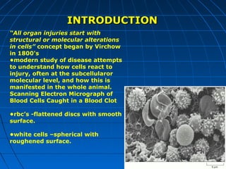
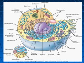
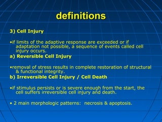
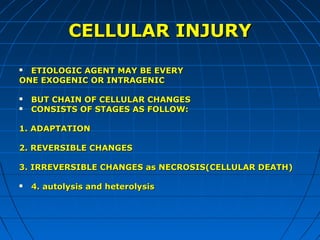
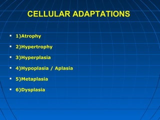
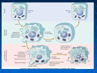
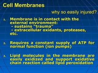
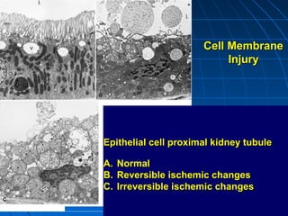
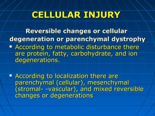
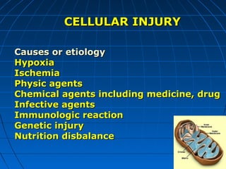
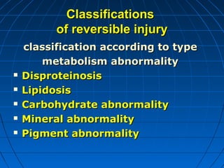
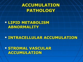
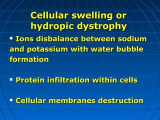
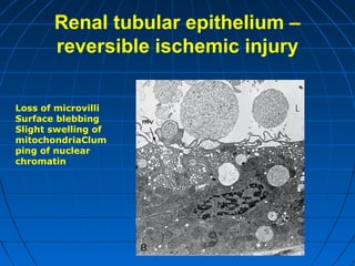
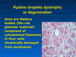
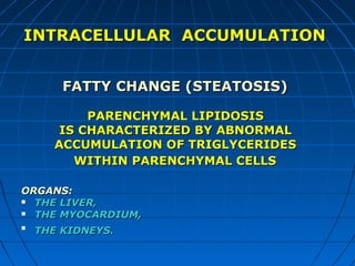
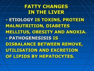
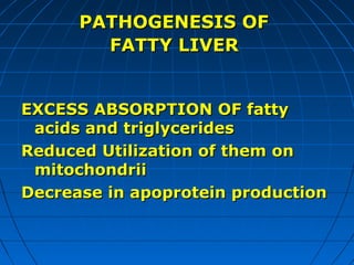
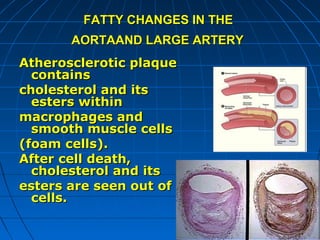
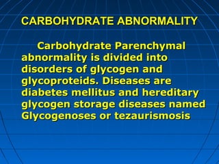
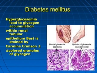
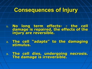
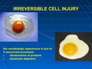
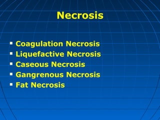
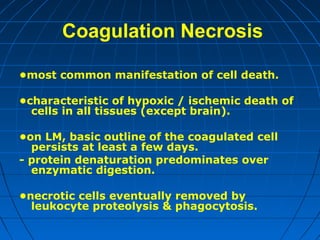
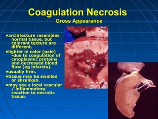
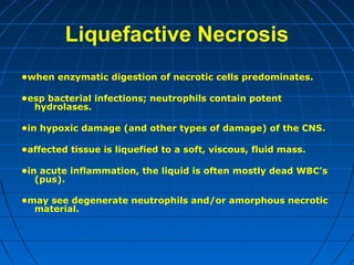
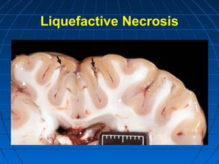
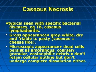
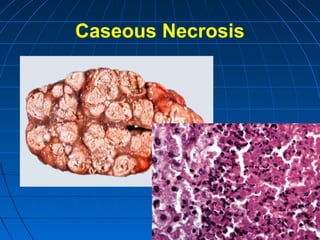
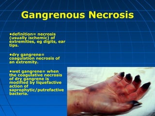
Cell & Tissue
Injury
Introduction






Every organism (body) consists
of organs
Every organ consists of tissues
Tissue consists of cells
Cell consists of ultrastructures
Introduction







There are tissues:
epithelial
mesenchymal tissue ( connective
tissue, fatty tissues, bones,
cartilages, muscles, vessels )
Nervous tissues
Bone marrow tissues
Lymphatic tissues
INTRODUCTION
“All organ injuries start with
structural or molecular alterations
in cells” concept began by Virchow
in 1800's
•modern study of disease attempts
to understand how cells react to
injury, often at the subcellularor
molecular level, and how this is
manifested in the whole animal.
Scanning Electron Micrograph of
Blood Cells Caught in a Blood Clot
•rbc’s -flattened discs with smooth
surface.
•white cells –spherical with
roughened surface.
INTRODUCTION
Structures of living systems are not
constant
They are destructured and restored
continuously
All living organisms absorb and
extract proteins, lipids (fats),
carbohydrates, and their
components as well as water, ions,
and pigments
definitions
1) Homeostasis
•cells maintain normal structure & function in response to
physiologic demands.
2) Cellular Adaptation
•as cells encounter stresses they undergo functional or
structural adaptations to maintain viability / homeostasis.
•respond to some stimuli by increasing or decreasing specific
organelle content.
•adaptive processes: atrophy, hypertrophy, hyperplasia and
metaplasia.
definitions
3) Cell Injury
•if limits of the adaptive response are exceeded or if
adaptation not possible, a sequence of events called cell
injury occurs.
a) Reversible Cell Injury
•removal of stress results in complete restoration of structural
& functional integrity.
b) Irreversible Cell Injury / Cell Death
•if stimulus persists or is severe enough from the start, the
cell suffers irreversible cell injury and death.
• 2 main morphologic patterns: necrosis & apoptosis.
CELLULAR INJURY
ETIOLOGIC AGENT MAY BE EVERY
ONE EXOGENIC OR INTRAGENIC





BUT CHAIN OF CELLULAR CHANGES
CONSISTS OF STAGES AS FOLLOW:

1. ADAPTATION
2. REVERSIBLE CHANGES
3. IRREVERSIBLE CHANGES as NECROSIS(CELLULAR DEATH)


4. autolysis and heterolysis
CELLULAR ADAPTATIONS


1)Atrophy



2)Hypertrophy



3)Hyperplasia



4)Hypoplasia / Aplasia



5)Metaplasia



6)Dysplasia
Cell Membranes
why so easily injured?
1.

2.

3.

Membrane is in contact with the
external environment:
- sustains “trauma”
- extracellular oxidants, proteases,
etc.
Requires a constant supply of ATP for
normal function (ion pumps)
Lipid molecules in the membrane are
easily oxidized and support oxidative
chain reaction called lipid peroxidation
Cell Membrane
Injury

Epithelial cell proximal kidney tubule
A. Normal
B. Reversible ischemic changes
C. Irreversible ischemic changes
CELLULAR INJURY
Reversible changes or cellular
degeneration or parenchymal dystrophy
 According to metabolic disturbance there
are protein, fatty, carbohydrate, and ion
degenerations.


According to localization there are
parenchymal (cellular), mesenchymal
(stromal- -vascular), and mixed reversible
changes or degenerations
CELLULAR INJURY
Causes or etiology
Hypoxia
Ischemia
Physic agents
Chemical agents including medicine, drug
Infective agents
Immunologic reaction
Genetic injury
Nutrition disbalance
CELLULAR INJURY









Mechanisms of reversible injury
Decomposition of membranes
Hyper infiltration of substances
(intracellular accumulation)
Unnatural syntheses
Disbalance of calcium metabolism
ATP depletion
Free radical- induced injury
induced injury
CELLULAR INJURY
Cellular injury depends on cell:
 1. type (myocardial cells dies in20- 30
min. ,but epidermis cells dies in
weeks,
after cause (etiologic agents ) acted.
 2.genetic makeup
 3.adaptability ( hepatic cells are more
adaptive cells, then neurons)
 4.status( normal or hypertrophic)
CELLULAR INJURY
Cellular injury depends on
injury:
 1. type (ischemia or infective
agent)
 2.its duration
 3.its severity
REVERSIBLE
Cellular injury









INTRACELLULAR RESPONSE INCLUDS
Aggregation of intramembranous particles
Endoplasmic reticulum swelling
Dispersion of ribosomes
Cell swelling
Clumping of nuclear chromatin
Mitochondrial swelling
Small densities within mitocondria
Mitochondrial Injury
Endoplasmic Reticulum Injury
Classifications
of reversible injury







classification according to type
metabolism abnormality
Disproteinosis
Lipidosis
Carbohydrate abnormality
Mineral abnormality
Pigment abnormality
ACCUMULATION
PATHOLOGY






LIPID METABOLISM
ABNORMALITY
INTRACELLULAR ACCUMULATION
STROMAL VASCULAR
ACCUMULATION
REVERSIBLE
Cellular injury






Intracellular accumulation
Cellular swelling or hydropic
dystrophy
Lipid accumulation
Glycogen accumulation
REVERSIBLE
Cellular injury
Classification according to cellular
disproteinosis
Cellular swelling or hydropic
(vacuole)
dystrophy or degeneration
 Hyaline droplet dystrophy or
degeneration
 Hyper keratinization

Cellular swelling or
hydropic dystrophy
Ions disbalance between sodium
and potassium with water bubble
formation




Protein infiltration within cells



Cellular membranes destruction
Renal tubular epithelium –
reversible ischemic injury
Loss of microvilli
Surface blebbing
Slight swelling of
mitochondriaClum
ping of nuclear
chromatin
Cellular swelling or
hydropic dystrophy
Diseases:
 Infective diseases
 Nephropathy
 Chronic glomerulonephritis
 Alcoholic disease
 Alzheimer disease
Cellular swelling or
hydropic dystrophy
Organs are as follow:





Kidney
Liver
Skin (epidermis)
Brain (neurons)
Cellular Swelling

•organ swollen with rounded edges.
•cut surface: tissue bulges and wet / heavy.
Cellular swelling or
hydropic dystrophy
The tubular vacuolization and
tubular dilation here
is a result of the
toxic effect of
ethyleneglycol
poisoning
Hyaline droplets dystrophy
or degeneration
Here are Mallory
bodies (the red
globular material)
composed of
cytoskeletal filaments
in liver cells
chronically damaged
from alcoholism
INTRACELLULAR ACCUMULATION
FATTY CHANGE (STEATOSIS)
PARENCHYMAL LIPIDOSIS
IS CHARACTERIZED BY ABNORMAL
ACCUMULATION OF TRIGLYCERIDES
WITHIN PARENCHYMAL CELLS
ORGANS:
 THE LIVER,
 THE MYOCARDIUM,


THE KIDNEYS.
FATTY CHANGES
IN THE LIVER
- ETIOLOGY IS TOXINS, PROTEIN
MALNUTRITION, DIABETES
MELLITUS, OBESITY AND ANOXIA.
- PATHOGENESISIS IS
DISBALANCE BETWEEN REMOVE,
UTILISATION AND EXCRETION
OF LIPIDS BY HEPATOCYTES.
PATHOGENESIS OF
FATTY LIVER
EXCESS ABSORPTION OF fatty
acids and triglycerides
Reduced Utilization of them on
mitochondrii
Decrease in apoprotein production
THE STAIN FOR LIPIDS IS
NAMED SUDAN THREE

Gross sample

Micro sample
h/e stained
FATTY LIVER

Hepatic liposis,
higher magnification.
The well-delineated
lipid filled
cytoplasmicvacuoles
causing swelling of
the hepatocytes,
usually pushing
nucleus to the
periphery of the cell.
Note, how the
vacuoles can be
single and large or
multiple and small.
FATTY CHANGES IN THE
HEART
ETIOLOGY HYPOXIA, INTOXICATION .
PATHOGENESIS LACK of OXYGEN LEAD TO
decreasing oxidative phosphorylation
anaerobic glycolysis decreasing ATP
synthesis mitochondria destruction
inhibition of fatty acid oxidation
toxins cause severe damage of
membranes and enzyme systems
Prolonged moderate hypoxia results in
focal intracellular fat deposits
APPEARANT BANDS OF
YELLOWED MYOCARDIUM
ALTERNATING WITH
BANDS OF DARKER, RED
BROWN UNINVOLVED HEART
TIGERET EFFECT´ ± TIGER
HEART

Severe fatty change is produced
by profound hypoxia with
diffused yellow±colored
myocardium
FATTY CHANGES IN THE
HEART
FATTY CHANGES IN THE
AORTAAND LARGE ARTERY
Atherosclerotic plaque
contains
cholesterol and its
esters within
macrophages and
smooth muscle cells
(foam cells).
After cell death,
cholesterol and its
esters are seen out of
cells.
CARBOHYDRATE ABNORMALITY
Carbohydrate Parenchymal
abnormality is divided into
disorders of glycogen and
glycoproteids. Diseases are
diabetes mellitus and hereditary
glycogen storage diseases named
Glycogenoses or tezaurismosis
Diabetes mellitus
Hyperglycosemia
lead to glycogen
accumulation
within renal
tubular
epithelium Best is
stained by
Carmine Crimson ±
±colored granules
of glycogen
Consequences of Injury
1.

2.

3.

No long term effects- - the cell
damage is repaired, the effects of the
injury are reversible.
The cell “adapts” to the damaging
stimulus.
The cell dies, undergoing necrosis.
The damage is irreversible.
IRREVERSIBLE CELL INJURY

the morphologic appearance is due to
2 concurrent processes:
1.
denaturation of proteins
2.
enzymatic digestion
Necrosis






Coagulation Necrosis
Liquefactive Necrosis
Caseous Necrosis
Gangrenous Necrosis
Fat Necrosis
Coagulation Necrosis
•most common manifestation of cell death.
•characteristic of hypoxic / ischemic death of
cells in all tissues (except brain).
•on LM, basic outline of the coagulated cell
persists at least a few days.
- protein denaturation predominates over
enzymatic digestion.
•necrotic cells eventually removed by
leukocyte proteolysis & phagocytosis.
Coagulation Necrosis
Gross Appearance
•architecture resembles
normal tissue, but
colorant texture are
different.
•lighter in color (pale)
-due to coagulation of
cytoplasmic proteins
and decreased blood
flow (eg infarcts).
•usually firm.
•tissue may be swollen
or shrunken.
•may see a local vascular
/ inflammatory
reaction to necrotic
tissue.
Coagulation Necrosis
Microscopic Appearance
•original cell shape &
tissue architecture is
Preserve die dead cells
resemble an eosinophilic
"shadow" of the original
cells.
•cytoplasm: increased
eosinophilia (H&E
stain)usually hyalinized
(homogeneous glassy
appearance) may be
mineralized.
a) Coagulation Necrosis
•nucleus:
1.
karyolysi
2.
pyknosis
3.
karyorrhexis
Karyorrhexis, lymphocytes, Lymphocyte nuclei have
fragmented (arrow). H&E stain.
Skeletal muscle
note coagulation necrosis of
myofibers characterized by
fragmentation and
hyalinization; also note
extensive mineralization
(blue-purple staining)
Liquefactive Necrosis
•when enzymatic digestion of necrotic cells predominates.
•esp bacterial infections; neutrophils contain potent
hydrolases.
•in hypoxic damage (and other types of damage) of the CNS.
•affected tissue is liquefied to a soft, viscous, fluid mass.
•in acute inflammation, the liquid is often mostly dead WBC’s
(pus).
•may see degenerate neutrophils and/or amorphous necrotic
material.
Liquefactive Necrosis
Caseous Necrosis
•typical seen with specific bacterial
diseases, eg TB, caseous
lymphadenitis.
 Gross appearance• grey-white, dry
and friable to pasty (caseous =
cheese like).
 Microscopic appearance• dead cells
persist as amorphous, coarsely
granular, eosinophilic debris.• don’t
retain cellular outline but don’t
undergo complete dissolution either.
Caseous Necrosis
Gangrenous Necrosis
•definition= necrosis
(usually ischemic) of
extremities, eg digits, ear
tips.
•dry gangrene=
coagulation necrosis of
an extremity.
•wet gangrene= when
the coagulative necrosis
of dry gangrene is
modified by liquefactive
action of
saprophytic/putrefactive
bacteria.
Fat Necrosis
•distinguished by its location in
body fat stores.

•etiology: inflammation (eg
pancreatitis), Vit E deficiency,
trauma, idiopathic
Fat Necrosis
Summary








All human disease occur because of
cell/tissue injury
Membranes-outer and mitochondrial
are key targets
Many early steps are reversible
Cell death follows going beyond a
point of no return -drop in pH, rise in
Ca2+
Thank you for your kind attention.

cell injury

  • 1.
  • 2.
    Introduction     Every organism (body)consists of organs Every organ consists of tissues Tissue consists of cells Cell consists of ultrastructures
  • 3.
    Introduction      There are tissues: epithelial mesenchymaltissue ( connective tissue, fatty tissues, bones, cartilages, muscles, vessels ) Nervous tissues Bone marrow tissues Lymphatic tissues
  • 4.
    INTRODUCTION “All organ injuriesstart with structural or molecular alterations in cells” concept began by Virchow in 1800's •modern study of disease attempts to understand how cells react to injury, often at the subcellularor molecular level, and how this is manifested in the whole animal. Scanning Electron Micrograph of Blood Cells Caught in a Blood Clot •rbc’s -flattened discs with smooth surface. •white cells –spherical with roughened surface.
  • 6.
    INTRODUCTION Structures of livingsystems are not constant They are destructured and restored continuously All living organisms absorb and extract proteins, lipids (fats), carbohydrates, and their components as well as water, ions, and pigments
  • 7.
    definitions 1) Homeostasis •cells maintainnormal structure & function in response to physiologic demands. 2) Cellular Adaptation •as cells encounter stresses they undergo functional or structural adaptations to maintain viability / homeostasis. •respond to some stimuli by increasing or decreasing specific organelle content. •adaptive processes: atrophy, hypertrophy, hyperplasia and metaplasia.
  • 8.
    definitions 3) Cell Injury •iflimits of the adaptive response are exceeded or if adaptation not possible, a sequence of events called cell injury occurs. a) Reversible Cell Injury •removal of stress results in complete restoration of structural & functional integrity. b) Irreversible Cell Injury / Cell Death •if stimulus persists or is severe enough from the start, the cell suffers irreversible cell injury and death. • 2 main morphologic patterns: necrosis & apoptosis.
  • 9.
    CELLULAR INJURY ETIOLOGIC AGENTMAY BE EVERY ONE EXOGENIC OR INTRAGENIC    BUT CHAIN OF CELLULAR CHANGES CONSISTS OF STAGES AS FOLLOW: 1. ADAPTATION 2. REVERSIBLE CHANGES 3. IRREVERSIBLE CHANGES as NECROSIS(CELLULAR DEATH)  4. autolysis and heterolysis
  • 10.
  • 12.
    Cell Membranes why soeasily injured? 1. 2. 3. Membrane is in contact with the external environment: - sustains “trauma” - extracellular oxidants, proteases, etc. Requires a constant supply of ATP for normal function (ion pumps) Lipid molecules in the membrane are easily oxidized and support oxidative chain reaction called lipid peroxidation
  • 13.
    Cell Membrane Injury Epithelial cellproximal kidney tubule A. Normal B. Reversible ischemic changes C. Irreversible ischemic changes
  • 14.
    CELLULAR INJURY Reversible changesor cellular degeneration or parenchymal dystrophy  According to metabolic disturbance there are protein, fatty, carbohydrate, and ion degenerations.  According to localization there are parenchymal (cellular), mesenchymal (stromal- -vascular), and mixed reversible changes or degenerations
  • 15.
    CELLULAR INJURY Causes oretiology Hypoxia Ischemia Physic agents Chemical agents including medicine, drug Infective agents Immunologic reaction Genetic injury Nutrition disbalance
  • 16.
    CELLULAR INJURY        Mechanisms ofreversible injury Decomposition of membranes Hyper infiltration of substances (intracellular accumulation) Unnatural syntheses Disbalance of calcium metabolism ATP depletion Free radical- induced injury induced injury
  • 17.
    CELLULAR INJURY Cellular injurydepends on cell:  1. type (myocardial cells dies in20- 30 min. ,but epidermis cells dies in weeks, after cause (etiologic agents ) acted.  2.genetic makeup  3.adaptability ( hepatic cells are more adaptive cells, then neurons)  4.status( normal or hypertrophic)
  • 18.
    CELLULAR INJURY Cellular injurydepends on injury:  1. type (ischemia or infective agent)  2.its duration  3.its severity
  • 19.
    REVERSIBLE Cellular injury        INTRACELLULAR RESPONSEINCLUDS Aggregation of intramembranous particles Endoplasmic reticulum swelling Dispersion of ribosomes Cell swelling Clumping of nuclear chromatin Mitochondrial swelling Small densities within mitocondria
  • 20.
  • 21.
  • 22.
    Classifications of reversible injury      classificationaccording to type metabolism abnormality Disproteinosis Lipidosis Carbohydrate abnormality Mineral abnormality Pigment abnormality
  • 23.
  • 24.
    REVERSIBLE Cellular injury    Intracellular accumulation Cellularswelling or hydropic dystrophy Lipid accumulation Glycogen accumulation
  • 25.
    REVERSIBLE Cellular injury Classification accordingto cellular disproteinosis Cellular swelling or hydropic (vacuole) dystrophy or degeneration  Hyaline droplet dystrophy or degeneration  Hyper keratinization 
  • 26.
    Cellular swelling or hydropicdystrophy Ions disbalance between sodium and potassium with water bubble formation   Protein infiltration within cells  Cellular membranes destruction
  • 27.
    Renal tubular epithelium– reversible ischemic injury Loss of microvilli Surface blebbing Slight swelling of mitochondriaClum ping of nuclear chromatin
  • 28.
    Cellular swelling or hydropicdystrophy Diseases:  Infective diseases  Nephropathy  Chronic glomerulonephritis  Alcoholic disease  Alzheimer disease
  • 29.
    Cellular swelling or hydropicdystrophy Organs are as follow:     Kidney Liver Skin (epidermis) Brain (neurons)
  • 30.
    Cellular Swelling •organ swollenwith rounded edges. •cut surface: tissue bulges and wet / heavy.
  • 31.
    Cellular swelling or hydropicdystrophy The tubular vacuolization and tubular dilation here is a result of the toxic effect of ethyleneglycol poisoning
  • 32.
    Hyaline droplets dystrophy ordegeneration Here are Mallory bodies (the red globular material) composed of cytoskeletal filaments in liver cells chronically damaged from alcoholism
  • 33.
    INTRACELLULAR ACCUMULATION FATTY CHANGE(STEATOSIS) PARENCHYMAL LIPIDOSIS IS CHARACTERIZED BY ABNORMAL ACCUMULATION OF TRIGLYCERIDES WITHIN PARENCHYMAL CELLS ORGANS:  THE LIVER,  THE MYOCARDIUM,  THE KIDNEYS.
  • 34.
    FATTY CHANGES IN THELIVER - ETIOLOGY IS TOXINS, PROTEIN MALNUTRITION, DIABETES MELLITUS, OBESITY AND ANOXIA. - PATHOGENESISIS IS DISBALANCE BETWEEN REMOVE, UTILISATION AND EXCRETION OF LIPIDS BY HEPATOCYTES.
  • 35.
    PATHOGENESIS OF FATTY LIVER EXCESSABSORPTION OF fatty acids and triglycerides Reduced Utilization of them on mitochondrii Decrease in apoprotein production
  • 36.
    THE STAIN FORLIPIDS IS NAMED SUDAN THREE Gross sample Micro sample h/e stained
  • 37.
    FATTY LIVER Hepatic liposis, highermagnification. The well-delineated lipid filled cytoplasmicvacuoles causing swelling of the hepatocytes, usually pushing nucleus to the periphery of the cell. Note, how the vacuoles can be single and large or multiple and small.
  • 38.
    FATTY CHANGES INTHE HEART ETIOLOGY HYPOXIA, INTOXICATION . PATHOGENESIS LACK of OXYGEN LEAD TO decreasing oxidative phosphorylation anaerobic glycolysis decreasing ATP synthesis mitochondria destruction inhibition of fatty acid oxidation toxins cause severe damage of membranes and enzyme systems
  • 39.
    Prolonged moderate hypoxiaresults in focal intracellular fat deposits APPEARANT BANDS OF YELLOWED MYOCARDIUM ALTERNATING WITH BANDS OF DARKER, RED BROWN UNINVOLVED HEART TIGERET EFFECT´ ± TIGER HEART Severe fatty change is produced by profound hypoxia with diffused yellow±colored myocardium
  • 40.
  • 41.
    FATTY CHANGES INTHE AORTAAND LARGE ARTERY Atherosclerotic plaque contains cholesterol and its esters within macrophages and smooth muscle cells (foam cells). After cell death, cholesterol and its esters are seen out of cells.
  • 42.
    CARBOHYDRATE ABNORMALITY Carbohydrate Parenchymal abnormalityis divided into disorders of glycogen and glycoproteids. Diseases are diabetes mellitus and hereditary glycogen storage diseases named Glycogenoses or tezaurismosis
  • 43.
    Diabetes mellitus Hyperglycosemia lead toglycogen accumulation within renal tubular epithelium Best is stained by Carmine Crimson ± ±colored granules of glycogen
  • 44.
    Consequences of Injury 1. 2. 3. Nolong term effects- - the cell damage is repaired, the effects of the injury are reversible. The cell “adapts” to the damaging stimulus. The cell dies, undergoing necrosis. The damage is irreversible.
  • 45.
    IRREVERSIBLE CELL INJURY themorphologic appearance is due to 2 concurrent processes: 1. denaturation of proteins 2. enzymatic digestion
  • 46.
  • 47.
    Coagulation Necrosis •most commonmanifestation of cell death. •characteristic of hypoxic / ischemic death of cells in all tissues (except brain). •on LM, basic outline of the coagulated cell persists at least a few days. - protein denaturation predominates over enzymatic digestion. •necrotic cells eventually removed by leukocyte proteolysis & phagocytosis.
  • 48.
    Coagulation Necrosis Gross Appearance •architectureresembles normal tissue, but colorant texture are different. •lighter in color (pale) -due to coagulation of cytoplasmic proteins and decreased blood flow (eg infarcts). •usually firm. •tissue may be swollen or shrunken. •may see a local vascular / inflammatory reaction to necrotic tissue.
  • 49.
    Coagulation Necrosis Microscopic Appearance •originalcell shape & tissue architecture is Preserve die dead cells resemble an eosinophilic "shadow" of the original cells. •cytoplasm: increased eosinophilia (H&E stain)usually hyalinized (homogeneous glassy appearance) may be mineralized. a) Coagulation Necrosis •nucleus: 1. karyolysi 2. pyknosis 3. karyorrhexis
  • 50.
    Karyorrhexis, lymphocytes, Lymphocytenuclei have fragmented (arrow). H&E stain.
  • 51.
    Skeletal muscle note coagulationnecrosis of myofibers characterized by fragmentation and hyalinization; also note extensive mineralization (blue-purple staining)
  • 52.
    Liquefactive Necrosis •when enzymaticdigestion of necrotic cells predominates. •esp bacterial infections; neutrophils contain potent hydrolases. •in hypoxic damage (and other types of damage) of the CNS. •affected tissue is liquefied to a soft, viscous, fluid mass. •in acute inflammation, the liquid is often mostly dead WBC’s (pus). •may see degenerate neutrophils and/or amorphous necrotic material.
  • 53.
  • 54.
    Caseous Necrosis •typical seenwith specific bacterial diseases, eg TB, caseous lymphadenitis.  Gross appearance• grey-white, dry and friable to pasty (caseous = cheese like).  Microscopic appearance• dead cells persist as amorphous, coarsely granular, eosinophilic debris.• don’t retain cellular outline but don’t undergo complete dissolution either.
  • 55.
  • 56.
    Gangrenous Necrosis •definition= necrosis (usuallyischemic) of extremities, eg digits, ear tips. •dry gangrene= coagulation necrosis of an extremity. •wet gangrene= when the coagulative necrosis of dry gangrene is modified by liquefactive action of saprophytic/putrefactive bacteria.
  • 57.
    Fat Necrosis •distinguished byits location in body fat stores. •etiology: inflammation (eg pancreatitis), Vit E deficiency, trauma, idiopathic
  • 58.
  • 59.
    Summary     All human diseaseoccur because of cell/tissue injury Membranes-outer and mitochondrial are key targets Many early steps are reversible Cell death follows going beyond a point of no return -drop in pH, rise in Ca2+
  • 60.
    Thank you foryour kind attention.