Downloaded 141 times


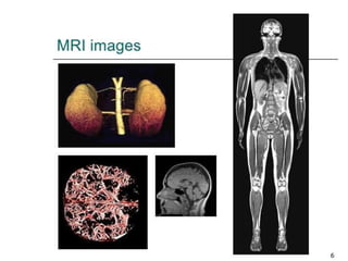


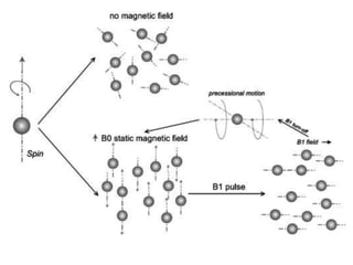
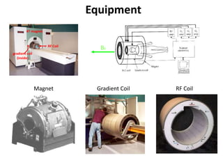
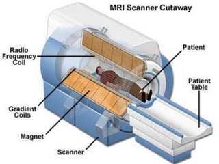

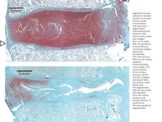

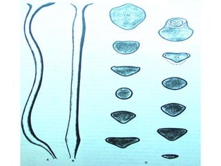
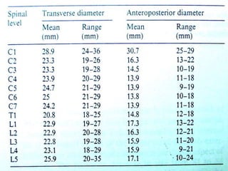
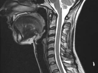
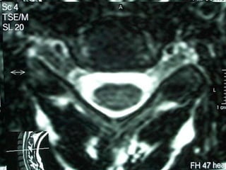

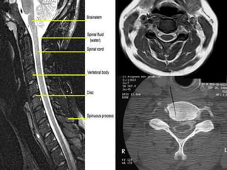
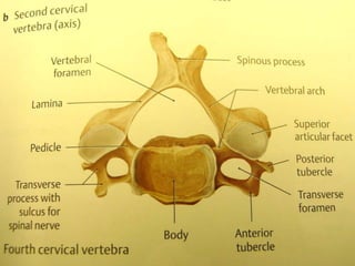
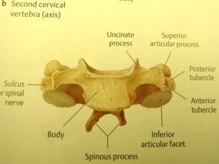
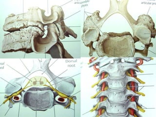
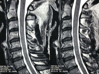
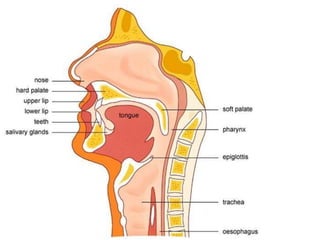
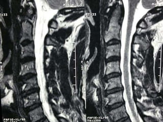
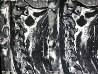
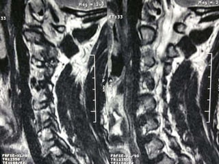
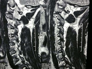
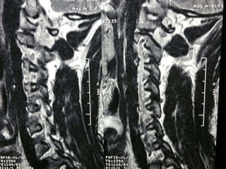
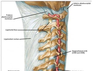
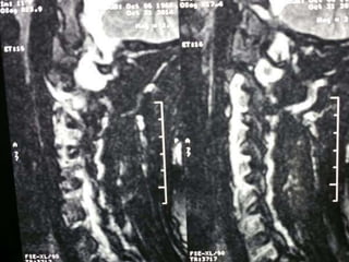
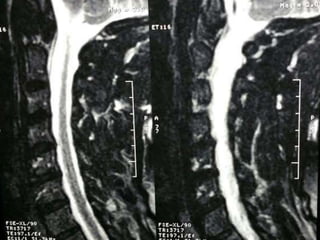
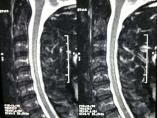
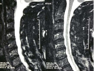
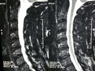
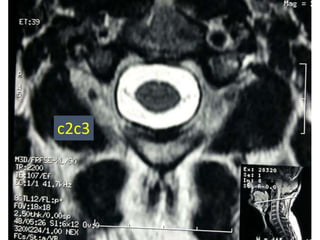
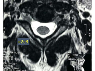
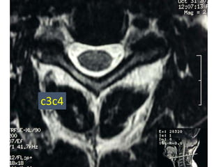
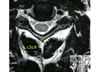
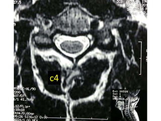
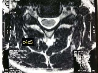
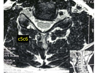
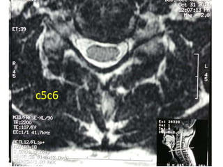
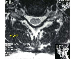
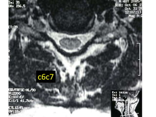
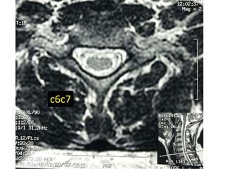
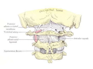

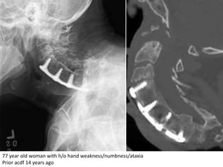
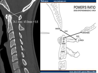
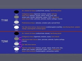
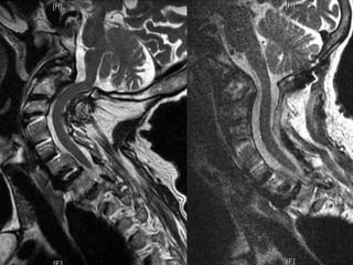

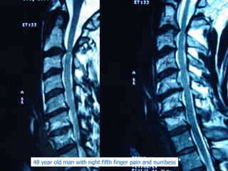
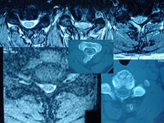
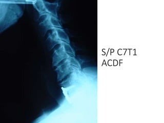

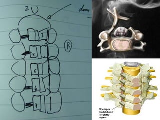
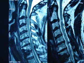
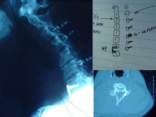

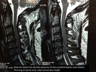
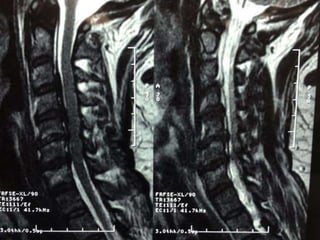
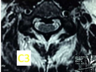
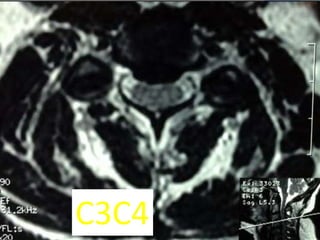
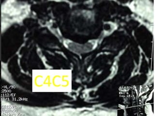
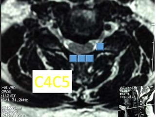
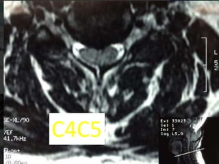
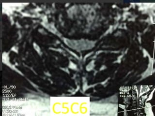
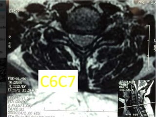
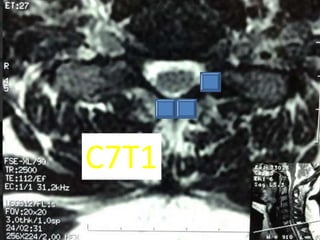
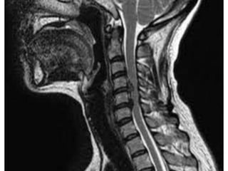
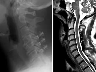
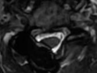


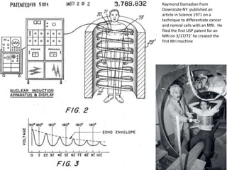

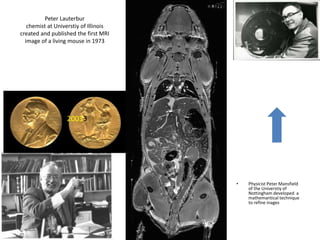
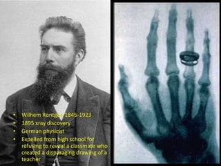
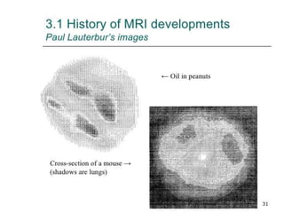

This document provides information on how to read a cervical MRI. It begins with background on MRI equipment such as the magnet, gradient coil, and RF coil. It then discusses the anatomy of intervertebral disks and how degeneration appears on MRI. The remainder of the document reviews several patient case examples, noting relevant imaging findings and clinical histories. Key details like disc height, spinal cord measurements, prior surgeries, and symptoms are highlighted for each case. A brief history of MRI development is also included, mentioning early pioneers like Damadian, Lauterbur, and Mansfield.


