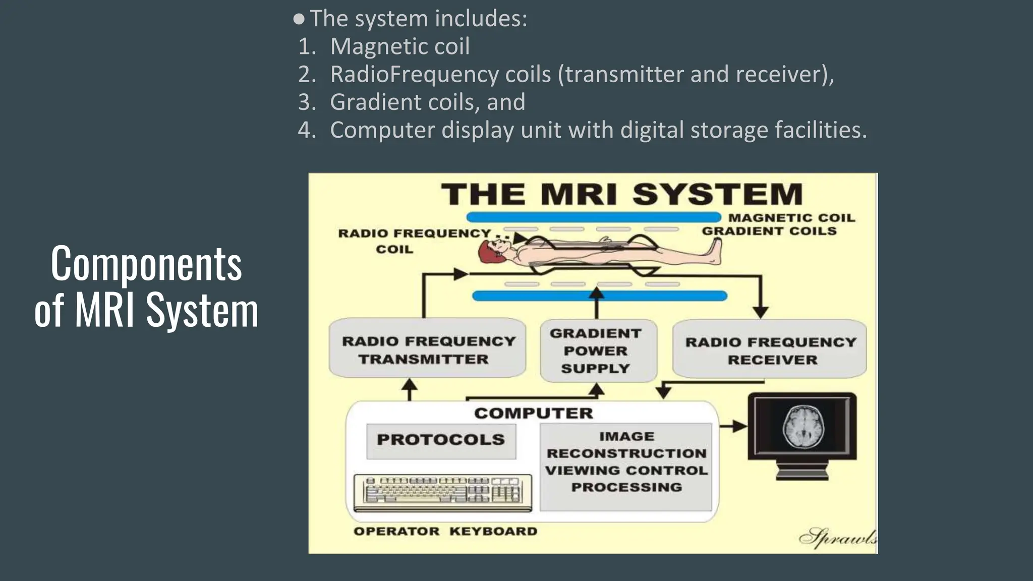
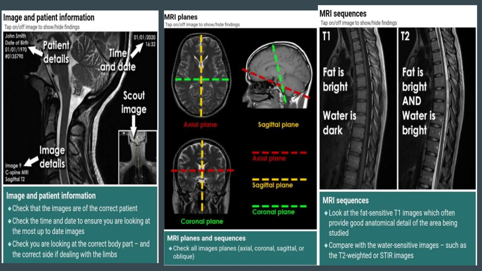
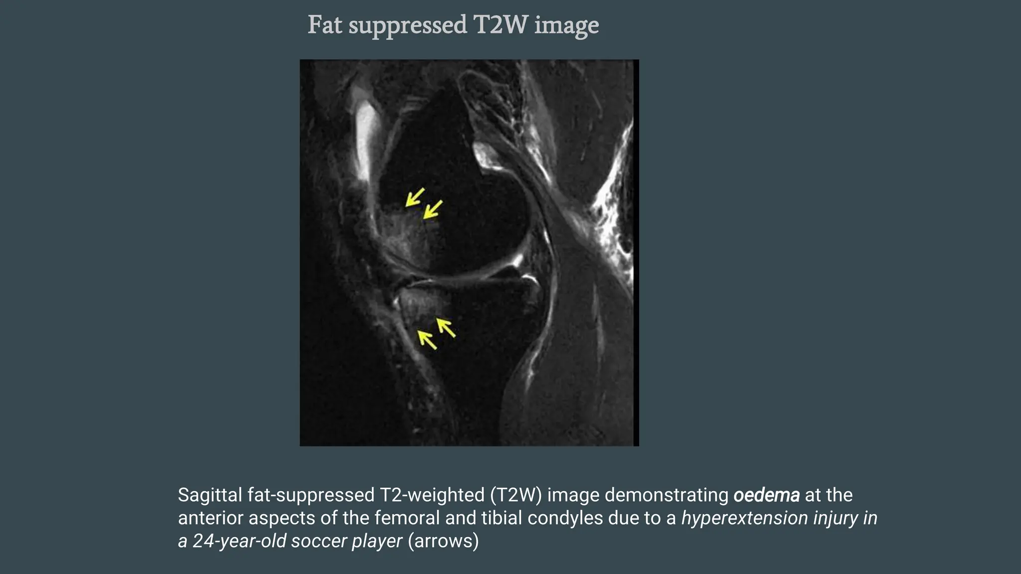
SlideShare a Scribd company logo
1 of 80
MRI
IN ORTHOPAEDICS
MODERATOR:DR.SANTOSH
PRESENTER:DR.MD AAMIR MALLIK
MAGNETIC
RESONANCE
IMAGING
(MRI)
●Introduction
●Components of MRI scanner system
●Basic principle of MRI
●What are MRI images?
●MRI signal production
●T1 vs T2 Images
●Specialised MRI sequences
●Contrast agents
●Systematic approach
●MRI safety and limitations
Introduction
●It is a technique that uses a magnetic field and radiofrequency
waves to create detailed images of the organs and tissues
within the body.
●Nikola tesla invented the rotating magnetic field in 1882 in
Hungary.
●All MRI machines are caliberated in “Tesla” units.
●The strength of a magnetic field is measured in Tesla or Gauss
units.
Components
of MRI System
●The system includes:
1. Magnetic coil
2. RadioFrequency coils (transmitter and receiver),
3. Gradient coils, and
4. Computer display unit with digital storage facilities.
Basic Principle
of MRI
●The ability of MRI to image body parts depends on the intrinsic spin of
atomic nuclei with an odd number of protons and/or neutrons (e.g.,
hydrogen), thus generating a magnetic moment. Atomic nuclei of tissues
placed within the main magnetic field from the usual random alignment of
their magnetic poles tend to align along the direction of that field.
●The application of RF pulses causes the nuclei to absorb energy and induces
resonance of particular sets of nuclei, which causes their orientation to the
magnetic field.
●The required frequency of the pulse is determined by the strength of the
magnetic field and the particular nucleus undergoing investigation.
●When the RF field is removed, the energy absorbed during the transition from a high to
low-energy state is subsequently released, and this can be recorded as an electrical
signal that provides the data from which digital images are derived.
●Signal intensity refers to the strength of the radio wave that a tissue emits after
excitation. The strength of this radio wave determines the degree of brightness of the
imaged structures.
MRI signal
production
(Principle)
What are MRI
Images?
●X-ray and CT images can be considered to be a map of density of tissues
in the body
●MRI images can be considered as a map of proton energy within tissues
of the body. A variety of MRI images can be produced which emphasise
different tissue types, in particular those that contain a large amount of fat
or water.
●Bright areas on an MRI image represent high ‘signal’ given off by protons
in the body during the scanning process.
●White areas on an X-ray or CT image = high density structures
●White areas on an MRI image = high signal intensity structures
Systematic
approach to
interpretation
of MRI images
●Check image and patient details over the film
●Check MRI planes
●Check MRI sequences(T1,T2,STIR)
●Check for abnormal MRI signal
●Compare with previous radiologic images(MRI/X-
ray/CT/USG)
●Co-relate with clinical findings
T1 vs T2
Images
●It’s all about FAT and WATER
●The two basic types of MRI images are T1-weighted
and T2-weighted images, often referred to as T1 and
T2 images.
●The timing of radiofrequency pulse sequences used to
make T1 images results in images which highlight fat
tissue within the body.
●The timing of radiofrequency pulse sequences used to
make T2 images results in images which highlight
fat AND water within the body.
●So, this makes things easy to remember.
●T1 images – 1 tissue type is bright – FAT
●T2 images – 2 tissue types are bright –
FAT and WATER
TISSUE T1 T2
Fluid Dark Bright(ww2)
Fat Bright Intermediate
Tendon/
ligament
Dark dark
Air Black Black
,
Specialised
MRI sequences
STIR(Short Tau
Inversion Recovery):
• STIR (Short Tau Inversion Recovery) images
are highly water-sensitive and the timing of
the pulse sequence used acts to suppress
signal coming from fatty tissues – so ONLY
WATER is bright
• A combination of standard T1 images and
STIR images can be compared to determine
the amount of fat or water within a body part
• In these MRI images abnormal signal is seen
in the vertebral bodies and intervertebral disc
• Abnormal low signal on the T1 image and
abnormal high signal on the STIR image –
indicates abnormal fluid
• These are typical appearances of
spondylodiscitis (also known as discitis)
Proton density weighted image
Preoperative magnetic resonance imaging depicted as a proton
density weighted coronal section showing a cartilage defect
Fat suppressed T2W image
Sagittal fat-suppressed T2-weighted (T2W) image demonstrating oedema at the
anterior aspects of the femoral and tibial condyles due to a hyperextension injury in
a 24-year-old soccer player (arrows)
Gradient echo(T2*) image
IDEAL gradient-recalled echo (GRE) image
shows bright synovial fluid (arrow)
MRI Arthrogram
•Fluid containing gadolinium has been injected into the
shoulder joint revealing displacement of the anterior glenoid
labrum in this patient with recurrent shoulder dislocation
•G = Glenoid of scapula
•H = Head of humerus
Advantages of
MRI
●Images can be obtained in any plane and provide
superb soft tissue contrast,anatomic detail and
simultaneous demonstration of bones and soft
tissues.As a result,it is the best single modality for
evaluating most types of musculoskeletal pathology.
●The most useful modality for detecting marrow
pathology(neoplastic marrow infiltration,bone
contusion,occult fracture,tumour).
●The test of choice for evaluating neurologic deficits
related to spinal trauma and neoplasm.
●Can be used in combination with contrast
agents(gadolinium)
●No ionising radiation.
Uses of MRI
●MRI excels in the display of soft tissue detail.It is widely
used in the diagnosis of sports injuries because of its ability
to differentiate b/w the different soft tissues and distinguish
partial tears of tendons and ligaments from complete tears.
●MRI is very sensitive for detecting changes and variations in
bone marrow. This is important in the diagnosis of bone
tumors,stress fractures and avascular necrosis.
●In some cases,MRI has replaced invasive diagnostic
procedures such as arthroscopy ,as in the detection of
meniscal tears.
●MRI is the best modality for evaluation of disc herniations
and other potential causes of nerve root impingement.
●MRI has the ability to stage neoplasms in bone and soft
tissues as well as evaluate the extent of tissue invasion
before surgery.It is more sensitive than bone scan for
detecting bone metastases,although bone scan is more
effective as a screening technique.
General Spine Anatomy
Intervertebral Disc complex has three components:
● Cartilaginous end plate
● Annulus fibrosus
● Nucleus pulposus
● intervertebral discs show intermediate signal intensity on T1-weighted images and
high signal intensity on T2-weighted images.The outer annulus appears
hypointense on T2.
● As the disc degenerates and as patients age, the signal intensity of the
nucleus pulposus decreases.
● The ligamentum flavum connects the lamina of adjacent vertebrae and
is seen as a hypointense band posterior to the dura.
● nerve roots have intermediate signal intensity and are surrounded by
high signal intensity fat on T1-weighted images and by high signal
intensity CSF on T2-weighted images..
MRI Spine indications:
▪Evaluation of intervertebral disk disease
▪Evaluation of nerve roots, posterior longitudinal
ligament and intervertebral foramen
▪Postoperative back pain (failed-back surgery)
▪Pyogenic and tuberculous infections
▪Epidural hematoma or abscess
▪Primary and metastatic tumors of the vertebral column
▪Spinal cord injury
▪Spinal cord tumors (extradural and intradural)
▪Abnormalities of thecal sac
▪Developmental deformities of the spinal cord and
vertebral column.
CERVICAL SPINE
AXIAL
VIEW
SAGITTAL VIEW
THORACIC SPINE
AXIAL
VIEW
SAGITTAL
VIEW
SAGITTAL IMAGE
Mid
sagittal
Parasagital
Foraminal sagital
Lumbar Spine
● The midsagittal
image should be
evaluated first,then
sequentially
evaluated toward
each side to assess
the facet joints and
neural foramina.
● sagittal images can
be concurrently
evaluated with the
axial sequence to
confirm laterality.
AXIAL
IMAGES
● the degree of
contribution of the
three primary
contributors to
spinal stenosis (disc
pathology, facet
arthropathy, and
ligamentum flavum
hypertrophy) should
be noted
● Herniation is defined as a
localized displacement of disc
contents beyond the borders
of the intervertebral disc
space
● disc material may include
● nucleus
● cartilage
● fragmented apophyseal bone
● annular tissue or a
combination
DISC DEGENERATION DISEASE
Disc protrusion Disc extrusion
Lumbar disc protrusion. Sagittal (A) and axial (B) T2-weighted images
showing a central disc protrusion at the L4-L5 level (arrow
on each).
Disc herniation
Lumbar Spinal Stenosis
● compression
of the neural
elements in
the spinal
canal, lateral
recesses, or
neural
foramina.
Spinal canal stenosis
TB SPINE
●Meniscal lesion
●Cruciate ligament tears
●Collateral ligament tears
●Cartilage lesions: arthritis,osteochondritis dissecans
●Inflammatory changes: Synovitis
●Changes in bone: oedema, necrosis, fracture
●Cysts : baker cyst
●Tumors
●Intra-articular loose bodies
●Soft tissue envelope: tear ,hematoma, tumor.
MRI Knee joint indications
ACL
● Course
● Composed of the antero-
medial and posterolateral
bundles.
● Intraarticular but
extrasynovial
● Should not show marked
increased signal on T2-
weighted imaging, but it
may have minimally
increased signal on T1-
weighted images because
of the presence of fatty
tissue
PCL
● PCL curves anteriorly
to insert on the
anterolateral aspect of
the medial femoral
condyle
● appears as a thicker,
darker, curved band
compared with the
ACL.
NORMAL MENISCI
low intensity signals on both T1w and T2w
• Mensici are best seen in sagittal view
• In central cuts-triangle,in peripheral cuts-Bow tie appearance
• Lesions-areas of increased signal
• Sensitivity and specificity of MRI >90%
▪ MENISCAL INJURIES:
Lesions are seen as areas of
increased signal and are
classified:
Grade 1- Globular signal
within the meniscus.
Grade 2- Linear signal within
the meniscus not reaching the
articular surface.
Grade 3- Linear signal within
the meniscus reaching the
articular surface. surface.
GRADE-1 GRADE-2 GRADE-3
● Most tears
involve the
medial
meniscus, but
most acute
tears involve
the lateral
meniscus
● chronic ACL
tears associated
with meniscus
tears.
posterior horn of the medial meniscus
medial meniscus and
displaced inferior to the
PCL (arrow-
head), exhibiting the
double-PCL sign
complete radial tear - empty meniscus sign
ACL TEARS
● H/o twisting or valgus
injury to the knee with a
planted foot and often
describes sensing a
“pop” inside the knee.
● adult ACL usually
avulses from its femoral
attachment or develops
an intrasubstance tear.
● The tendon does not
have to dissociate
completely to become
incompetent. In skeletally immature patients,
the ACL may remain intact and avulse a fragment of
bone off of the attachment.
normal
acute
avulsio
n
chronic
ACL
Tear
● sagittal fat-suppressed proton-density image shows
advanced degeneration (marked thickening) of the
ACL
Segond fracture
PCL Tear
▪ Seen as a curving band of
fibrous tissue appearing
homogeneously hypointense
on sagittal MRI series.
▪ Discontinuity of the ligament
or fluid signal within its
substance indicates a tear.
Partial PCL tear Complete PCL tear PCL avulsion
MRI Shoulder
NORMAL SHOULDER MRI
ROTATOR CUFF TEAR
•MR abnormalities
• Full-thickness tear: high SI on
T2WI
• Direct signs
Tendon discontinuity
Fluid signal in tendon gap
Retraction of
musculotendinous junction
• Associated findings
Subacromial/ subdeltoid
bursal fluid
Muscle atrophy
SUPRASPINATUS TEAR
Biceps tendon tear
Subscapularis Tendon Tear
•MRI:
• Detachment from lesser tuberosity
• Increased SI, thin/thick
• Contrast over the lesser tuberosity
Adhesive capsulitis (Frozen shoulder)
• Inflammatory process ->
progressive capsular retraction, scar
tissue
MRI
• Complete obliteration of the fat
triangle under the coracoid process
(subcoracoid triangle sign)
• Scar tissue
• Thickening of the CHL
• Axillary recess thickening
• Arthrography:
• Decreased joint capacity
• Small capsular recesses
• Serrated appearance of capsular
attachments
.
MRI Hip indications
• Avascular necrosis of femoral head
• Transient osteoporosis of the hip
• Occult fractures
• Evaluation of the acetabular labrum
• Avascular necrosis is the most
frequent indication for hip imaging.
Even when initial roentgenograms
show normal images MRI may be
positive for AVN.
• In T1-weighted images,intensity is
decreased in areas affected by
AVN. The surrounding ischemic
bone is seen as a low-signal band.
• In T2 images, a second inner band
of high intensity is seen which is
the “double line” sign diagnostic of
AVN
AVN of femoral head
Transient osteoporosis
• self-limited process of uncertain etiology related to reflex
sympathetic dystrophy and regional migratory
osteoporosis.
• MRI shows diffuse edema in the femoral head, extending
into the intertrochanteric region.
• This is seen as a low signal area onT1 when compared to the
normal bone marrow,
• whereas on T2
it becomes hyperintense.
Occult fractures
• MRI changes are apparent immediately, with linear areas of low
signal easily seen in the fatty marrow on T1- weighted images, and
surrounding edema seen with T2- weighted images
Contraindications
of MRI
1. Intra cerebral aneurysm clips
2. Internal hearing aids
3. Middle ear prosthesis
4. Cardiac pacemakers
5. 1st trimester pregnancy
6. Metallic orbital foreign bodies
Orthopaedic implants and prosthetics are
safe on MRI
Limitations of
MRI
●Fractures on the posterior elements of the
spine are difficult to detect with MRI(more
cortical bone)
●Assessment of fracture healing
●Hardware (depending on type,may produce
severe artifact,obscuring adjacent tissues.
●More time consuming than CT
●Claustrophobia
●Even the slight movements of the patient on
the examination bed can distort the MRI
image.
●More costlier than USG,CT.
MRI safety
warnings
REFERENCES
●Fundamentals of musculoskeletal imaging-
Lynn.N.Mckinnis
●Essential orthopaedics;Imaging of the
Musculoskeletal system-Mark.D.Miller
●Turek’s Orthopaedics;Imaging in
Orthopaedics
●Essential orthopaedics-Manish kumar
varshney
●www.radiologymasterclass.co.uk
“THANK YOU”

More Related Content

Similar to mri in orthopaedics its importance and understanding

Imaging in orthopaedics
Imaging  in  orthopaedicsImaging  in  orthopaedics
Imaging in orthopaedicsBipulBorthakur
 
diagnosticimaginginoromaxillofacialsurgery-141214161540-conversion-gate02.pptx
diagnosticimaginginoromaxillofacialsurgery-141214161540-conversion-gate02.pptxdiagnosticimaginginoromaxillofacialsurgery-141214161540-conversion-gate02.pptx
diagnosticimaginginoromaxillofacialsurgery-141214161540-conversion-gate02.pptxSamarElgendi
 
Introduction to musculoskeletal radiology
Introduction to musculoskeletal radiologyIntroduction to musculoskeletal radiology
Introduction to musculoskeletal radiologySubhanjan Das
 
Neuroimaging Lecture
Neuroimaging LectureNeuroimaging Lecture
Neuroimaging Lecturetest
 
Imaging in orthopaedics
Imaging  in  orthopaedicsImaging  in  orthopaedics
Imaging in orthopaedicsBipulBorthakur
 
1. MRI interpretation.docx
1. MRI interpretation.docx1. MRI interpretation.docx
1. MRI interpretation.docxssuserc88386
 
MRI in Orthopibohuvuvuvuvuvuvyvyvaedics.pptx
MRI in Orthopibohuvuvuvuvuvuvyvyvaedics.pptxMRI in Orthopibohuvuvuvuvuvuvyvyvaedics.pptx
MRI in Orthopibohuvuvuvuvuvuvyvyvaedics.pptxAarshRathod2
 
Radiology & Medical Imaging
Radiology & Medical ImagingRadiology & Medical Imaging
Radiology & Medical ImagingMadhuka Perera
 
Foundations of Diagnostic Imaging for Physical Therapist
Foundations of Diagnostic Imaging for Physical TherapistFoundations of Diagnostic Imaging for Physical Therapist
Foundations of Diagnostic Imaging for Physical TherapistDana Tew
 
Weightbearig CT Scans For Foot Ankle Surgery.pptx
Weightbearig CT Scans For Foot Ankle Surgery.pptxWeightbearig CT Scans For Foot Ankle Surgery.pptx
Weightbearig CT Scans For Foot Ankle Surgery.pptxAhmed Ashour dr.
 

Similar to mri in orthopaedics its importance and understanding (20)

Imaging in orthopaedics
Imaging  in  orthopaedicsImaging  in  orthopaedics
Imaging in orthopaedics
 
diagnosticimaginginoromaxillofacialsurgery-141214161540-conversion-gate02.pptx
diagnosticimaginginoromaxillofacialsurgery-141214161540-conversion-gate02.pptxdiagnosticimaginginoromaxillofacialsurgery-141214161540-conversion-gate02.pptx
diagnosticimaginginoromaxillofacialsurgery-141214161540-conversion-gate02.pptx
 
Introduction to musculoskeletal radiology
Introduction to musculoskeletal radiologyIntroduction to musculoskeletal radiology
Introduction to musculoskeletal radiology
 
Neuroimaging Lecture
Neuroimaging LectureNeuroimaging Lecture
Neuroimaging Lecture
 
Imaging in orthopaedics
Imaging  in  orthopaedicsImaging  in  orthopaedics
Imaging in orthopaedics
 
Imaging in sports injury
Imaging in sports injuryImaging in sports injury
Imaging in sports injury
 
Mri knee
Mri kneeMri knee
Mri knee
 
1. MRI interpretation.docx
1. MRI interpretation.docx1. MRI interpretation.docx
1. MRI interpretation.docx
 
MRI in orthopaedics
MRI in orthopaedicsMRI in orthopaedics
MRI in orthopaedics
 
MRI Brain
MRI BrainMRI Brain
MRI Brain
 
MRI in Orthopibohuvuvuvuvuvuvyvyvaedics.pptx
MRI in Orthopibohuvuvuvuvuvuvyvyvaedics.pptxMRI in Orthopibohuvuvuvuvuvuvyvyvaedics.pptx
MRI in Orthopibohuvuvuvuvuvuvyvyvaedics.pptx
 
MRI.pptx
MRI.pptxMRI.pptx
MRI.pptx
 
Ct and mri,ppt
Ct and mri,pptCt and mri,ppt
Ct and mri,ppt
 
Radiology & Medical Imaging
Radiology & Medical ImagingRadiology & Medical Imaging
Radiology & Medical Imaging
 
Foundations of Diagnostic Imaging for Physical Therapist
Foundations of Diagnostic Imaging for Physical TherapistFoundations of Diagnostic Imaging for Physical Therapist
Foundations of Diagnostic Imaging for Physical Therapist
 
MRI basics
MRI basicsMRI basics
MRI basics
 
Brain imaging in psychiatry
Brain imaging in psychiatryBrain imaging in psychiatry
Brain imaging in psychiatry
 
Oncology imaging
Oncology imagingOncology imaging
Oncology imaging
 
Weightbearig CT Scans For Foot Ankle Surgery.pptx
Weightbearig CT Scans For Foot Ankle Surgery.pptxWeightbearig CT Scans For Foot Ankle Surgery.pptx
Weightbearig CT Scans For Foot Ankle Surgery.pptx
 
3D-MRI Evaluation of the Anterolateral Ligament: An Evaluation of ACL Deficie...
3D-MRI Evaluation of the Anterolateral Ligament: An Evaluation of ACL Deficie...3D-MRI Evaluation of the Anterolateral Ligament: An Evaluation of ACL Deficie...
3D-MRI Evaluation of the Anterolateral Ligament: An Evaluation of ACL Deficie...
 

Recently uploaded

Best Rate (Hyderabad) Call Girls Jahanuma ⟟ 8250192130 ⟟ High Class Call Girl...
Best Rate (Hyderabad) Call Girls Jahanuma ⟟ 8250192130 ⟟ High Class Call Girl...Best Rate (Hyderabad) Call Girls Jahanuma ⟟ 8250192130 ⟟ High Class Call Girl...
Best Rate (Hyderabad) Call Girls Jahanuma ⟟ 8250192130 ⟟ High Class Call Girl...astropune
 
Russian Escorts Girls Nehru Place ZINATHI 🔝9711199012 ☪ 24/7 Call Girls Delhi
Russian Escorts Girls  Nehru Place ZINATHI 🔝9711199012 ☪ 24/7 Call Girls DelhiRussian Escorts Girls  Nehru Place ZINATHI 🔝9711199012 ☪ 24/7 Call Girls Delhi
Russian Escorts Girls Nehru Place ZINATHI 🔝9711199012 ☪ 24/7 Call Girls DelhiAlinaDevecerski
 
Lucknow Call girls - 8800925952 - 24x7 service with hotel room
Lucknow Call girls - 8800925952 - 24x7 service with hotel roomLucknow Call girls - 8800925952 - 24x7 service with hotel room
Lucknow Call girls - 8800925952 - 24x7 service with hotel roomdiscovermytutordmt
 
Call Girls Service Surat Samaira ❤️🍑 8250192130 👄 Independent Escort Service ...
Call Girls Service Surat Samaira ❤️🍑 8250192130 👄 Independent Escort Service ...Call Girls Service Surat Samaira ❤️🍑 8250192130 👄 Independent Escort Service ...
Call Girls Service Surat Samaira ❤️🍑 8250192130 👄 Independent Escort Service ...CALL GIRLS
 
Bangalore Call Girls Nelamangala Number 7001035870 Meetin With Bangalore Esc...
Bangalore Call Girls Nelamangala Number 7001035870  Meetin With Bangalore Esc...Bangalore Call Girls Nelamangala Number 7001035870  Meetin With Bangalore Esc...
Bangalore Call Girls Nelamangala Number 7001035870 Meetin With Bangalore Esc...narwatsonia7
 
Aspirin presentation slides by Dr. Rewas Ali
Aspirin presentation slides by Dr. Rewas AliAspirin presentation slides by Dr. Rewas Ali
Aspirin presentation slides by Dr. Rewas AliRewAs ALI
 
VIP Call Girls Indore Kirti 💚😋 9256729539 🚀 Indore Escorts
VIP Call Girls Indore Kirti 💚😋  9256729539 🚀 Indore EscortsVIP Call Girls Indore Kirti 💚😋  9256729539 🚀 Indore Escorts
VIP Call Girls Indore Kirti 💚😋 9256729539 🚀 Indore Escortsaditipandeya
 
VIP Call Girls Pune Vrinda 9907093804 Short 1500 Night 6000 Best call girls S...
VIP Call Girls Pune Vrinda 9907093804 Short 1500 Night 6000 Best call girls S...VIP Call Girls Pune Vrinda 9907093804 Short 1500 Night 6000 Best call girls S...
VIP Call Girls Pune Vrinda 9907093804 Short 1500 Night 6000 Best call girls S...Miss joya
 
♛VVIP Hyderabad Call Girls Chintalkunta🖕7001035870🖕Riya Kappor Top Call Girl ...
♛VVIP Hyderabad Call Girls Chintalkunta🖕7001035870🖕Riya Kappor Top Call Girl ...♛VVIP Hyderabad Call Girls Chintalkunta🖕7001035870🖕Riya Kappor Top Call Girl ...
♛VVIP Hyderabad Call Girls Chintalkunta🖕7001035870🖕Riya Kappor Top Call Girl ...astropune
 
High Profile Call Girls Coimbatore Saanvi☎️ 8250192130 Independent Escort Se...
High Profile Call Girls Coimbatore Saanvi☎️  8250192130 Independent Escort Se...High Profile Call Girls Coimbatore Saanvi☎️  8250192130 Independent Escort Se...
High Profile Call Girls Coimbatore Saanvi☎️ 8250192130 Independent Escort Se...narwatsonia7
 
Book Paid Powai Call Girls Mumbai 𖠋 9930245274 𖠋Low Budget Full Independent H...
Book Paid Powai Call Girls Mumbai 𖠋 9930245274 𖠋Low Budget Full Independent H...Book Paid Powai Call Girls Mumbai 𖠋 9930245274 𖠋Low Budget Full Independent H...
Book Paid Powai Call Girls Mumbai 𖠋 9930245274 𖠋Low Budget Full Independent H...Call Girls in Nagpur High Profile
 
💎VVIP Kolkata Call Girls Parganas🩱7001035870🩱Independent Girl ( Ac Rooms Avai...
💎VVIP Kolkata Call Girls Parganas🩱7001035870🩱Independent Girl ( Ac Rooms Avai...💎VVIP Kolkata Call Girls Parganas🩱7001035870🩱Independent Girl ( Ac Rooms Avai...
💎VVIP Kolkata Call Girls Parganas🩱7001035870🩱Independent Girl ( Ac Rooms Avai...Taniya Sharma
 
Bangalore Call Girls Marathahalli 📞 9907093804 High Profile Service 100% Safe
Bangalore Call Girls Marathahalli 📞 9907093804 High Profile Service 100% SafeBangalore Call Girls Marathahalli 📞 9907093804 High Profile Service 100% Safe
Bangalore Call Girls Marathahalli 📞 9907093804 High Profile Service 100% Safenarwatsonia7
 
Bangalore Call Girl Whatsapp Number 100% Complete Your Sexual Needs
Bangalore Call Girl Whatsapp Number 100% Complete Your Sexual NeedsBangalore Call Girl Whatsapp Number 100% Complete Your Sexual Needs
Bangalore Call Girl Whatsapp Number 100% Complete Your Sexual NeedsGfnyt
 
Call Girls Service Jaipur Grishma WhatsApp ❤8445551418 VIP Call Girls Jaipur
Call Girls Service Jaipur Grishma WhatsApp ❤8445551418 VIP Call Girls JaipurCall Girls Service Jaipur Grishma WhatsApp ❤8445551418 VIP Call Girls Jaipur
Call Girls Service Jaipur Grishma WhatsApp ❤8445551418 VIP Call Girls Jaipurparulsinha
 
(👑VVIP ISHAAN ) Russian Call Girls Service Navi Mumbai🖕9920874524🖕Independent...
(👑VVIP ISHAAN ) Russian Call Girls Service Navi Mumbai🖕9920874524🖕Independent...(👑VVIP ISHAAN ) Russian Call Girls Service Navi Mumbai🖕9920874524🖕Independent...
(👑VVIP ISHAAN ) Russian Call Girls Service Navi Mumbai🖕9920874524🖕Independent...Taniya Sharma
 
(Rocky) Jaipur Call Girl - 9521753030 Escorts Service 50% Off with Cash ON De...
(Rocky) Jaipur Call Girl - 9521753030 Escorts Service 50% Off with Cash ON De...(Rocky) Jaipur Call Girl - 9521753030 Escorts Service 50% Off with Cash ON De...
(Rocky) Jaipur Call Girl - 9521753030 Escorts Service 50% Off with Cash ON De...indiancallgirl4rent
 
Call Girl Number in Vashi Mumbai📲 9833363713 💞 Full Night Enjoy
Call Girl Number in Vashi Mumbai📲 9833363713 💞 Full Night EnjoyCall Girl Number in Vashi Mumbai📲 9833363713 💞 Full Night Enjoy
Call Girl Number in Vashi Mumbai📲 9833363713 💞 Full Night Enjoybabeytanya
 
Call Girls Ludhiana Just Call 9907093804 Top Class Call Girl Service Available
Call Girls Ludhiana Just Call 9907093804 Top Class Call Girl Service AvailableCall Girls Ludhiana Just Call 9907093804 Top Class Call Girl Service Available
Call Girls Ludhiana Just Call 9907093804 Top Class Call Girl Service AvailableDipal Arora
 

Recently uploaded (20)

Best Rate (Hyderabad) Call Girls Jahanuma ⟟ 8250192130 ⟟ High Class Call Girl...
Best Rate (Hyderabad) Call Girls Jahanuma ⟟ 8250192130 ⟟ High Class Call Girl...Best Rate (Hyderabad) Call Girls Jahanuma ⟟ 8250192130 ⟟ High Class Call Girl...
Best Rate (Hyderabad) Call Girls Jahanuma ⟟ 8250192130 ⟟ High Class Call Girl...
 
Russian Escorts Girls Nehru Place ZINATHI 🔝9711199012 ☪ 24/7 Call Girls Delhi
Russian Escorts Girls  Nehru Place ZINATHI 🔝9711199012 ☪ 24/7 Call Girls DelhiRussian Escorts Girls  Nehru Place ZINATHI 🔝9711199012 ☪ 24/7 Call Girls Delhi
Russian Escorts Girls Nehru Place ZINATHI 🔝9711199012 ☪ 24/7 Call Girls Delhi
 
Lucknow Call girls - 8800925952 - 24x7 service with hotel room
Lucknow Call girls - 8800925952 - 24x7 service with hotel roomLucknow Call girls - 8800925952 - 24x7 service with hotel room
Lucknow Call girls - 8800925952 - 24x7 service with hotel room
 
Call Girls Service Surat Samaira ❤️🍑 8250192130 👄 Independent Escort Service ...
Call Girls Service Surat Samaira ❤️🍑 8250192130 👄 Independent Escort Service ...Call Girls Service Surat Samaira ❤️🍑 8250192130 👄 Independent Escort Service ...
Call Girls Service Surat Samaira ❤️🍑 8250192130 👄 Independent Escort Service ...
 
Bangalore Call Girls Nelamangala Number 7001035870 Meetin With Bangalore Esc...
Bangalore Call Girls Nelamangala Number 7001035870  Meetin With Bangalore Esc...Bangalore Call Girls Nelamangala Number 7001035870  Meetin With Bangalore Esc...
Bangalore Call Girls Nelamangala Number 7001035870 Meetin With Bangalore Esc...
 
Aspirin presentation slides by Dr. Rewas Ali
Aspirin presentation slides by Dr. Rewas AliAspirin presentation slides by Dr. Rewas Ali
Aspirin presentation slides by Dr. Rewas Ali
 
VIP Call Girls Indore Kirti 💚😋 9256729539 🚀 Indore Escorts
VIP Call Girls Indore Kirti 💚😋  9256729539 🚀 Indore EscortsVIP Call Girls Indore Kirti 💚😋  9256729539 🚀 Indore Escorts
VIP Call Girls Indore Kirti 💚😋 9256729539 🚀 Indore Escorts
 
VIP Call Girls Pune Vrinda 9907093804 Short 1500 Night 6000 Best call girls S...
VIP Call Girls Pune Vrinda 9907093804 Short 1500 Night 6000 Best call girls S...VIP Call Girls Pune Vrinda 9907093804 Short 1500 Night 6000 Best call girls S...
VIP Call Girls Pune Vrinda 9907093804 Short 1500 Night 6000 Best call girls S...
 
♛VVIP Hyderabad Call Girls Chintalkunta🖕7001035870🖕Riya Kappor Top Call Girl ...
♛VVIP Hyderabad Call Girls Chintalkunta🖕7001035870🖕Riya Kappor Top Call Girl ...♛VVIP Hyderabad Call Girls Chintalkunta🖕7001035870🖕Riya Kappor Top Call Girl ...
♛VVIP Hyderabad Call Girls Chintalkunta🖕7001035870🖕Riya Kappor Top Call Girl ...
 
High Profile Call Girls Coimbatore Saanvi☎️ 8250192130 Independent Escort Se...
High Profile Call Girls Coimbatore Saanvi☎️  8250192130 Independent Escort Se...High Profile Call Girls Coimbatore Saanvi☎️  8250192130 Independent Escort Se...
High Profile Call Girls Coimbatore Saanvi☎️ 8250192130 Independent Escort Se...
 
Book Paid Powai Call Girls Mumbai 𖠋 9930245274 𖠋Low Budget Full Independent H...
Book Paid Powai Call Girls Mumbai 𖠋 9930245274 𖠋Low Budget Full Independent H...Book Paid Powai Call Girls Mumbai 𖠋 9930245274 𖠋Low Budget Full Independent H...
Book Paid Powai Call Girls Mumbai 𖠋 9930245274 𖠋Low Budget Full Independent H...
 
💎VVIP Kolkata Call Girls Parganas🩱7001035870🩱Independent Girl ( Ac Rooms Avai...
💎VVIP Kolkata Call Girls Parganas🩱7001035870🩱Independent Girl ( Ac Rooms Avai...💎VVIP Kolkata Call Girls Parganas🩱7001035870🩱Independent Girl ( Ac Rooms Avai...
💎VVIP Kolkata Call Girls Parganas🩱7001035870🩱Independent Girl ( Ac Rooms Avai...
 
Bangalore Call Girls Marathahalli 📞 9907093804 High Profile Service 100% Safe
Bangalore Call Girls Marathahalli 📞 9907093804 High Profile Service 100% SafeBangalore Call Girls Marathahalli 📞 9907093804 High Profile Service 100% Safe
Bangalore Call Girls Marathahalli 📞 9907093804 High Profile Service 100% Safe
 
Bangalore Call Girl Whatsapp Number 100% Complete Your Sexual Needs
Bangalore Call Girl Whatsapp Number 100% Complete Your Sexual NeedsBangalore Call Girl Whatsapp Number 100% Complete Your Sexual Needs
Bangalore Call Girl Whatsapp Number 100% Complete Your Sexual Needs
 
Call Girls Service Jaipur Grishma WhatsApp ❤8445551418 VIP Call Girls Jaipur
Call Girls Service Jaipur Grishma WhatsApp ❤8445551418 VIP Call Girls JaipurCall Girls Service Jaipur Grishma WhatsApp ❤8445551418 VIP Call Girls Jaipur
Call Girls Service Jaipur Grishma WhatsApp ❤8445551418 VIP Call Girls Jaipur
 
(👑VVIP ISHAAN ) Russian Call Girls Service Navi Mumbai🖕9920874524🖕Independent...
(👑VVIP ISHAAN ) Russian Call Girls Service Navi Mumbai🖕9920874524🖕Independent...(👑VVIP ISHAAN ) Russian Call Girls Service Navi Mumbai🖕9920874524🖕Independent...
(👑VVIP ISHAAN ) Russian Call Girls Service Navi Mumbai🖕9920874524🖕Independent...
 
(Rocky) Jaipur Call Girl - 9521753030 Escorts Service 50% Off with Cash ON De...
(Rocky) Jaipur Call Girl - 9521753030 Escorts Service 50% Off with Cash ON De...(Rocky) Jaipur Call Girl - 9521753030 Escorts Service 50% Off with Cash ON De...
(Rocky) Jaipur Call Girl - 9521753030 Escorts Service 50% Off with Cash ON De...
 
Call Girl Number in Vashi Mumbai📲 9833363713 💞 Full Night Enjoy
Call Girl Number in Vashi Mumbai📲 9833363713 💞 Full Night EnjoyCall Girl Number in Vashi Mumbai📲 9833363713 💞 Full Night Enjoy
Call Girl Number in Vashi Mumbai📲 9833363713 💞 Full Night Enjoy
 
Call Girls Ludhiana Just Call 9907093804 Top Class Call Girl Service Available
Call Girls Ludhiana Just Call 9907093804 Top Class Call Girl Service AvailableCall Girls Ludhiana Just Call 9907093804 Top Class Call Girl Service Available
Call Girls Ludhiana Just Call 9907093804 Top Class Call Girl Service Available
 
Escort Service Call Girls In Sarita Vihar,, 99530°56974 Delhi NCR
Escort Service Call Girls In Sarita Vihar,, 99530°56974 Delhi NCREscort Service Call Girls In Sarita Vihar,, 99530°56974 Delhi NCR
Escort Service Call Girls In Sarita Vihar,, 99530°56974 Delhi NCR
 

mri in orthopaedics its importance and understanding

  • 2. MAGNETIC RESONANCE IMAGING (MRI) ●Introduction ●Components of MRI scanner system ●Basic principle of MRI ●What are MRI images? ●MRI signal production ●T1 vs T2 Images ●Specialised MRI sequences ●Contrast agents ●Systematic approach ●MRI safety and limitations
  • 3. Introduction ●It is a technique that uses a magnetic field and radiofrequency waves to create detailed images of the organs and tissues within the body. ●Nikola tesla invented the rotating magnetic field in 1882 in Hungary. ●All MRI machines are caliberated in “Tesla” units. ●The strength of a magnetic field is measured in Tesla or Gauss units.
  • 4. Components of MRI System ●The system includes: 1. Magnetic coil 2. RadioFrequency coils (transmitter and receiver), 3. Gradient coils, and 4. Computer display unit with digital storage facilities.
  • 5. Basic Principle of MRI ●The ability of MRI to image body parts depends on the intrinsic spin of atomic nuclei with an odd number of protons and/or neutrons (e.g., hydrogen), thus generating a magnetic moment. Atomic nuclei of tissues placed within the main magnetic field from the usual random alignment of their magnetic poles tend to align along the direction of that field. ●The application of RF pulses causes the nuclei to absorb energy and induces resonance of particular sets of nuclei, which causes their orientation to the magnetic field. ●The required frequency of the pulse is determined by the strength of the magnetic field and the particular nucleus undergoing investigation.
  • 6. ●When the RF field is removed, the energy absorbed during the transition from a high to low-energy state is subsequently released, and this can be recorded as an electrical signal that provides the data from which digital images are derived. ●Signal intensity refers to the strength of the radio wave that a tissue emits after excitation. The strength of this radio wave determines the degree of brightness of the imaged structures.
  • 8. What are MRI Images? ●X-ray and CT images can be considered to be a map of density of tissues in the body ●MRI images can be considered as a map of proton energy within tissues of the body. A variety of MRI images can be produced which emphasise different tissue types, in particular those that contain a large amount of fat or water. ●Bright areas on an MRI image represent high ‘signal’ given off by protons in the body during the scanning process. ●White areas on an X-ray or CT image = high density structures ●White areas on an MRI image = high signal intensity structures
  • 9. Systematic approach to interpretation of MRI images ●Check image and patient details over the film ●Check MRI planes ●Check MRI sequences(T1,T2,STIR) ●Check for abnormal MRI signal ●Compare with previous radiologic images(MRI/X- ray/CT/USG) ●Co-relate with clinical findings
  • 10.
  • 11.
  • 12. T1 vs T2 Images ●It’s all about FAT and WATER ●The two basic types of MRI images are T1-weighted and T2-weighted images, often referred to as T1 and T2 images. ●The timing of radiofrequency pulse sequences used to make T1 images results in images which highlight fat tissue within the body. ●The timing of radiofrequency pulse sequences used to make T2 images results in images which highlight fat AND water within the body. ●So, this makes things easy to remember. ●T1 images – 1 tissue type is bright – FAT ●T2 images – 2 tissue types are bright – FAT and WATER TISSUE T1 T2 Fluid Dark Bright(ww2) Fat Bright Intermediate Tendon/ ligament Dark dark Air Black Black
  • 13. ,
  • 14.
  • 15.
  • 16.
  • 18. STIR(Short Tau Inversion Recovery): • STIR (Short Tau Inversion Recovery) images are highly water-sensitive and the timing of the pulse sequence used acts to suppress signal coming from fatty tissues – so ONLY WATER is bright • A combination of standard T1 images and STIR images can be compared to determine the amount of fat or water within a body part • In these MRI images abnormal signal is seen in the vertebral bodies and intervertebral disc • Abnormal low signal on the T1 image and abnormal high signal on the STIR image – indicates abnormal fluid • These are typical appearances of spondylodiscitis (also known as discitis)
  • 19. Proton density weighted image Preoperative magnetic resonance imaging depicted as a proton density weighted coronal section showing a cartilage defect
  • 20. Fat suppressed T2W image Sagittal fat-suppressed T2-weighted (T2W) image demonstrating oedema at the anterior aspects of the femoral and tibial condyles due to a hyperextension injury in a 24-year-old soccer player (arrows)
  • 21. Gradient echo(T2*) image IDEAL gradient-recalled echo (GRE) image shows bright synovial fluid (arrow)
  • 22. MRI Arthrogram •Fluid containing gadolinium has been injected into the shoulder joint revealing displacement of the anterior glenoid labrum in this patient with recurrent shoulder dislocation •G = Glenoid of scapula •H = Head of humerus
  • 23. Advantages of MRI ●Images can be obtained in any plane and provide superb soft tissue contrast,anatomic detail and simultaneous demonstration of bones and soft tissues.As a result,it is the best single modality for evaluating most types of musculoskeletal pathology. ●The most useful modality for detecting marrow pathology(neoplastic marrow infiltration,bone contusion,occult fracture,tumour). ●The test of choice for evaluating neurologic deficits related to spinal trauma and neoplasm. ●Can be used in combination with contrast agents(gadolinium) ●No ionising radiation.
  • 24. Uses of MRI ●MRI excels in the display of soft tissue detail.It is widely used in the diagnosis of sports injuries because of its ability to differentiate b/w the different soft tissues and distinguish partial tears of tendons and ligaments from complete tears. ●MRI is very sensitive for detecting changes and variations in bone marrow. This is important in the diagnosis of bone tumors,stress fractures and avascular necrosis. ●In some cases,MRI has replaced invasive diagnostic procedures such as arthroscopy ,as in the detection of meniscal tears. ●MRI is the best modality for evaluation of disc herniations and other potential causes of nerve root impingement. ●MRI has the ability to stage neoplasms in bone and soft tissues as well as evaluate the extent of tissue invasion before surgery.It is more sensitive than bone scan for detecting bone metastases,although bone scan is more effective as a screening technique.
  • 25. General Spine Anatomy Intervertebral Disc complex has three components: ● Cartilaginous end plate ● Annulus fibrosus ● Nucleus pulposus ● intervertebral discs show intermediate signal intensity on T1-weighted images and high signal intensity on T2-weighted images.The outer annulus appears hypointense on T2.
  • 26. ● As the disc degenerates and as patients age, the signal intensity of the nucleus pulposus decreases. ● The ligamentum flavum connects the lamina of adjacent vertebrae and is seen as a hypointense band posterior to the dura. ● nerve roots have intermediate signal intensity and are surrounded by high signal intensity fat on T1-weighted images and by high signal intensity CSF on T2-weighted images..
  • 27.
  • 28.
  • 29. MRI Spine indications: ▪Evaluation of intervertebral disk disease ▪Evaluation of nerve roots, posterior longitudinal ligament and intervertebral foramen ▪Postoperative back pain (failed-back surgery) ▪Pyogenic and tuberculous infections ▪Epidural hematoma or abscess ▪Primary and metastatic tumors of the vertebral column ▪Spinal cord injury ▪Spinal cord tumors (extradural and intradural) ▪Abnormalities of thecal sac ▪Developmental deformities of the spinal cord and vertebral column.
  • 33. Lumbar Spine ● The midsagittal image should be evaluated first,then sequentially evaluated toward each side to assess the facet joints and neural foramina. ● sagittal images can be concurrently evaluated with the axial sequence to confirm laterality.
  • 34.
  • 35.
  • 36. AXIAL IMAGES ● the degree of contribution of the three primary contributors to spinal stenosis (disc pathology, facet arthropathy, and ligamentum flavum hypertrophy) should be noted
  • 37. ● Herniation is defined as a localized displacement of disc contents beyond the borders of the intervertebral disc space ● disc material may include ● nucleus ● cartilage ● fragmented apophyseal bone ● annular tissue or a combination
  • 39. Disc protrusion Disc extrusion
  • 40. Lumbar disc protrusion. Sagittal (A) and axial (B) T2-weighted images showing a central disc protrusion at the L4-L5 level (arrow on each).
  • 42.
  • 43.
  • 44. Lumbar Spinal Stenosis ● compression of the neural elements in the spinal canal, lateral recesses, or neural foramina.
  • 47. ●Meniscal lesion ●Cruciate ligament tears ●Collateral ligament tears ●Cartilage lesions: arthritis,osteochondritis dissecans ●Inflammatory changes: Synovitis ●Changes in bone: oedema, necrosis, fracture ●Cysts : baker cyst ●Tumors ●Intra-articular loose bodies ●Soft tissue envelope: tear ,hematoma, tumor. MRI Knee joint indications
  • 48. ACL ● Course ● Composed of the antero- medial and posterolateral bundles. ● Intraarticular but extrasynovial ● Should not show marked increased signal on T2- weighted imaging, but it may have minimally increased signal on T1- weighted images because of the presence of fatty tissue
  • 49.
  • 50. PCL ● PCL curves anteriorly to insert on the anterolateral aspect of the medial femoral condyle ● appears as a thicker, darker, curved band compared with the ACL.
  • 51. NORMAL MENISCI low intensity signals on both T1w and T2w • Mensici are best seen in sagittal view • In central cuts-triangle,in peripheral cuts-Bow tie appearance • Lesions-areas of increased signal • Sensitivity and specificity of MRI >90%
  • 52. ▪ MENISCAL INJURIES: Lesions are seen as areas of increased signal and are classified: Grade 1- Globular signal within the meniscus. Grade 2- Linear signal within the meniscus not reaching the articular surface. Grade 3- Linear signal within the meniscus reaching the articular surface. surface.
  • 54.
  • 55. ● Most tears involve the medial meniscus, but most acute tears involve the lateral meniscus ● chronic ACL tears associated with meniscus tears. posterior horn of the medial meniscus
  • 56. medial meniscus and displaced inferior to the PCL (arrow- head), exhibiting the double-PCL sign
  • 57. complete radial tear - empty meniscus sign
  • 58. ACL TEARS ● H/o twisting or valgus injury to the knee with a planted foot and often describes sensing a “pop” inside the knee. ● adult ACL usually avulses from its femoral attachment or develops an intrasubstance tear. ● The tendon does not have to dissociate completely to become incompetent. In skeletally immature patients, the ACL may remain intact and avulse a fragment of bone off of the attachment.
  • 59.
  • 60.
  • 61.
  • 63. ● sagittal fat-suppressed proton-density image shows advanced degeneration (marked thickening) of the ACL Segond fracture
  • 64. PCL Tear ▪ Seen as a curving band of fibrous tissue appearing homogeneously hypointense on sagittal MRI series. ▪ Discontinuity of the ligament or fluid signal within its substance indicates a tear.
  • 65. Partial PCL tear Complete PCL tear PCL avulsion
  • 68. ROTATOR CUFF TEAR •MR abnormalities • Full-thickness tear: high SI on T2WI • Direct signs Tendon discontinuity Fluid signal in tendon gap Retraction of musculotendinous junction • Associated findings Subacromial/ subdeltoid bursal fluid Muscle atrophy SUPRASPINATUS TEAR
  • 70. Subscapularis Tendon Tear •MRI: • Detachment from lesser tuberosity • Increased SI, thin/thick • Contrast over the lesser tuberosity
  • 71. Adhesive capsulitis (Frozen shoulder) • Inflammatory process -> progressive capsular retraction, scar tissue MRI • Complete obliteration of the fat triangle under the coracoid process (subcoracoid triangle sign) • Scar tissue • Thickening of the CHL • Axillary recess thickening • Arthrography: • Decreased joint capacity • Small capsular recesses • Serrated appearance of capsular attachments .
  • 72. MRI Hip indications • Avascular necrosis of femoral head • Transient osteoporosis of the hip • Occult fractures • Evaluation of the acetabular labrum
  • 73. • Avascular necrosis is the most frequent indication for hip imaging. Even when initial roentgenograms show normal images MRI may be positive for AVN. • In T1-weighted images,intensity is decreased in areas affected by AVN. The surrounding ischemic bone is seen as a low-signal band. • In T2 images, a second inner band of high intensity is seen which is the “double line” sign diagnostic of AVN AVN of femoral head
  • 74. Transient osteoporosis • self-limited process of uncertain etiology related to reflex sympathetic dystrophy and regional migratory osteoporosis. • MRI shows diffuse edema in the femoral head, extending into the intertrochanteric region. • This is seen as a low signal area onT1 when compared to the normal bone marrow, • whereas on T2 it becomes hyperintense.
  • 75. Occult fractures • MRI changes are apparent immediately, with linear areas of low signal easily seen in the fatty marrow on T1- weighted images, and surrounding edema seen with T2- weighted images
  • 76. Contraindications of MRI 1. Intra cerebral aneurysm clips 2. Internal hearing aids 3. Middle ear prosthesis 4. Cardiac pacemakers 5. 1st trimester pregnancy 6. Metallic orbital foreign bodies Orthopaedic implants and prosthetics are safe on MRI
  • 77. Limitations of MRI ●Fractures on the posterior elements of the spine are difficult to detect with MRI(more cortical bone) ●Assessment of fracture healing ●Hardware (depending on type,may produce severe artifact,obscuring adjacent tissues. ●More time consuming than CT ●Claustrophobia ●Even the slight movements of the patient on the examination bed can distort the MRI image. ●More costlier than USG,CT.
  • 79. REFERENCES ●Fundamentals of musculoskeletal imaging- Lynn.N.Mckinnis ●Essential orthopaedics;Imaging of the Musculoskeletal system-Mark.D.Miller ●Turek’s Orthopaedics;Imaging in Orthopaedics ●Essential orthopaedics-Manish kumar varshney ●www.radiologymasterclass.co.uk