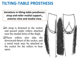
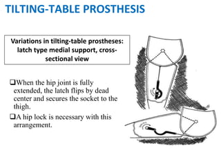
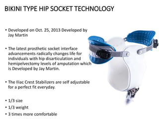
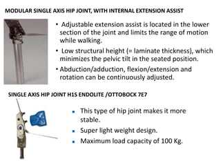
This document discusses advances in hip disarticulation prostheses. It begins by describing hip disarticulation amputation and challenges with prosthetic fitting at this level. It then covers the evolution of prosthetic designs including traditional tilting-table models, the seminal Canadian design, and more recent designs incorporating lightweight materials and anatomical shaping. Key components like the socket, hip joint, and suspension methods are examined. The document emphasizes ongoing efforts to improve mobility, comfort and long-term prosthetic use for individuals with hip disarticulation amputations.
Prosthetic management of different levels of amputationAamirSiddiqui56
In this presentation, i have covered all the basics about levels of amputation. I have mentioned the different levels of amputation and their prosthetic management. Beneficial for those who are in the field of P & O.
presentation is about Orthosis and prosthesis. It gives Classification of Orthosis. It describes structure, function, Indication and uses of Orthosis. Also describes different types of Prostheses, their parts and function.
Prosthetic management of symes and partial foot amputationSmita Nayak
prosthetic management of partial foot and syme's amputation is a very challenging task. Now a days the availability of advanced technology some how fulfilling the need of the amputee but not the fully.
Over the past decade, technology and research have greatly expanded the functionality and aesthetics of prosthetic feet. Today, amputees have a wide array of feet from which to choose. Various models are designed for activities ranging from walking, dancing and running to cycling, golfing, swimming and even snow skiing.
A complete description of the lower limb orthosis is available in the following presentation with an in depth understanding of the same.It covers the ankle foot orthosis,Knee orthosis the knee ankle foot orthosis and hip orthosis.
In this presentation detailed discussion about the amputation and syme amputation and biomechanics are there. also alignment of symes prosthesis is discussed.
Disarticulation prosthesis
Individuals with knee and hip disarticulation wear prostheses that include the same distal components as prostheses for lower levels.
Any prosthetic foot can be used with either an endoskeleton or exoskeleton shank. The major distinction, therefore, is in the proximal portion of the prostheses.
Plastic molded to provide weightbearing on the ipsilateral ischial tuberosity and buttocks. The person with transpelvic amputation who does not retain the ipsilateral tuberosity or iliac crest has a socket with a higher proximal Trimline, sometimes encompassing the lower thorax.
In this presentation, I have added evidence based practice ankle joints which are frequently used in orthotic treatment. Hope it reaches to every person out there seeking information regarding the same.
Beneficial for those, who are in the field of P & O.
I have drafted types of orthotic knee joints and their indications. Advancement of orthotic knee joints and their mechanisms.
Advantages & Disadvantages of orthotic knee joints.
Hope this is beneficial for you all.
The patellar-tendon-bearing socket is made of laminated or molded plastic that provides an intimate fit over the entire area of the amputated portion of the limb, including the distal end. The anterior wall of the socket is high enough to encompass the distal half of the patella while the posterior wall rises slightly higher than the apex of the in- ward bulge of the socket at the level of the patellar tendon. The medial and lateral walls are slightly higher than the anterior wall to control mediolateral forces applied to the stump, thereby enhancing stability, in this plane. The basic PTB socket also incorporates a soft insert liner
Provide:
The PTB socket provides total contact with the stump.
This feature helps to prevent edema, provides some additional support area (although the end of the stump cannot tolerate much pressure), and probably provides better sensory feedback to the wearer because of the greater area of contact between the amputated limb and the socket
Air cushion Socket
One of the designs, the PTB air-cushion socket, consists of an elastic inner sleeve (stockinette impregnated with silicone rubber) within a rigid outer shell and cap. A sealed chamber between these two walls contains air at atmospheric pressure. Distal support is provided by the tension of the sleeve itself and by compression of the air sealed in the chamber.
The advantages of the air-cushion socket are that is offers a possibility for higher distal loading,
Maintains distal contact even when limb volume changes slightly,
Lessens skin damage due to stretching of skin against an unyielding surface.
The increased distal loading may enhance circulation by decreasing the need for more constrictive proximal loading.
Two major difficulties are that it is complicated to fabricate and, once completed, modifications are very difficult.
Other Socket Variations
In addition to the variations-described above, which relate primarily to weight-bearing characteristics.
Other variations of the basic PTB socket involve the upward extension of the anterior, medial, and lateral walls to provide al- ternate means of suspension.
Since these designs relate to suspension, they are described in the section that follows.
Supracondylar Suprapetellar System
The basic PTB prosthesis is suspended by means of a supracondylar cuff ,which is attached to the socket in the posteromedial and posterolateral areas, and encircles the thigh just above the femoral epicondyles and patella
. The supracondylar cuff serves-primarily to retain the prosthesis on the stump.
It provides only slight assistance for mediolateral stability but does resist forces that tend to force the knee into hyperextension.
Most amputees, with the possible exception of those with very-short or painful stumps, find the stump-socket pressures well within their tolerance with this type of suspension.
The supracondylar/suprapatellar (SC/SP) suspension system has similarly high medial and lateral.
New Directions in Targeted Therapeutic Approaches for Older Adults With Mantl...i3 Health
i3 Health is pleased to make the speaker slides from this activity available for use as a non-accredited self-study or teaching resource.
This slide deck presented by Dr. Kami Maddocks, Professor-Clinical in the Division of Hematology and
Associate Division Director for Ambulatory Operations
The Ohio State University Comprehensive Cancer Center, will provide insight into new directions in targeted therapeutic approaches for older adults with mantle cell lymphoma.
STATEMENT OF NEED
Mantle cell lymphoma (MCL) is a rare, aggressive B-cell non-Hodgkin lymphoma (NHL) accounting for 5% to 7% of all lymphomas. Its prognosis ranges from indolent disease that does not require treatment for years to very aggressive disease, which is associated with poor survival (Silkenstedt et al, 2021). Typically, MCL is diagnosed at advanced stage and in older patients who cannot tolerate intensive therapy (NCCN, 2022). Although recent advances have slightly increased remission rates, recurrence and relapse remain very common, leading to a median overall survival between 3 and 6 years (LLS, 2021). Though there are several effective options, progress is still needed towards establishing an accepted frontline approach for MCL (Castellino et al, 2022). Treatment selection and management of MCL are complicated by the heterogeneity of prognosis, advanced age and comorbidities of patients, and lack of an established standard approach for treatment, making it vital that clinicians be familiar with the latest research and advances in this area. In this activity chaired by Michael Wang, MD, Professor in the Department of Lymphoma & Myeloma at MD Anderson Cancer Center, expert faculty will discuss prognostic factors informing treatment, the promising results of recent trials in new therapeutic approaches, and the implications of treatment resistance in therapeutic selection for MCL.
Target Audience
Hematology/oncology fellows, attending faculty, and other health care professionals involved in the treatment of patients with mantle cell lymphoma (MCL).
Learning Objectives
1.) Identify clinical and biological prognostic factors that can guide treatment decision making for older adults with MCL
2.) Evaluate emerging data on targeted therapeutic approaches for treatment-naive and relapsed/refractory MCL and their applicability to older adults
3.) Assess mechanisms of resistance to targeted therapies for MCL and their implications for treatment selection
Ozempic: Preoperative Management of Patients on GLP-1 Receptor Agonists Saeid Safari
Preoperative Management of Patients on GLP-1 Receptor Agonists like Ozempic and Semiglutide
ASA GUIDELINE
NYSORA Guideline
2 Case Reports of Gastric Ultrasound
Prix Galien International 2024 Forum ProgramLevi Shapiro
June 20, 2024, Prix Galien International and Jerusalem Ethics Forum in ROME. Detailed agenda including panels:
- ADVANCES IN CARDIOLOGY: A NEW PARADIGM IS COMING
- WOMEN’S HEALTH: FERTILITY PRESERVATION
- WHAT’S NEW IN THE TREATMENT OF INFECTIOUS,
ONCOLOGICAL AND INFLAMMATORY SKIN DISEASES?
- ARTIFICIAL INTELLIGENCE AND ETHICS
- GENE THERAPY
- BEYOND BORDERS: GLOBAL INITIATIVES FOR DEMOCRATIZING LIFE SCIENCE TECHNOLOGIES AND PROMOTING ACCESS TO HEALTHCARE
- ETHICAL CHALLENGES IN LIFE SCIENCES
- Prix Galien International Awards Ceremony
Factory Supply Best Quality Pmk Oil CAS 28578–16–7 PMK Powder in Stockrebeccabio
Factory Supply Best Quality Pmk Oil CAS 28578–16–7 PMK Powder in Stock
Telegram: bmksupplier
signal: +85264872720
threema: TUD4A6YC
You can contact me on Telegram or Threema
Communicate promptly and reply
Free of customs clearance, Double Clearance 100% pass delivery to USA, Canada, Spain, Germany, Netherland, Poland, Italy, Sweden, UK, Czech Republic, Australia, Mexico, Russia, Ukraine, Kazakhstan.Door to door service
Hot Selling Organic intermediates
Couples presenting to the infertility clinic- Do they really have infertility...Sujoy Dasgupta
Dr Sujoy Dasgupta presented the study on "Couples presenting to the infertility clinic- Do they really have infertility? – The unexplored stories of non-consummation" in the 13th Congress of the Asia Pacific Initiative on Reproduction (ASPIRE 2024) at Manila on 24 May, 2024.
Title: Sense of Taste
Presenter: Dr. Faiza, Assistant Professor of Physiology
Qualifications:
MBBS (Best Graduate, AIMC Lahore)
FCPS Physiology
ICMT, CHPE, DHPE (STMU)
MPH (GC University, Faisalabad)
MBA (Virtual University of Pakistan)
Learning Objectives:
Describe the structure and function of taste buds.
Describe the relationship between the taste threshold and taste index of common substances.
Explain the chemical basis and signal transduction of taste perception for each type of primary taste sensation.
Recognize different abnormalities of taste perception and their causes.
Key Topics:
Significance of Taste Sensation:
Differentiation between pleasant and harmful food
Influence on behavior
Selection of food based on metabolic needs
Receptors of Taste:
Taste buds on the tongue
Influence of sense of smell, texture of food, and pain stimulation (e.g., by pepper)
Primary and Secondary Taste Sensations:
Primary taste sensations: Sweet, Sour, Salty, Bitter, Umami
Chemical basis and signal transduction mechanisms for each taste
Taste Threshold and Index:
Taste threshold values for Sweet (sucrose), Salty (NaCl), Sour (HCl), and Bitter (Quinine)
Taste index relationship: Inversely proportional to taste threshold
Taste Blindness:
Inability to taste certain substances, particularly thiourea compounds
Example: Phenylthiocarbamide
Structure and Function of Taste Buds:
Composition: Epithelial cells, Sustentacular/Supporting cells, Taste cells, Basal cells
Features: Taste pores, Taste hairs/microvilli, and Taste nerve fibers
Location of Taste Buds:
Found in papillae of the tongue (Fungiform, Circumvallate, Foliate)
Also present on the palate, tonsillar pillars, epiglottis, and proximal esophagus
Mechanism of Taste Stimulation:
Interaction of taste substances with receptors on microvilli
Signal transduction pathways for Umami, Sweet, Bitter, Sour, and Salty tastes
Taste Sensitivity and Adaptation:
Decrease in sensitivity with age
Rapid adaptation of taste sensation
Role of Saliva in Taste:
Dissolution of tastants to reach receptors
Washing away the stimulus
Taste Preferences and Aversions:
Mechanisms behind taste preference and aversion
Influence of receptors and neural pathways
Impact of Sensory Nerve Damage:
Degeneration of taste buds if the sensory nerve fiber is cut
Abnormalities of Taste Detection:
Conditions: Ageusia, Hypogeusia, Dysgeusia (parageusia)
Causes: Nerve damage, neurological disorders, infections, poor oral hygiene, adverse drug effects, deficiencies, aging, tobacco use, altered neurotransmitter levels
Neurotransmitters and Taste Threshold:
Effects of serotonin (5-HT) and norepinephrine (NE) on taste sensitivity
Supertasters:
25% of the population with heightened sensitivity to taste, especially bitterness
Increased number of fungiform papillae
MANAGEMENT OF ATRIOVENTRICULAR CONDUCTION BLOCK.pdfJim Jacob Roy
Cardiac conduction defects can occur due to various causes.
Atrioventricular conduction blocks ( AV blocks ) are classified into 3 types.
This document describes the acute management of AV block.
Anti ulcer drugs and their Advance pharmacology ||
Anti-ulcer drugs are medications used to prevent and treat ulcers in the stomach and upper part of the small intestine (duodenal ulcers). These ulcers are often caused by an imbalance between stomach acid and the mucosal lining, which protects the stomach lining.
||Scope: Overview of various classes of anti-ulcer drugs, their mechanisms of action, indications, side effects, and clinical considerations.
Title: Sense of Smell
Presenter: Dr. Faiza, Assistant Professor of Physiology
Qualifications:
MBBS (Best Graduate, AIMC Lahore)
FCPS Physiology
ICMT, CHPE, DHPE (STMU)
MPH (GC University, Faisalabad)
MBA (Virtual University of Pakistan)
Learning Objectives:
Describe the primary categories of smells and the concept of odor blindness.
Explain the structure and location of the olfactory membrane and mucosa, including the types and roles of cells involved in olfaction.
Describe the pathway and mechanisms of olfactory signal transmission from the olfactory receptors to the brain.
Illustrate the biochemical cascade triggered by odorant binding to olfactory receptors, including the role of G-proteins and second messengers in generating an action potential.
Identify different types of olfactory disorders such as anosmia, hyposmia, hyperosmia, and dysosmia, including their potential causes.
Key Topics:
Olfactory Genes:
3% of the human genome accounts for olfactory genes.
400 genes for odorant receptors.
Olfactory Membrane:
Located in the superior part of the nasal cavity.
Medially: Folds downward along the superior septum.
Laterally: Folds over the superior turbinate and upper surface of the middle turbinate.
Total surface area: 5-10 square centimeters.
Olfactory Mucosa:
Olfactory Cells: Bipolar nerve cells derived from the CNS (100 million), with 4-25 olfactory cilia per cell.
Sustentacular Cells: Produce mucus and maintain ionic and molecular environment.
Basal Cells: Replace worn-out olfactory cells with an average lifespan of 1-2 months.
Bowman’s Gland: Secretes mucus.
Stimulation of Olfactory Cells:
Odorant dissolves in mucus and attaches to receptors on olfactory cilia.
Involves a cascade effect through G-proteins and second messengers, leading to depolarization and action potential generation in the olfactory nerve.
Quality of a Good Odorant:
Small (3-20 Carbon atoms), volatile, water-soluble, and lipid-soluble.
Facilitated by odorant-binding proteins in mucus.
Membrane Potential and Action Potential:
Resting membrane potential: -55mV.
Action potential frequency in the olfactory nerve increases with odorant strength.
Adaptation Towards the Sense of Smell:
Rapid adaptation within the first second, with further slow adaptation.
Psychological adaptation greater than receptor adaptation, involving feedback inhibition from the central nervous system.
Primary Sensations of Smell:
Camphoraceous, Musky, Floral, Pepperminty, Ethereal, Pungent, Putrid.
Odor Detection Threshold:
Examples: Hydrogen sulfide (0.0005 ppm), Methyl-mercaptan (0.002 ppm).
Some toxic substances are odorless at lethal concentrations.
Characteristics of Smell:
Odor blindness for single substances due to lack of appropriate receptor protein.
Behavioral and emotional influences of smell.
Transmission of Olfactory Signals:
From olfactory cells to glomeruli in the olfactory bulb, involving lateral inhibition.
Primitive, less old, and new olfactory systems with different path
ARTIFICIAL INTELLIGENCE IN HEALTHCARE.pdfAnujkumaranit
Artificial intelligence (AI) refers to the simulation of human intelligence processes by machines, especially computer systems. It encompasses tasks such as learning, reasoning, problem-solving, perception, and language understanding. AI technologies are revolutionizing various fields, from healthcare to finance, by enabling machines to perform tasks that typically require human intelligence.
The prostate is an exocrine gland of the male mammalian reproductive system
It is a walnut-sized gland that forms part of the male reproductive system and is located in front of the rectum and just below the urinary bladder
Function is to store and secrete a clear, slightly alkaline fluid that constitutes 10-30% of the volume of the seminal fluid that along with the spermatozoa, constitutes semen
A healthy human prostate measures (4cm-vertical, by 3cm-horizontal, 2cm ant-post ).
It surrounds the urethra just below the urinary bladder. It has anterior, median, posterior and two lateral lobes
It’s work is regulated by androgens which are responsible for male sex characteristics
Generalised disease of the prostate due to hormonal derangement which leads to non malignant enlargement of the gland (increase in the number of epithelial cells and stromal tissue)to cause compression of the urethra leading to symptoms (LUTS
Lung Cancer: Artificial Intelligence, Synergetics, Complex System Analysis, S...Oleg Kshivets
RESULTS: Overall life span (LS) was 2252.1±1742.5 days and cumulative 5-year survival (5YS) reached 73.2%, 10 years – 64.8%, 20 years – 42.5%. 513 LCP lived more than 5 years (LS=3124.6±1525.6 days), 148 LCP – more than 10 years (LS=5054.4±1504.1 days).199 LCP died because of LC (LS=562.7±374.5 days). 5YS of LCP after bi/lobectomies was significantly superior in comparison with LCP after pneumonectomies (78.1% vs.63.7%, P=0.00001 by log-rank test). AT significantly improved 5YS (66.3% vs. 34.8%) (P=0.00000 by log-rank test) only for LCP with N1-2. Cox modeling displayed that 5YS of LCP significantly depended on: phase transition (PT) early-invasive LC in terms of synergetics, PT N0—N12, cell ratio factors (ratio between cancer cells- CC and blood cells subpopulations), G1-3, histology, glucose, AT, blood cell circuit, prothrombin index, heparin tolerance, recalcification time (P=0.000-0.038). Neural networks, genetic algorithm selection and bootstrap simulation revealed relationships between 5YS and PT early-invasive LC (rank=1), PT N0—N12 (rank=2), thrombocytes/CC (3), erythrocytes/CC (4), eosinophils/CC (5), healthy cells/CC (6), lymphocytes/CC (7), segmented neutrophils/CC (8), stick neutrophils/CC (9), monocytes/CC (10); leucocytes/CC (11). Correct prediction of 5YS was 100% by neural networks computing (area under ROC curve=1.0; error=0.0).
CONCLUSIONS: 5YS of LCP after radical procedures significantly depended on: 1) PT early-invasive cancer; 2) PT N0--N12; 3) cell ratio factors; 4) blood cell circuit; 5) biochemical factors; 6) hemostasis system; 7) AT; 8) LC characteristics; 9) LC cell dynamics; 10) surgery type: lobectomy/pneumonectomy; 11) anthropometric data. Optimal diagnosis and treatment strategies for LC are: 1) screening and early detection of LC; 2) availability of experienced thoracic surgeons because of complexity of radical procedures; 3) aggressive en block surgery and adequate lymph node dissection for completeness; 4) precise prediction; 5) adjuvant chemoimmunoradiotherapy for LCP with unfavorable prognosis.
2. • Hip disarticulation is the surgical removal of the
entire lower limb by transection through the hip
joint.
• Trying to overcome the loss of three weight-bearing
joints, rather than one or two.
• Not routinely seen in the average clinical practice.
• Reduced mobility and increased energy expenditure
during gait.
• Prosthesis fitting is therefore limited to motivated
and physiologically vigorous individuals and even
then a significant number don’t become long term
user.
3. CAUSES OF HIP DISARTICULATION AMPUTATION
1. Malignant musculoskeletal tumors (most often in younger patients)
2. Limb ischemia (perivascular disease and complications to diabetes)
3. Trauma (such severe traumas often result in the death of the patient)
4. Severe lower limbs infections (chronic skin or bone infection)
5. medical negligence
Most Prosthetist have little experience with this type of amputation-Only
20% of hip amputees use a prosthetic leg full-time (i.e. 8 to 12 hr./day)
From these 20%, only a small minority use a prosthetic leg without a cane
or crutch
This small minority of full time users without walking aids consists
primarily of the young patients with malignant tumors.
There is a persistent belief within the medical community that most
middle aged hip-disarticulation amputees will ambulate with crutches or a
wheelchair only!!!
4. LEVEL OF HIP DISARTICULATION
Modified hip
disarticulation
True Hip
disarticulation
Transpelvic amputation
Hemipelvectomy
5. PROSTHETIC MANAGEMENT FOR HIP DISARTICULATION
Due to energy requirement for prosthetic use is as high as 200% of normal
human ambulation. There is a high rejection rate of prosthetic use at this level
of this amputation.
There is a lack of muscles power available to control hip, knee, and ankle joint
which results fixed and slow cadence.
Only those patient who has developed sufficient balance are likely to wear
prosthesis for ambulation.
Usually the mobility with crutches and the sound leg is much faster and
requires less energy expenditure than using a prosthesis
Selection of appropriate component is challenge for prosthetist
Less lever arm
6.
7. BIOMECHANICS OF H.D.PROSTHESIS
• The Canadian design HD prosthesis was introduced by Colin McLaurin and the
biomechanics of this prosthesis were clarified by Radcliffe in 1957.
• The high level prosthesis is stabilized by the ground reaction force, which occurs
during walking.
• For example when standing in the prosthesis, the person’s weight bearing line
falls posterior to the hip joint, anterior to the knee joint and anterior to the ankle
joint.
Knee Joint
Knee Axis
SEF
Knee Joint
Knee Axis SEF
Single
Equivalent
Force
(Projection of
CG)
10. ALIGNMENT OF H.D. PROSTHESIS
5° to 10°
external rotation
The length of the prosthesis is 12 mm shorter than the sound side, so that the foot can clear the
floor during midswing.
Single-axis knee is placed 15 mm posterior and a dynamic response midfoot is placed 20 mm
anterior to the bisection.
11. Evolution of hip Disarticulation Prosthesis
Prior to 1954 consisted of a moulded leather socket with a
laterally placed locking hip joint called a tilting-table prosthesis.
Later, "Canadian" design was introduced by McLaurin in
1954 with the use of free hip, knee, and ankle joints.This is now
the standard for prosthetic fitting worldwide, and locking joints
are very rarely necessary.
A molded plastic socket encloses the ischial tuberosity for
weight bearing, extends over the crest of the ilium to provide
suspension during swing phase.
On this concept, Lynquist proposed a fitting concept for a
Canadian type plastic socket. Which proved to be effective for
achieving comfortable weight bearing via the soft tissues, even
though there is no bony structure on the involved side of these
amputation.
12. THE SAUCER-TYPE PROSTHESIS
It is essentially a short above-knee leg
with a saucer-shaped socket on top of
the thigh.
Suspension is by means of a single-
axis joint and pelvic band and may
include straps that pass over the
shoulder.
This type is most suitable for short
femur amputations because adequate
stability is difficult to achieve without
the additional bone structure.
13. In accord with common practice with above-knee legs, the hip
joint is placed well forward, thus providing some measure of
stability.
A lock may or may not be used at the hip joint, if a lock is
used, it is of the semiautomatic type.
The lock provides stability (at some loss of function), but it
offers mechanical difficulties because all the loads are funneled
through the relatively small joint.
14. TILTING-TABLE PROSTHESIS
• The traditional device prior to 1954 consisted of a
molded leather socket with a laterally placed
locking hip joint called a tilting-table prosthesis
• A belt around the pelvis and often with a strap
over the shoulder.
• The socket is articulated on the thigh section with
a metal joint lateral to the acetabulum.
• Because it is extremely difficult to make a hip joint
strong enough to bear the entire load, contact
between the socket and the medial edge of the
thigh section is essential in weight-bearing, and
this is equally important when a lock is used.
• Without a lock, the wearer has little control over
the limb, most of the stability during the stance
phase being afforded by friction between the
socket and the thigh section.
15. A strap is fastened to the socket
and passed under rollers attached
near the medial brim of the thigh.
These rollers also take the
downward thrust of the socket, and
a metal track may be attached to
the socket for the rollers to bear
upon.
Variations in tilting-table prostheses:
strap and-roller medial support.,
anterior view and medial view.
TILTING-TABLE PROSTHESIS
16. When the hip joint is fully
extended, the latch flips by dead
center and secures the socket to the
thigh.
A hip lock is necessary with this
arrangement.
Variations in tilting-table prostheses:
latch type medial support, cross-
sectional view
TILTING-TABLE PROSTHESIS
17. Variations in tilting-table
prostheses: hip joint below
socket. anterior view and medial
view.
The walking function is identical,
but the hip joint has been lowered
to a position beneath the socket
where a full-width bearing may be
made much lighter.
Because of the position of the joint
directly below the center of
gravity, however, a lock must be
used.
TILTING-TABLE PROSTHESIS
18. This design uses a track-and-roller
mechanism in which the center of
rotation is a few inches lower and
anterior to the acetabulum.
But binding of the rollers on the
tracks prevented free motion, but it is
worth noting since in principle it is
almost identical to the present
Canadian type, and it seems to be
designed with a view toward
improving function.
Variations in tilting-table
prostheses: track and- roller joint.
TILTING-TABLE PROSTHESIS
19. CANADIAN HIP DISARTICULATION PROSTHESIS
FOR DECADES SURGEONS AND
PROSTHETIST STRUGGLED WITH THE
CHALLENGE TO FIND A GOOD
PROSTHETIC FIT.
The first experimental prosthesis
employed a four-link mechanism
The socket was plastic and the thigh
section aluminum alloy.
It was intended that a posterior
strap be used to lock the leg in full
extension, but initial trials indicated
adequate stability without a lock.
20. THE CANADIAN-TYPE HIP-DISARTICULATION
PROSTHESIS: FINAL DESIGN
• To extend the front link to include the
knee joint and to replace the rear link
with a simple rubber stop to prevent
hyperextension.
• This final configuration, permitted the use
of a single broad joint without locks.
• The most apparent difficulty was the
tendency for too long and too slow a
stride, and thus the elastic webbing was
added to restrain hip flexion.
• The ischial seat is nearly always available
for direct weight bearing, and the areas
for taking pressure elsewhere are large.
• If the socket is extended in the form of a
band across the back of the pelvis and
around to the opposite iliac crest, then
three points of the in nominate bones are
firmly gripped.
22. SOCKET TECHNOLOGY FOR HIP DISARTICULATION
In addition to previous technologies ,
by using ultralight materials for the
socket, its weight drops to less than
one-quarter that of traditional sockets.
Reduced weight translates directly into
less energy expenditure.
The new socket design further differs
from traditional models in that it
actually consists of two parts the ultra
light frame and a flexible inner socket.
This softer inner socket increases
comfort and improves the hygienic
properties of the prosthesis.
23. This new design “locks” the prosthesis
Onto the wearer on three planes: front-
to-back, side-to-side, and top-to-
bottom.
The intimate fit resulting from this
approach to suspension prevents
pistoning.
Achieving this LOCK requires the
socket closely around the anterior
pubic bone, the posterior sacrum, the
ischial tuberosity, and the ileac crest.
24.
25. CANADIAN-TYPE HIP-DISARTICULATION SOCKET
This is bucket type socket design.
Hip disarticulation socket design, which would encapsulate the Iliac crest,
ischium and ischial ramus.
It provide lateral stability using three point pressure system.
Anteroposterior stability.
45 degree inclination angle from posterior and anterior side .
Joint attached at anterodistal end.
26. UCLA Anatomical HIP-DISARTICULATION SOCKET(1980)
Hip disarticulation socket design, which would
encapsulate the ischium and ischial ramus in a
more anatomical contour than previous socket
designs, might produce an improved prosthetic
fitting.
Provide more comfort.
This was an attempt at a more anatomical socket
contours detailing the ischial ramus angle and the
medial inclination of the ischium were included in
the cast.
Thus the medial brim need not extend as high or
contain as much of the ischial ramus.
The initial concept for a hip disarticulation socket
was a one-piece polyethylene design with a
laminated frame to which the hip joint would be
attached.
27. Dycor´s Roller Track socket (1997)
Positioning the hip joint in the centre of the
socket rather than anteriorly enhances toe
clearance during swing phase because the knee
swings forward and upward rather than forward
and downward.
Hip rotation occurs through a laterally mounted
single axis joint with extension stop and flexion
bias.
Vertical loads are supported by a wheel attached
to the thigh pylon component trans versing a
hemispherical track built into the socket.
Strength and weight is enhanced because the
supporting structure is always directly under and
opposed to the dynamic applied load.
28. • The Glenrose socket design is made of three layers
and encompasses the affected side and iliac crest,
reducing the band passing around the sound side
between the iliac crest and the greater trochanter.
• The basic design is similar to the traditional North-
western University Diagonal Socket. The major
differences between this design are aggressiveness
of the trim lines and improved suspension method
presented below.
• The first layer is made of a soft thermoplastic
elastomer. This layer provides a soft flexible
transition on the medial wall, a high friction
surface to aid suspension, and a flexible, forgiving
trimline above the iliac crest.
• The butterfly shape allows movement of the iliac
crest suspension without buckling of the material.
• This piece is difficult to fabricate and requires a
skilled technician to accomplish.
GLENROSE SEMIFLEXIBLE SOCKET DESIGN
29. DIAGONAL SOCKET DESIGN
• It has the opening for donning located
on the sound side.
• The socket margin on the amputated
side is no higher than the level of the
anterior superior iliac spine.
• The rigid anterior and posterior
sections of the socket rise diagonally
from this level to cover the ilium on the
sound side, because both anterior and
posterior sections of the socket
contribute support, antero-posterior
movement of the socket on the pelvis is
minimized, reducing a painful pressure
on the inferior ramus.
• In addition the lateral support necessary
to prevent the stump from falling out of
the socket is more directly and firmly
achieved.
The Glenrose Socket Design,
anterior view. (A) Semi-flexible
layer; (B) Semi-rigid layer; (C)
Rigid layer.
30. SILICON FRAME SOCKET
• Silicone Frame Socket This ischial ramus
containment socket fastens the pelvis
diagonally on both sides between the iliac
crests and the ischial tuberosity.
• This technique has been realized by Udo
Danske. A silicone liner in the shape of a
swimsuit is fashioned in order to spread
out the forces and protect the skin. Then a
two-piece rigid frame fastening both iliac
crest is fabricated.
• Here iliac crest and spine are left
uncovered Posteriorly, the socket is
trimmed so that the frame lies on the
sacrum but lumbar vertebrae are left free.
32. BIKINI TYPE HIP SOCKET TECHNOLOGY
• Developed on Oct. 25, 2013 Developed by
Jay Martin
• The latest prosthetic socket interface
advancements radically changes life for
individuals with hip disarticulation and
hemipelvectomy levels of amputation which
is Developed by Jay Martin.
• The Iliac Crest Stabilizers are self adjustable
for a perfect fit everyday.
• 1/3 size
• 1/3 weight
• 3 times more comfortable
33. • Instead of encapsulating the entire pelvis with a thick bulky bucket,
they designed lightweight, bikini socket and iliac crest stabilizers
provide a more direct biomechanical link between the device and
its user, resulting in superior control, comfort, and functional
outcomes.
• The open air design makes wearing the prosthetic significantly
lighter, cooler, and more breathable.
34. PROSTHETIC HIP JOINT
• Functions of the Prosthetic HIP Joint
•Mobility
•Stability
•Durability
•Comfort
35. • The hip joint is screwed to the socket lamination
plate with the double hinged plate.
• For sitting, using a lever.
• Flexion and extension are adjusted by sliding the
extension stop bumper on the tube.
• Hip rotation is adjustable.
MODULAR SINGLE AXIS HIP JOINT, WITH LOCK
MODULAR SINGLE AXIS HIP JOINT, WITH EXTENSION ASSIST
Instead of the lock, the joint has an extension
assist with lateral latex bands for stride
control and a built-in adjustable extension
stop bumper.
The extension assist limits the range of motion
of the joint while walking.
36. MODULAR SINGLE AXIS HIP JOINT, WITH INTERNAL EXTENSION ASSIST
• Adjustable extension assist is located in the lower
section of the joint and limits the range of motion
while walking.
• Low structural height (= laminate thickness), which
minimizes the pelvic tilt in the seated position.
• Abduction/adduction, flexion/extension and
rotation can be continuously adjusted.
SINGLE AXIS HIP JOINT H1S ENDOLITE /OTTOBOCK 7E7
This type of hip joint makes it more
stable.
Super light weight design.
Maximum load capacity of 100 Kg.
37. POLYCENTRIC PROSTHETIC HIP JOINT
Features:
• Tracks hip movement to optimize gait.
• The hip folds anteriorly to ensure good sitting comfort
and Cosmesis.
• 130 degree flexion/extension
• Alignment allows adjustment in all planes
HYDRAULIC POLYCENTRIC PROSTHETIC HIP JOINT
• harmoniously damping joint movements in both the swing
and the stance phase with the goal of allowing prosthesis
wearer to achieve a gait pattern that comes close to the
physiological model.
• controlled heel strike
• Significant reduction of pelvic tilt, harmonious hip extension
• Controlled and smooth rollover on the prosthesis under full
load
• Small, lightweight and suitable for a body weight up to 125
kilograms
38. HELIX 3D PROSTHETIC HIP JOINT
• The joint consists of a so-called spatial four-axis
mechanism with hydraulic stance and swing phase
control.
• less sudden pelvic movements during weight transfer.
• Support for swing phase initiation
• Control of hip movements during the swing phase
• Three-dimensional movements in terms of the
relationship between hip joint extension / flexion and
transversal pelvic rotation
• Produces a three-dimensional hip movement & promotes
a symmetrical and natural gait pattern.
• Reduce the risk of falling and thereby to increase
functional safety.
• Makes a large flexion angle possible, to facilitate everyday
situations like putting on shoes or getting into a car.
39. The four-axis polycentric structure of the described hip joint consists
of two ball joints and two single-axis connections, with the rear axis
tilted in relation to the structure. Ball joints form the two anterior
connections. Specialised hydraulics control the level of stance and
swing phase resistance in this hip joint .
40. Produces a three-dimensional hip movement & promotes a symmetrical and natural gait
pattern
Three-dimensional movements in terms of the relationship between hip joint extension /
flexion and transversal pelvic rotation
41. KNEE JOINTS FOR HD PROSTHESIS
Single axis knees are not recommended for HD prosthesis. True but, a well aligned single axis knee
works very well in an HD prosthesis
Most widely utilized due to its light weight,
low cost, and excellent durability.
Friction resistance is often eliminated
For the Canadian hip disarticulation design,
more sophisticated mechanisms have proved
their value and are gradually becoming more
common.
SINGLE-AXIS CONSTANT-FRICTION
WEIGHT ACTIVATED SAFETY KNEE JOINT
Second most frequently utilized component.
Because there is very little increase in cost or weight and
reliability has been good
Missteps causing up to 15 degrees of knee flexion will not
result in knee buckle, which makes gait training less difficult
for the patient and therapist.
42. Slightly heavier than the other, this component offers maximum stance-phase stability.
Because the stability is inherent in the multi linkage design, it does not erode as the knee
mechanism wears during use.
Good toe clearance
Any fluid-control mechanism (hydraulic or pneumatic) results in a smoother gait.
• In addition, a more rapid cadence is also possible.
• The preferred mechanism has separate knee flexion and extension resistance adjustments.
• In essence, the limb steps forward more rapidly.
• As the shank moves into extension, the fluid resistance at the knee transmits the momentum
up to the thigh segment and pushes the hip joint forward into flexion
POLYCENTRIC KNEE JOINT (WITH OR WITHOUT FLUID CONTROL
MECHANISM )
43. Microprocessor controlled knee joint
• Assists the user in maintaining knee stability on a variety of surfaces.
• Reduces the risk of falls.
• Adjustable flexion extension
• Microprocessor controlled SNS resistance.
• Step over step stair climbing
44. Increased security and confidence/ Stumble Control.
Reduces conscious effort and stress for user to maintain knee
stability.
Step over step stair climbing
45. TUBE CLAMP ADAPTER
Adjustable Offset
Adapter (Titanium)
Adjustable
Height
Adapter
(Titanium)
(Aluminum)
Angled
Adapter for
HD
Prosthesis
(Titanium)
Male
Adapter
(Titanium)
The adapter is
available with three
angles that is
10°,20°,30°.
It establishes the
connection between
the pyramid adapter
in the knee joint and
the tube of the
anteriorly located hip
joint.
46. FOOT MECHANISMS FOR H.D. PROSTHESIS
• Dynamic response feet are commonly chosen for their lightweight
design. Because of the slowed gait of the hip disarticulation patient,
only in the more active patient can true dynamic responsiveness be
observed.
• An inexpensive solid ankle cushioned heel foot with a soft heel
cushion can also be used to increase knee stability.
• Although single-axis and multiaxial feet may be used to increase
stability, they add substantial weight to the distal end of the limb.
Recommended for the Canadian hip disarticulation
design due to its moderate weight, low cost, and
excellent durability.
The heel is composed of a foam wedge that provides
cushioning in the heel section during heel strike.
Used on an amputee’s initial prosthesis, when the
potential functional level of an amputee has yet to be
determined.
SACH FOOT
47. SINGLE AXIS FOOT
• In those cases where slightly more knee stability is
desired
• The single axis foot includes bumpers, which control
ankle flexion.
• This allows the prosthetic forefoot to contact the
floor quickly during after heel strike. heavier than
more basic feet, such as the SACH.
MULTI AXIAL FOOT
•Multi axial feet have liabilities similar to the
single-axis versions but add extra degrees of
freedom in hind foot inversion/ eversion and
transverse rotation.
• In addition to accommodating uneven ground,
absorbing some of the torque of walking, and
protecting the patient's skin from shear stresses.
48. FLEXIBLE-ENDOSKELETON FOOT
• The solid-ankle flexible-endoskeleton (SAFE) foot
inaugurated a class that could be termed "flexible-
keel" designs.
• Offers some transverse rotation as well.
DYNAMIC-RESPONSE FEET
o Provide a active push-off
o This foot typically returns 90% of stored energy and is recommended for a
wide range of age, activity and amputation levels.
o a combination of creative design and innovative lightweight construction
technology
o controlled movements help the user build more confidence in the
prosthesis.
49.
50. Prosthetic Rehabilitation Is To Aid The Amputee To Gain Independence At The
Highest Level They Can, With The Most Efficient Gait Possible