Total elbow arthroplasty
•
116 likes•14,520 views

This document discusses total elbow arthroplasty. It provides an overview of the different types of elbow implants, including fully constrained, semi-constrained, and unconstrained designs. Semi-constrained implants are most commonly used. Patient selection criteria and contraindications are outlined. Post-operative care involves restricting motion and weight-bearing initially. Common complications include instability, polyethylene wear, osteolysis, loosening, and infection. Revision surgery may be needed in cases of painful or failed elbow replacements.

Recommended
More Related Content
What's hot
What's hot (20)
Viewers also liked
Viewers also liked (20)
Similar to Total elbow arthroplasty
Similar to Total elbow arthroplasty (20)
Recently uploaded
Recently uploaded (20)
Total elbow arthroplasty
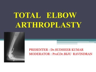
- 1. TOTAL ELBOW ARTHROPLASTY PRESENTER : Dr.SUDHEER KUMAR MODERATOR : Prof.Dr.BIJU RAVINDRAN
- 2. INTRODUCTION Elbow arthroplasty has been described in multiple forms over time. Semi-constrained total elbow arthroplasty, in particular, has a well-studied track record However, the procedure is associated with a relatively high complication rate Not durable as replacements of the hip, knee, or shoulder In particular, the excessive loads placed on the device by high-demand patients is a common cause of failure.
- 3. GOAL OF TOTAL ELBOW ARTHROPLASTY Restore functional mechanics of elbow Relief of pain Restoration of motion Stability
- 4. IMPLANT TYPES • Depending on rigidity of fixation of humeral component to ulnar component • FULLY CONSTRAINED • SEMI CONSTRAINED • UNCONSTRAINED
- 5. FULLY CONSTRAINED • Metal to metal hinge with PMMA cement fixation • Rarely used now as they have tendency to loosen & breakage • Eg: stanmoore.Dee,Mckee.GSB1 & Mazas designs
- 6. SEMI CONSTRAINED • 2 or 3 part prosthesis • Metal to high density polyethylene articulation connected with locking pin or snap-fit device • Have built in Valgus & varus laxity for side to side dissipation of forces • Eg: Coonrad, Mayo, Tri-axial, Schlein, AHSC Pritchard-walker
- 7. UNCONSTRAINED • 2 part prosthesis • Metal to high density polyethylene articulation without locking pin or snap-fit link • Design consists of - stems for humeral component or resurfacing devices • Parts unlinked in a attempt to anatomically duplicates the articular surface of the elbow • Require normal intact ligaments, anterior capsule & appropriate static alignment
- 8. PATIENT SELECTION It is the most definitive functional procedure for END STAGE ELBOW ARTHRITIS • End stage-Rheumatoid Arthritis with radiological evidence of joint destruction • Acute unreconstructable fracture > 60 age • Bilateral elbow ankylosis • Bony or fibrous ankylosis with elbow in poor functioning position
- 9. Contd… • As a revision of failed elbow arthroplasty • Loss of bone stock caused by tumour/trauma • End stage –Osteoarthritis • Post traumatic arthritis • Nonunion of distal humerus • Hemophilic arthropathy
- 10. IMPLANT SELECTION Depends to a great extend on the Fate of capsuloligamentous structures about the elbow Integrity of the musculature Amount of bone remaining at the elbow joint • Resurfacing /unconstrained prosthetic designs-patients with a more stable joint with more bone remaining • Semi-constrained- patients with extensive injury to ligaments & capsule of joint, atrophic musculature & loss of considerable bone stock
- 11. CONTRAINDICATIONS • Active or recent elbow Sepsis(absolute) • Poor soft tissue envelope • Non restorable function of Biceps & Triceps • Poor patient compliance with activity & weight lifting restrictions • Flaccid paralysis of upper extremity • Young vigorous patient with a post traumatic destroyed elbow • Neuropathic elbow joint • Ankylosis of Ipsilateral shoulder
- 12. PRE-OP PLANNING • Routine AP & lateral radiographs Assess humeral bow & medullary canal size in lateral projection Note size & angulation of the ulnar medullary canal In both projection • Templates are available for all the varying size prostheses & very useful • Ulnar nerve examination-document if any degree of impairment noted • Elbow aspiration & culture to rule out joint sepsis –if infection is suspected or rheumatoid elbow is red/hot/swollen
- 13. COONRAD-MORREY PROSTHESIS • Semi-constrained hinged prosthesis • High-molecular-weight polyethylene bushing & titanium humeral and ulnar components. • Designed with 7 * of rotary and side-to-side laxity. • Humeral and ulnar stems match the shapes of the medullary canals. • The triangular humeral stem is flattened near the base at the inferior flatter and wider portion of the medullary canal of the humerus.
- 14. • The large medullary stem enhances rigid fixation. • Its long stem, contour & distal anterior flange increase resistance to torque. • Careful bone removal in the intercondylar area of the humerus is necessary to allow a tight fit of the humeral prosthesis. • Prosthesis usually is inserted with the elbow fully flexed • Components also can be inserted separately and then joined. • Right & left prostheses are available as trial components.
- 18. POST OP IMAGING • AP and lateral films are obtained as baseline reference • used for future comparison to follow up studies to document stability of prosthesis. • Initial radiographs are scrutinized for: • Alignment ▫ Humeral and ulnar stems should align with the long axis of the bone ▫ Humeral and ulnar components show normal articulation without evidence of dislocation
- 19. • Peri-prosthetic fractures • Cement technique ▫ Cement should coat the prosthesis stems without extravasation into soft tissues ▫ Intraoperative fractures or osteopenic bone increase risk of cement leak which may damage radial or ulnar nerves.
- 20. POST OP CARE The extremity is elevated overnight with elbow above the shoulder. The drains & compressive dressing are removed the day after surgery. A light dressing is then applied & passive elbow flexion and extension are allowed as tolerated. A collar and cuff are used & instructions in the activities of daily living are provided by an occupational therapist.
- 21. Active elbow extension must be avoided for 3 months until the triceps heals. Strengthening exercises are avoided. Patient is encouraged to avoid lifting more than 5 lb for the first 3 months after surgery Thereafter, lifting is restricted to 10 pounds
- 22. RESULTS • RATING SYSTEM OF MORREY used 3 criteria to rate TEA Xray appearance pain motion 1.GOOD RESULT No radiographic change at the bone cement /prosthesis interface No pain >90* of flexion 60* of pronation & supination
- 23. 2.FAIR RESULTS • > 1mm of widening of any bone cement prosthesis interface • Mild pain • Between 50* & 90* of flexion & extension • Less than 40* of pronation & supination 3.POOR RESULTS • >2mm of widening of any bone cement prosthesis interface • Pain that significantly limits activity • Less than 50* of flexion & extension • Revision of elbow arthroplasty
- 25. PAINFUL OR FAILED ELBOW ARTHROPLASTY • Evaluated with serial conventional radiographs • May be due to a variety of problems. These include: • Instability • Polyethylene problems • Osteolysis • Loosening • Infection • Fractures • Heterotopic ossification
- 27. INSTABILITY • Instability occurs in the form of dislocation or subluxation • Most common complication requiring revision of unconstrained prostheses • reported to occur in between 9% and 10% of total elbow arthroplasties INTRA-OP PRECAUTIONS • Appropriate tensioning of the medial & lateral ligament complexes • preservation of the anterior capsule and triceps
- 28. INSTABILITY • Instability in SEMI-CONSTRAINED TEA is usually caused by wear of the polyethylene bushings or disengagement of the linkage pin. • Proper prosthesis alignment is crucial in linked arthroplasty. • Malalignment can cause abnormal forces to be generated across the joint which can lead to early hardware failure.
- 29. Polyethylene Problems • Polyethylene components are prone to wear after long term usage. • Wear and the subsequent debris that it produces initiate synovitis and foreign body reaction, which can ultimately lead to severe bone loss. • In many linked arthroplasty prostheses models, bushings are made of polyethylene. • A bushing is defined as a fixed or removable cylindrical lining used to reduce friction between the humeral and ulnar components.
- 30. Polyethylene Problems • Bushing wear is determined on an AP radiograph. METHOD: • A line was drawn parallel to the yoke of the humeral component, & another line was drawn parallel to the medial or lateral surface of the ulnar component. INFERENCE : • An angle of intersection > 7 * indicates excessive tolerance of the bushings due to polyethylene deformation
- 31. Polyethylene Problems • Certain factors are associated with the development of bushing wear. They are • younger patient age, • male sex, • Post traumatic arthritis, • Pre operative elbow deformity, • Supra condylar nonunion, • high activity levels
- 32. Osteolysis • Refers to both focal and linear periprosthetic bone loss adjacent to any joint replacement • Results from a foreign body response to particulate debris from the wear of arthroplasty components and cement • Particles are taken up by macrophages & giant cells which may release cytokines that initiate a cascade reaction ultimately resulting in osteolysis
- 33. • Osteolysis is usually asymptomatic. • Follow-up radiographs are used to identify the process early • Thinned cortex and weakened bone places patients at risk of pathologic fracture, prosthesis subsidence and loosening • Large amounts of particulate may dissolve in joint fluid, staining the fluid and synovium a dark metallic color. This is called metallosis
- 34. Aseptic Loosening • Failure of the bond between an implant and bone in the absence of infection. • It is the most frequent cause of long-term implant failure RISK FACTORS: patients who continue to use their elbow in strenuous activities and heavy lifting constrained, linked prosthesis types. • Presents with insidious onset of activity related pain in a previously well functioning prosthesis.
- 35. Aseptic Loosening Radiographic signs of loosening include: Progressive and extensive widening of interfaces between bone-cement, bone-prosthesis, or cement-prosthesis Fragmentation or fracture of cement Migration/subsidence of prosthetic components Bead shedding in porous-coated prostheses
- 36. Marked loosening of humeral stem with migration antero-laterally beyond the humeral cortex.
- 37. INFECTION • Infection rates after TEA remain greater than those for total hip or knee arthroplasties. • Reason: relative paucity of soft tissue coverage in the elbow compared to the other joints. • Typical infecting organisms are Staphylococcus aureus , Staphylococcus epidemidis
- 38. INFECTION • Risk factors : oral steroid use, diabetes mellitus, distant osteomyelitis, infected prostheses at other sites & H/O septic arthritis in the affected elbow. Clinical signs and symptoms may consist of • Pain • Decreasing range of motion • Fever • Night sweats • Chills • Erythema • Draining sinus tract
- 39. Radiographic features of infection include: • Progressive and extensive widening of interfaces between bone-cement, bone-prosthesis or cement-prosthesis • Periosteal bone formation • Periprosthetic bone resorption • Soft tissue or periprosthetic gas
- 40. Infected total elbow arthroplasty. There is a large joint effusion (red arrow), distended olecranon bursa (O), and irregular interface widening about the ulnar stem (green arrow).
- 41. INFECTION • Infection of elbow replacements requires aggressive treatment with Antibiotics Surgical management may consist of: • Irrigation and debridement ▫ Considered when infection is acute ▫ May require repeat procedures, placing patient at risk for soft-tissue damage • Resection arthroplasty ▫ Hardware removed and antibiotic impregnated cement space placed ▫ Eventually followed by revision arthroplasty
- 42. • Cement spacers are temporary prostheses made of antibiotic impregnated methylmethacralate. • The cement is prepared in the surgical site, mixed with antibiotics, and formed by the surgeon into forms resembling the components of the elbow arthroplasty. Advantages : • Provide local dispersal of antibiotics to the infected joint area • Maintain length • Minimize dead space • Preserve soft-tissue planes • Facilitate ease of revision arthroplasty
- 43. PERIPROSTHETIC FRACTURES • The rate of periprosthetic fractures after elbow arthroplasty vary from 6% to 22% • Classified using Mayo classification of periprosthetic fractures after elbow arthroplasty Fractures are classified according to • location • bone quality • whether component is stable or loose • Classification of the humerus and ulna are determined separately.
- 44. Mayo classification of periprosthetic fractures after elbow arthroplasty