Downloaded 764 times
![Epidemiology
Hernias comprise approximately 7% of all
surgical outpatient visits.
Male: female ratio is 8:1.
They affect 1-3% of young children.
In men, the incidence rises from 11 per
10,000 person-years, aged 16-24 years,
200 per 10,000 person-years, aged 75
years or above.[1]
](https://image.slidesharecdn.com/herniaandherniorrhaphy-131117154227-phpapp01/85/Hernia-and-herniorrhaphy-4-320.jpg)


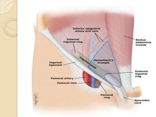
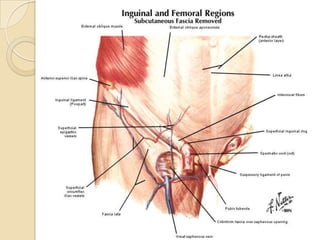
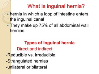
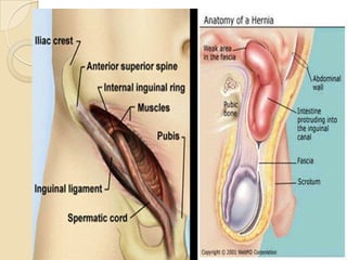

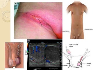


This document provides an overview of inguinal hernias. It defines a hernia as the protrusion of an organ through a weakness in the muscle wall. It then discusses the epidemiology, types, anatomy, etiology, pathogenesis, signs and symptoms, diagnosis, treatment and complications of inguinal hernias. The treatment options covered are herniotomy, herniorrhaphy, and laparoscopic repair techniques like the Lichtenstein method. The prognosis is typically good, though there is a small risk of recurrence.


![ONFH[AVN HIP] -TRIPLE REGIME -A NOVAL SURGICAL CONCEPT .pptx](https://cdn.slidesharecdn.com/ss_thumbnails/onfhavnhip2026koaconcalicutdrgokuldevdrmashraf-260210064517-213ec005-thumbnail.jpg?width=640&height=640&fit=bounds)


