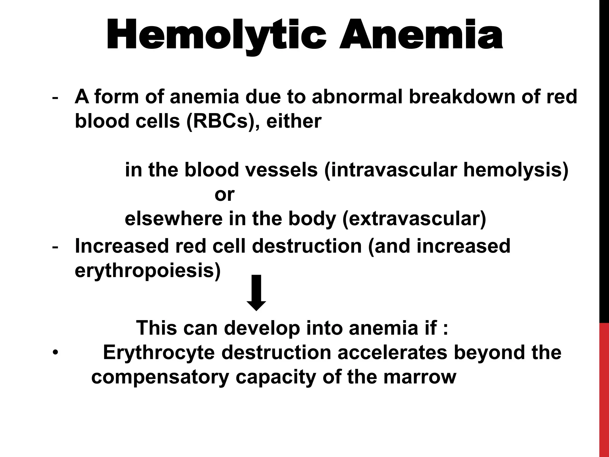
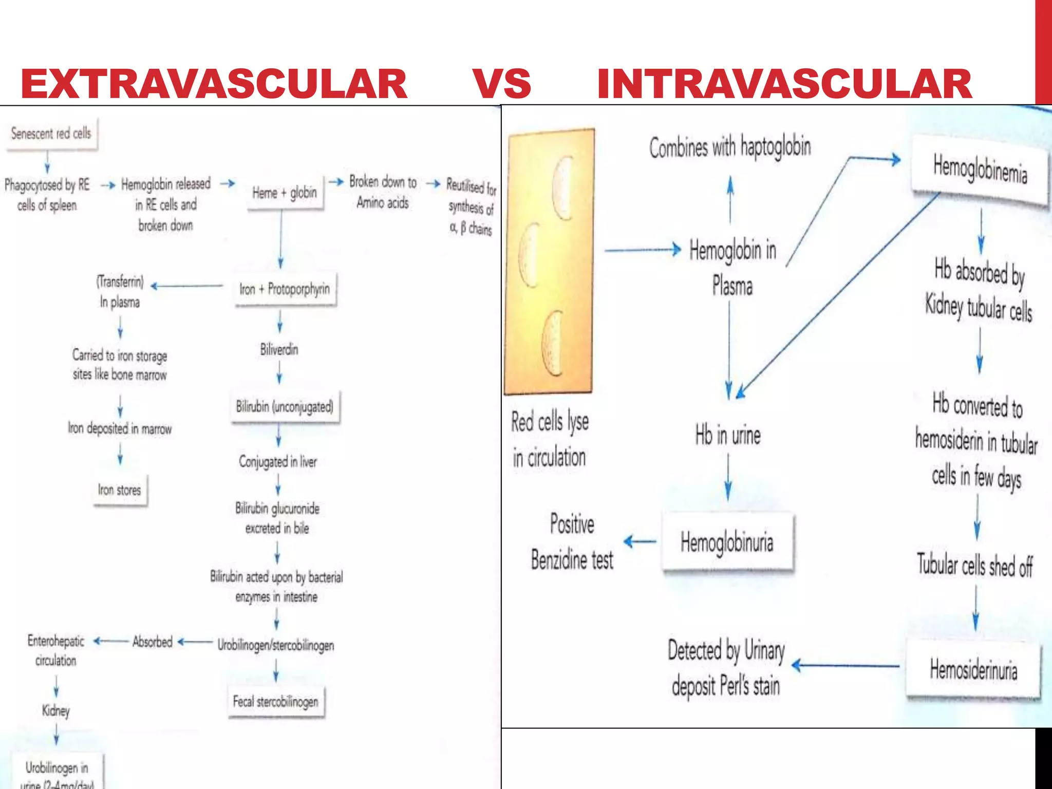
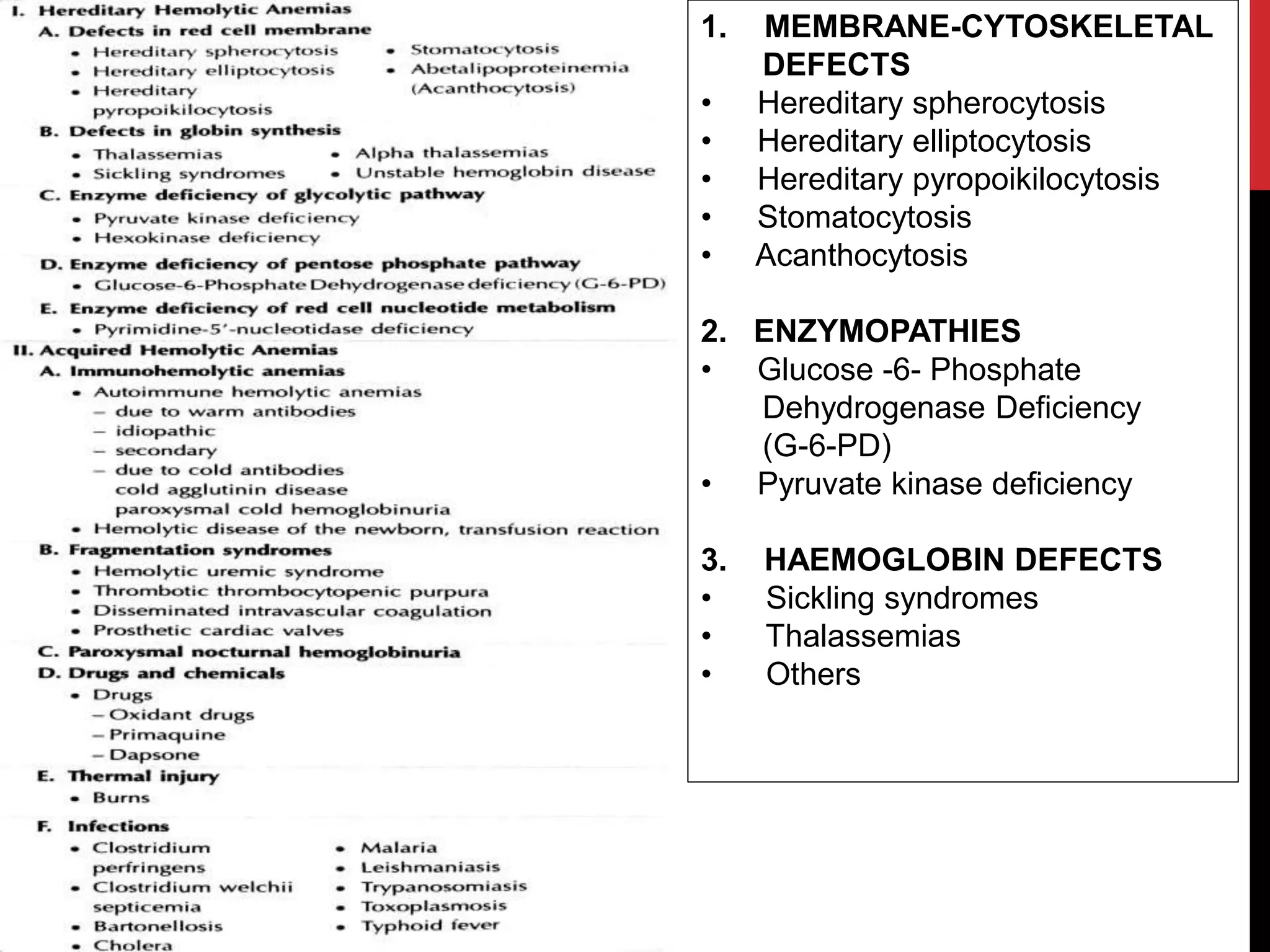
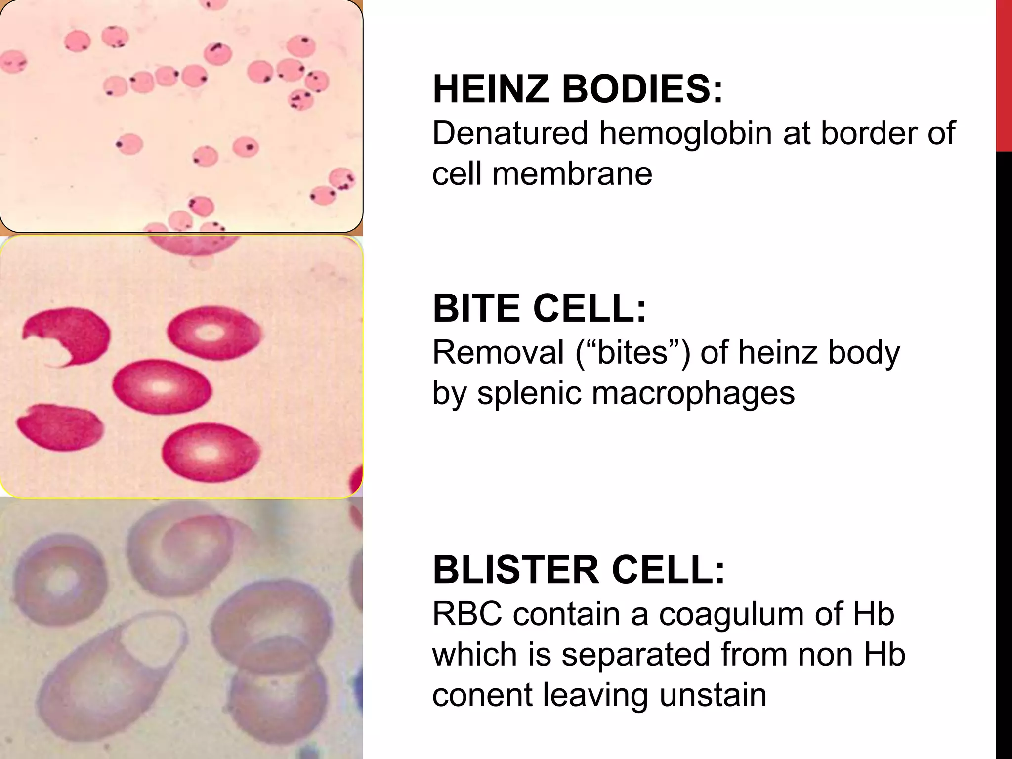
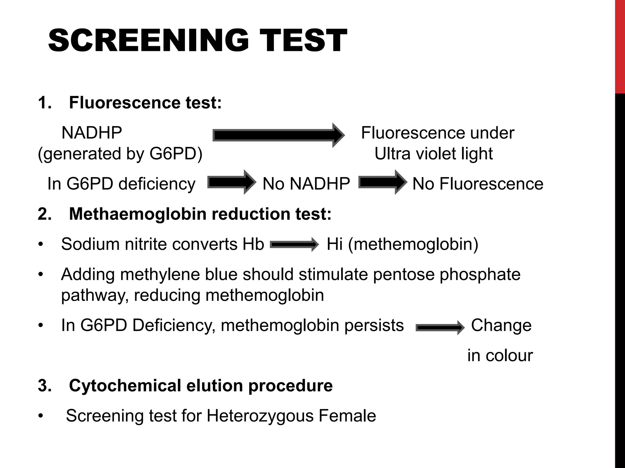
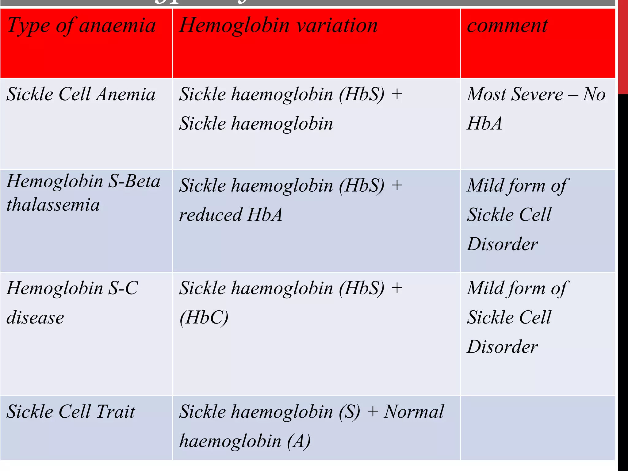
Hereditary hemolytic anemia can develop due to abnormal breakdown of red blood cells either in the blood vessels (intravascular hemolysis) or elsewhere in the body (extravascular). This document discusses the different types of hereditary hemolytic anemias including membrane-cytoskeletal defects like hereditary spherocytosis, enzymopathies like glucose-6-phosphate dehydrogenase deficiency, and hemoglobin defects like sickle cell disease. The causes, pathophysiology, clinical manifestations, and diagnostic testing for each condition are summarized.
2. Hemolytic Anemia
- A form of anemia due to abnormal breakdown of red
blood cells (RBCs), either
in the blood vessels (intravascular hemolysis)
or
elsewhere in the body (extravascular)
- Increased red cell destruction (and increased
erythropoiesis)
This can develop into anemia if :
• Erythrocyte destruction accelerates beyond the
compensatory capacity of the marrow
7. Hereditary spherocytosis
- Autosomal-dominant,
- most common disorder of the red cell membrane (1:2000).
• Deficiency of spectrin,
• combined deficiency of spectrin and ankyrin ,
• mutations of ( ankyrin, α or β-spectrin, protein 4.2,band 3)
- Influx of Na+ 10 times the normal rate
- Increased cytoplasmic viscosity
Gene Locus
ANK1 8p11.2
SPTB 14q22-q23
SPTA 1q21
SLC4A1 17q21-q22
EPB42 15q15
8. MOLECULAR PATHOPHYSIOLOGY
Deficiency in spectrin, ankyrin, protein3
Lipid bilayer skeleton uncoupling
Membrane loss in the form of
microvesicles
Surface area deficiency
Spherocytosis
Impaired passage through splenic cord
Sequestration
9. VARIOUS PRESENTATIONS
HS in neonate
• Hemolytic d/s of newborn
• Prolonged neonatal jaundice
• May require exchange transfusion.
• Anemia progressing to Congestive Cardiac Failure
• Hydrops fetalis (rare)
• Palpable spleen
• Investigate parents
HEMOLYTIC CRISIS
• Precipitated by viral infection : Infectious mononucleosis
• Exercise induced
• Anemia, jaundice, vomiting, abdominal pain, tender spleen
• May happen also during recovery phase of aplastic crisis
10. APLASTIC CRISIS
• Less common, more serious
• Parvovirus B19 infection
• Fever, chills
• Vomiting, diarrhea, myalgias
• Slapped cheek apearance
• Following this - sudden pallor, jaundice, weakness
MEGALOBLASTIC CRISIS
• Due to a secondary folate deficiency
• In patients recovering from aplastic crisis
• Hence supplement 1mg/day of F.A. to children with HS
11. Normal or decreasedHb
> 8%Retics
60-87 flMCV
normalMCH
> 36g/dlMCHC
Normoblastic erythroid hyperplasia,
increased iron storage
BM
increasedOsmotic fragility
increasedIndirect bilirubin
increasedLDH
decreasedhaptoglobin
RBCs small and lack the central pallorPB Smear
Laboratory findings
12.
13. 1. OSMOTIC FRAGILITY TEST
- More commonly used
0
20
40
60
80
100
0.3 0.4 0.5 0.6
NaCl (%of normal saline)
%Hemolysis
Normal HS
2. FLOWCYTOMETRY – Based on EMA (eosin-5-maleimide)
- High specificity and sensitivity
14. TREATMENT GOAL:
• If Hb > 10 gm/dl and retics < 10% - No Rx
• If severe anemia, poor growth, aplastic crises and
age < 2 yrs – Transfusion
• If Hb < 10 gm/dl and retics > 10 % or massive
spleen- Splenectomy
• Folic acid- 1 mg/day
15. Hereditary Elliptocytosis
- Autosomal dominant
- Defect in the horizontal membran protein
interaction:
* Decreased association of spectrin dimers to form tetramers
* Defect in band 4.1
* Abnormalities in glycophorin C, abnormal anion
transport(band 3) with increased affinity to ankyrin
- The cells are abnormally permeable to Na+
- For Diagnosis : >25% RBCs are Elliptocytes on smear
(vary from 20-90%)
: OFT is normal
17. Hereditary Pyropoikilocytosis
- Autosomal recessive , closely related to HE
- Presents in infancy as severe HA with extreme
poikilocytosis
- Two defects :
1. Related to a deficiency in α-spectrin
2. The presence of mutant spectrin that prevents
association of heterodimers to tetradimers
19. Hereditary Stomatocytosis
- Autosomal dominant HA ,
- Deficiency of membrane protein STOMATIN on band 7
- Erythrocytes exhibit
increased Na+ and K+ permeability.
Increased rate of leak
constant tendency to swell and burst
Osmotic mechanical lysis
Mild to moderate anemia
Reticulocytosis- 5-20%
MCV – 110-150fl, MCHC reduced
20. OHS (Over hydrated stomatocytes):
RBC membrane is abnormally
permeable to Na+ , K+
(net gain of Na+ >net loss of K+)
the capacity of cation pump is
exceeded
Water enters the cell ------>
STOMATOCYTES(central slit like
pallor)
DHS
(Dehydrated stomatocytes) :
The net loss of intracellular K+
exceeds the passive Na+ influx
decreased water and cation content --
--> Xerocyte
* Variants :
21. Acanthocytosis
(Abnormal lipid composition)
- Acquired or inherited abnormalities of the membrane
lipids
- Liver disease, abetalipoproteinemia
- Lipid of the membrane exchange with plasma lipids
- Acquiring excess lipids cause abnormal shapes.
- Sequestered in spleen
22. 2. Enzyme deficiencies
- RBCs require constant energy to maintain
biconcave disc shape and hemoglobin in
reduced form.
- Without adequate energy, red cells lyse and/or
deform.
- An inherited deficiency in one of the erythrocyte
enzymes can compromise the integrity of the
cell membrane or Hb and cause hemolysis.
23. G6PD deficiency
- The most common erythrocyte enzyme disorder
- G6PD deficient cells more resistant to malarial parasites.
- Synthesis of G6PD determined by X chromosome.
- Usually only males affected.
- Heterozygous females - usually not symptomatic
Lyon’s hypothesis
unless random inactivation of X chromosome (rarely)
- The majority of people have no clinical expression of the
deficiency unless they have neonatal jaundice, exposed to
oxidative drugs, or have severe infections .
25. PRECIPITATING FACTORS
Antimalarials: primaquine, quinine, chloroquine
Antibiotics - nitrofuantoin, furazolidine,
cotrimoxazole, Nalidixic acid,
Chloramphenicol,
Others :
Vitamin K – large doses
Naphthalene (moth balls)
Benzene, Methylene blue
Probenecid
Acetyl salicylic acid (aspirin)
Fava beans
Septicemia and viral hepatitis in def. pts
Diabetic ketoacidosis
26. - Favism :
Sudden severe hemolytic
episode that develops in some
G6PD deficient individuals
after ingestion of fava beans
Signs :
- malaise, nausia, vomiting, abdominal pain, tremor, fever.
- Hemoglobinurea, jaundice
- Severe favism affects children between the ages of 2-5 years
Signs and symptoms
27. G6PD Variants can be classified into :
Class I mutations (G6PD Group A-) :
• A moderate form of the disease
• RBCs contain unstable G6PD enzyme, but normal activity in younger
RBCs and reticulocytes.
• Accordingly, only older RBCs are hemolysed in a hemolytic episode.
Class II mutations (G6PD Mediterranean):
• More severe (all RBCs are affected)
• G6PD enzyme shows normal stability but, very low activity in all RBCs.
Class III
• It is often associated with chronic non spherocytic anemia
(occurs even in absence of oxidative stress).
Indian Scenario:
G6PD Mediterranean - Severe def with HA, Favism, Neonatal jaundice
G6PD Orissa – Drug induced hemolysis
G6PD Kerala-kalyan - Drug induced hemolysis
29. BITE CELL:
Removal (“bites”) of heinz body
by splenic macrophages
HEINZ BODIES:
Denatured hemoglobin at border of
cell membrane
BLISTER CELL:
RBC contain a coagulum of Hb
which is separated from non Hb
conent leaving unstain
30. SCREENING TEST
1. Fluorescence test:
NADHP Fluorescence under
(generated by G6PD) Ultra violet light
In G6PD deficiency No NADHP No Fluorescence
2. Methaemoglobin reduction test:
• Sodium nitrite converts Hb Hi (methemoglobin)
• Adding methylene blue should stimulate pentose phosphate
pathway, reducing methemoglobin
• In G6PD Deficiency, methemoglobin persists Change
in colour
3. Cytochemical elution procedure
• Screening test for Heterozygous Female
31. NEVER ASSESS G6PD LEVELS
DURING ACUTE HEMOLYSIS.
WHY?
• During acute hemolysis- all deficient cells have been
hemolysed
• Young cells will be in circulation
• Young surviving cells may have normal levels of the
enzyme
• Hence falsely normal during acute episode
• Assess 2-4 months later
• Deficient G6PD levels will be evident
• Usually affected have <10% of normal level
32. PYRUVATE KINASE DEFICIENCY
- Autosomal recessive , > 180 mutations in the PK gene
- Clinically significant HA are associated with the
homozygous & double heterozygotes
- Single heterozygotes are asymptomatic
- Anemia – moderate, Retic count- 5-15%
- PS – Moderate Anisocytosis (Prickle cells, echinocytes)
- Moderate increased S.bilirubin
- Diagnosis: 1. Autohemolysis test
2. Quantitative assay
- Acquired PK deficiency is seen in some leukemias &
myelodysplastic disorders
33. PATHOPHYSIOLOGY
Energy producing reaction is prevented
failure of cation pumps
potassium loss, calcium
& sodium gain
dehydration
(echinocytes)
sequestration in splenic cord
and phagocytosis
34. SICKLE CELL DISEASE
(DREPANOCYTOSIS)
• Sickle-cell disease
(SCD), or sickle-cell
anaemia, is an
autosomal recessive
genetic blood disorder
characterized by red
blood cell that assume
an abnormal, rigid, sickle
shape.
35. Type of anaemia Hemoglobin variation comment
Sickle Cell Anemia Sickle haemoglobin (HbS) +
Sickle haemoglobin
Most Severe – No
HbA
Hemoglobin S-Beta
thalassemia
Sickle haemoglobin (HbS) +
reduced HbA
Mild form of
Sickle Cell
Disorder
Hemoglobin S-C
disease
Sickle haemoglobin (HbS) +
(HbC)
Mild form of
Sickle Cell
Disorder
Sickle Cell Trait Sickle haemoglobin (S) + Normal
haemoglobin (A)
36. PATHOPHYSIOLOGY
Mutation => beta globin- 6 - Glu Val.
• RBC typically live 90–120 days, sickle cells only 10–20 days
Deoxy HbS (polymerised)
Ca2+ influx, K+ leakage
stiff,viscous sickle cell
venocclusion Decreased RBC survival
microinfarctions,ischemic pains anemia,jaundice,
autoinfarct spleen gallstones,leg ulcers
37. • Gene defect is
known mutation of a single
nucleotide.
• The person that receives the
defective gene from both
parents will develop
Sickle-cell disease.
• The person who receives
only one defective gene from
either one of his parents will
develop Sickle-cell trait.
GENETICS
38.
39. COMMON SYMPTOMS
• Abdominal pain
• Bone pain
• Breathlessness
• Delayed growth and puberty
• Fever
• Jaundice
• Rapid heart rate
• Susceptibility to infections.
40. CLINICAL MANIFESTATIONS
VASO-OCLUSIVE CRISIS
• Ischemia, Pain, Necrosis
• Often leads to organ damage
SPLENIC SQUESTRATION CRISIS
• Acute, painful enlargements of the spleen, caused by
intrasplenic trapping of red cells
• Caused by intrasplenic trapping of red cells
• Die within 1-2 hours due to circulatory failure
• Autosplenectomy
APLASTIC CRISIS
Paravirus B19
• Divides in RBCs precursors and destroys them
• Stops erythropoiesis for two or three days
• Causes reticulocytopenia
• Disappears within one week with management & BT
HEMOLYTIC CRISIS- Common in patients with G6PD def.
41. DIAGNOSIS
Laboratory findings
• RBC’s: 5-50 % sickled
• Low hemoglobin; 7-10 gm/dl;
• HbA; 0%; HbS 85-98%
• Increased reticulocytes: 10-25%,
• Increased platelet, and leukocyte counts;
• Sickle forms on the peripheral smear
Routine neonatal screening programs:
- DNA from fetal cell for mutation
42. SICKLING
TEST
Normal RBC Sickled RBC
*Positive sickling test associated
with a normal haemoglobin is
likely to indicate a patient with
sickle cell trait.
Positive Test
HbS
Negative Test
HbA
This test is simple and quick,
used to identify the presence
of HbS.
Sickle Solubility Test (SST)
• A rapid and inexpensive technique used to screen for the presence of
sickling hemoglobins.
• A positive result must be confirmed by another method
(HPLC or electrophoresis)
43. HEMOGLOBIN
ELECTROPHORESIS TEST
• Haemoglobin electrophoresis will differentiate between homozygous and
heterozygous conditions.
• Hemoglobin types have different electrical charges and move at different
speeds.
*HbSS: Is less negative
by 2 compared to HbA .
Migrates slower than HbA
*HbAS: Has both HbA
and HbS.
Shows 2 bands
44. NEWBORN SCREENING
• In newborns who carry the sickle cell gene ,
fetal hemoglobin F will predominate, but a
small amount of hemoglobin S will also be
present.
• It is performed via the most sensitive Hb isoelectric focusing or
HPLC fractionation and identifies the specific types of hemoglobin
present.
DNA analysis
• This test is used to investigate alterations and mutations in the genes
that produce hemoglobin components.
• It may be performed to determine whether someone has one or two
copies of the Hb S mutation or has two different gene mutations.
• Genetic testing is most often used for prenatal testing: The usual
tests offered are chorionic villus sampling (CVS) or amniocentesis
“14 to 16 weeks”.
•There also may be a small amount of
hemoglobin A if they have sickle cell trait.
45. COMPLICATIONS
*Hand-Foot syndrome Pain,
Fever, Swelling.
*Overwhelming post-splenectomy
infection (OPSI) treated
with antibiotics and supportive care.
*Acute chest Syndrome
Chest pain, Shortness of breath,
Fever.
*Stroke Learning problems,
Long term disability, Brain
damage, Paralysis, Death.
*cholelithiasis (gall stones) &
Cholecytitis
Nausea, Vomiting, Jaundice,
Sweating, Clay-coloured stool.
46. COMPLICATIONS
*Retinopathy Blindness.
* Sickle cell nephropathy
Chronic renal failure.
*Pulmonary hypertension
Fatigue, Shortness of breath.
*In pregnancy spontaneous
abortion.
*Aseptic bone necrosis.
*Priapism Damage to the
Penis and Impotence.
47. MANAGEMENT
Blood transfusions:
• Acute chest crisis
• Decreases the risk for strokes
• Desferioxamine: iron chelator
Folic acid daily intake, Penicillin ,Malaria chemoprophylaxis
Hydroxyurea.
• Reactivates fetal Hb production
• Decreases severity of attacks
• Increases life span
• More effective with Erythropoietin.
Bone marrow transplant during childhood.
5-HMF(HydroxyMethylFurfural) - This natural compound binds
to red blood cells and increases their oxygen. This helps prevent
the red blood cells from sickling
48. THALASSEMIA
• Thalassemia is a group of inherited
disorders of hemoglobin synthesis
characterized by a reduced or absent
one or more of the globin chains of
adult hemoglobin.
• They characterised by varying
degrees of ineffective hematopoiesis
and increased hemolysis
50. GENETIC TYPES OF
THALASSEMIA :
There are two basic groups of thalassemia.
Alpha ( )Thalassemia
In alpha-thalassemia, the alpha genes are deleted; loss of one
gene (α-/α) or both genes (α-/α-) from each chromosome 16 may
occur, in association with the production of some or no alpha globin
chains
Beta ( )Thalassemia
In beta-thalassemia defective production usually results from
disabling point mutations causing no (β0) or reduced (β-) beta chain
production.
51. GENETIC BASIS OF
ALPHA THALASSEMIA
Encoding genes on chromosome 16 (short arm)
Each cell has 4 copies of the alpha globin gene
• Each gene responsible for ¼ production of alpha globin
4 possible mutation states:
• Loss of ONE gene silent carrier
• Loss of TWO genes thalassemia minor (trait)
• Loss of THREE genes Hemoglobin H
• Accumulation of beta chains
• Association of beta chains in groups of 4 Hemoglobin H
• Loss of FOUR genes Hemoglobin Barts
• NO alpha chains produced ∴ only gamma chains present
• Association of 4 gamma chains Hemoglobin Barts
52. CLINICAL OUTCOMES
Silent carriers
• asymptomatic
Alpha Thalassemia trait
• no anemia /mild anemia
• microcytosis -unusually small red blood cells due to fewer Hb in RBC
Hb H disease
• microcytosis & hemolysis (breakdown of RBC)
- results in severe anemia
• bone deformities
• splenomegaly (enlargement of spleen)
• “severe and life threatening”
• Golf ball inclusions on micrscopy
Bart’s Hydrops fetalis
• Hb Bart’s
• fatal hydrops fetalis
- fluid build-up in fetal compartments, leads to death occurs in utero
53. BETA THALASSEMIA
• Encoding genes on chromosome 11 (short arm)
• Each cell contains 2 copies of beta globin gene
• beta globin protein level = alpha globin protein level
• Suppression of gene more likely than deletion
• 2 mutations: beta-+-thal / beta-0-thal
• “Loss” of ONE gene thalassemia minor (trait)
• “Loss” of BOTH gene complex picture
• 2 beta-+-thal thalassemia intermedia / thalassemia major
• 2 beta-0-thal thalassemia major
• Excess of alpha globin chains
Beta Thalassemia: deficient/absent beta subunits
Commonly found in Mediterranean, Middle East, Asia, and
Africa
55. CLINICAL OUTCOMES
• Beta Thalassemia minor (trait)
• asymptomatic
• microcytosis
• minor anemia
• Elevated HbA2 >3.4%
• Beta Thalassemia intermedia .
• symptoms similar to Cooley’s Anemia but less severe
• Beta Thalassemia major (Cooley’s Anemia)
• most severe form
• moderate to severe anemia
• intramedullary hemolysis (RBC die before full development)
• peripheral hemolysis & splenomegaly
• skeletal abnormalities (overcompensation by bone marrow)
• congestive heart failure,pulmonary hypertension
57. SIGNS & SYMPTOMS
Beta Thalassaemia Minor :
Usually no signs or symptoms
except for a mild persistent anemia not responding to
hematinics.
BetaThalassaemia Major : manifests after 6 months
1. Pallor- fatigue, irritability
2. Growth retardation.
3. Recurrent infections
4. Bony abnormalities specially of the facial
bones,hemolytic facies, caput quadtratum
5. Enlarged spleen and liver.
6. Delayed sexual development
7. Features of complications .
58.
59. • Complete Blood Count (CBC) with red cell
indices and Peripheral Blood Film (PBF)
Examination and reticulocyte count.
• Hb low, RBC count and Hct decreased
• Thalassemics have uniform microcytosis
without increase in RDW
• PS findings
- Microcytic, hypochromic, with
anisocytosis,
- Polychromasia, moderate basophilic
stippling, and
- Fragmented erythrocytes, target cells,
large number of normoblasts
LAB DIAGNOSIS
60. LAB DIAGNOSIS ..CONT.
Osmotic fragility test : decreased
Urinary urobilinogen: increased
(Ehrlich test)
Stool examination: dark stools,
increased stercobilinogen.
Radiological changes: seen after 1
year
• X-ray of metacarpals,ribs, vertebra
show thinning of cortex
• X-ray of skull shows “hair on end
appearance”
• Generalised skeletal osteoporosis
61. Thalassemia trait Iron deficiency
anemia
RDW Normal(11.5-14.5) high
RBC count High relative to
hematocrit,Hb
levels
low
Thalassemia major Iron deficiency
anemia
serum Iron
levels
Serum iron high
Sr.ferritin high
TIBC decreased
Transferrin
saturation:
increased
Low
Low
Increased
decreased
63. COMPLICATIONS
Complications can be grouped as
(1) transfusion-transmitted infections,
(2) transfusional iron overload,
(3) toxicities of iron chelation therapy, and
(4) bacterial infections
CAUSES OF DEATH
• Congestive heart failure
• Arrhythmia
• Sepsis secondary to increased susceptability to
infection post spleenectomy
• Multiorgan failure due to hemochromatosis
64. MANAGEMENT OF THALASSEMIA
Comprehensive management includes the
following:
• Confirmation of the diagnosis
• Correction of anemia– Packed red cell transfusions
• Removal of excess iron– Chelation Therapy
• Management of complications – Endocrine and
Cardiac complications
• Pharmacological methods to increase gamma chain synthesis
•Supportive care
• Curative Treatment– Stem Cell Transplantation
• Future treatment– Gene replacement therapy.