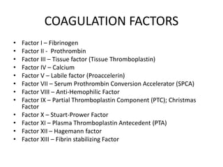
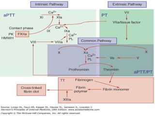
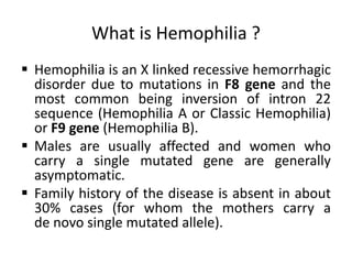
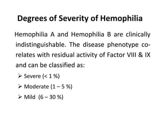
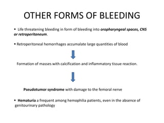
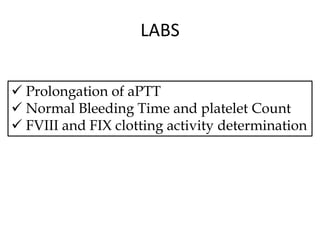
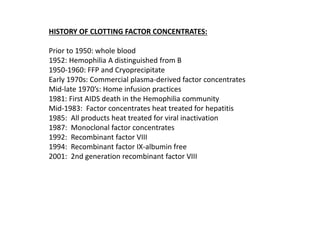
This document provides information on hemophilia, including the objectives, coagulation factors, types and severity of hemophilia, clinical manifestations, complications, labs, treatment including factor replacement therapy and management in special situations. It discusses the basic concepts of hemophilia, how to approach cases, calculate factor requirements, lifestyle modifications, and management during situations like surgery, dental procedures, delivery, and menstruation in hemophiliacs.
1. Hemostasis involves vasoconstriction, platelet plug formation, coagulation, and fibrinolysis.
2. Tests like bleeding time, prothrombin time, activated partial thromboplastin time, and thrombin time are used to assess the coagulation pathway.
3. Hemophilia is an inherited bleeding disorder caused by factor VIII or IX deficiency that impairs hemostasis and causes easy bruising and prolonged bleeding.
Hemophilia is a common hereditary coagulation disorder due to deficiency or reduce activity of clotting factor VIII or clotting factor IX.
This disorder is a X- linked recessive disorder.
Types:
Hemophilia A- deficiency of clotting factors VIII
Hemophilia B- deficiency of clotting factors IX
Hemophilia C- deficiency of clotting factors XI
Parahaemophilia- deficiency of clotting factor V
Causes of hemophilia
Hemophilia has a sex-linked recessive inheritance.
In most cases Hemophilia caused by a mutation in a gene that encodes for one of the clotting factors .
Since the hemophilia gene is located on the X chromosome, Hemophilia usually occurs in males, and Female is the carrier of hemophilia.
Diagnosis
Complete blood cell count
Coagulation studies
FVIII assay
Normal values for FVIII assays are 50-150%. Values in hemophilia are as follows:
Mild: >5%
Moderate: 1-5%
Severe: <1%
Treatment of Hemophilia
Other Types of Treatment
Desmopressin (DDAVP)
Antifibrinolytic Medicines
Vaccinations- hepatitis A and B.
Gene Therapy
Gene Therapy
New Drugs for Hemophilia treatment
Bangladesh perspectives
Bangladesh would have 10800 hemophiliacs.
But, initially the patients does not concern about hemophilia.
Patients are usually diagnosed only after bleeding episode and sometimes the episode are causes serious consequences.
Conclusion
Primary diagnosis with the success of gene therapy and availability of the new bioengineered products the prospect of the hemophiliacs will be brighter in near future.
This document discusses hemophilia, an inherited bleeding disorder caused by mutations in genes coding for blood clotting factors. It describes the history and genetics of the disease, including that hemophilia is X-linked recessive and mostly affects males. The main types are hemophilia A caused by factor VIII deficiency and hemophilia B caused by factor IX deficiency. Symptoms range from mild to severe bleeding. Treatment involves replacement of the missing clotting factor through therapies like fresh or concentrated plasma.
This document provides an overview of hemophilia, including:
- Hemophilia is an inherited bleeding disorder caused by deficiencies in coagulation factor VIII or IX.
- It affects males more often than females and symptoms include prolonged bleeding from cuts or wounds.
- Diagnosis involves screening tests and measuring coagulation factor levels. Severity depends on factor level.
- Treatment involves replacing the missing clotting factor through infusions or using bypassing agents for those who develop inhibitors against factor VIII or IX.
- Bypassing agents work by activating the clotting process without using the deficient factor. The choice of agent depends on factors like the treatment phase and dosing considerations.
- Monitoring is needed to track
Hemophilia is a genetic bleeding disorder caused by deficiencies in clotting factors VIII or IX. It is inherited in an X-linked recessive pattern and primarily affects males. Symptoms range from easy bruising to spontaneous internal bleeding that can be life-threatening. Treatment involves replacement of the missing clotting factor through infusions to prevent or treat bleeding episodes. Repeated bleeding can lead to chronic joint damage over time without proper management.
Hemophilia is a genetic bleeding disorder caused by deficiencies in clotting factors VIII or IX. The main symptoms are prolonged bleeding after injury or surgery and bleeding into joints or muscles. There are three main types - A, B, and C - defined by which clotting factor is deficient. Treatment involves replacing the missing clotting factor through infusions of plasma-derived or recombinant factor concentrates. Management also focuses on preventing bleeding episodes and complications through measures like RICE, immobilization, exercise, and infection control.
This document discusses hemophilia, including:
1. Hemophilia is an inherited bleeding disorder caused by deficiencies in coagulation factor VIII or IX.
2. There are two main types - Hemophilia A caused by factor VIII deficiency and Hemophilia B caused by factor IX deficiency.
3. Treatment involves replacing the missing clotting factor through products derived from plasma or recombinant sources to control bleeding and prevent joint damage. Regular prophylactic treatment starting in early childhood has been shown to significantly reduce long-term joint complications.
I apologize, I do not see any faces in the provided text. The document appears to be about hemophilia, a genetic bleeding disorder. It discusses causes, classifications by severity, management strategies, and dental considerations for treating patients with hemophilia. However, there are no images included that I could identify faces in.
This document discusses aplastic anemia, a bone marrow failure syndrome characterized by pancytopenia. It defines aplastic anemia as a failure of the bone marrow to produce sufficient blood cells, resulting in low red blood cells, white blood cells, and platelets. The causes include stem cell defects, bone marrow suppression by drugs or radiation, bone marrow infiltration by cancer cells, and immune-mediated destruction of hematopoietic stem cells. Aplastic anemia is evaluated based on symptoms of anemia, bleeding, and infections, along with low blood counts and a hypoplastic bone marrow on biopsy with less than 25% cellularity.
Hemophilia is a bleeding disorder that slows down the blood clotting process. People who have hemophilia often have longer bleeding after an injury or surgery. People who have severe hemophilia have spontaneous bleeding into the joints and muscles. Hemophilia occurs more commonly in males than in females.The two most common types of hemophilia are hemophilia A (also known as classic hemophilia) and hemophilia B (also known as Christmas disease). People who have hemophilia A have low levels of a blood clotting factor called factor eight (FVIII). People who have hemophilia B have low levels of factor nine (FIX).
The two types of hemophilia are caused by permanent gene changes (mutations) in different genes. Mutations in the FVIII gene cause hemophilia A. Mutations in the FIX gene cause hemophilia B. Proteins made by these genes have an important role in the blood clotting process. Mutations in either gene keep clots from forming when there is an injury, causing too much bleeding that can be difficult to stop
Hemophilia A is the most common type of this condition. One in 5,000 to 10,000 males worldwide have hemophilia A. Hemophilia B is less common, and it affects 1 in 20,000 to 34,500 males worldwide.
1) Hemophilia is a bleeding disorder caused by deficiencies in clotting factors VIII or IX, resulting in prolonged bleeding from minor injuries or surgery.
2) There are two main types - type A from a factor VIII deficiency is most common, type B from a factor IX deficiency is called Christmas disease.
3) Treatment involves replacing the missing clotting factor, though some may develop inhibitors requiring additional therapies like immunosuppression. Nursing focuses on education, prevention of injury, and management of bleeding episodes.
This document provides an overview of hemophilia, a genetic bleeding disorder caused by a defect in blood clotting factors. It discusses the physical, mental, and emotional characteristics of hemophilia. The genetic source is a defect on the X chromosome, so it primarily affects males. Treatment involves replacing the missing clotting factor through concentrates from donor blood or produced in a lab. Some interesting facts noted are that Queen Victoria passed it to descendants and the life expectancy was previously only 11 years.
Usman Ghani, a 7-year-old boy, presented with pallor, easy fatigability, and bruising. Investigations revealed very low blood counts and a bone marrow biopsy showed hypoplastic/aplastic bone marrow. He was diagnosed with aplastic anemia. Aplastic anemia is a condition where the bone marrow fails to produce sufficient new blood cells, leading to pancytopenia. Treatment involves blood transfusions, antibiotics, growth factors, immunosuppressive drugs, and stem cell transplantation depending on the severity of the condition.
Haemophilia is a rare inherited bleeding disorder caused by deficiencies in clotting factors VIII (hemophilia A) or IX (hemophilia B). The document discusses the history, types, signs and symptoms, inheritance, and treatment of hemophilia. It was first described in 1803 and is often called the "disease of kings" because Queen Victoria was a carrier who passed it on to descendants across Europe. Hemophilia A is the most common type and is treated with clotting factor replacement therapy or desmopressin.
Disseminated intravascular coagulation (DIC) is a syndrome characterized by widespread activation of coagulation that can occur as a result of various underlying conditions. It results from an imbalance between coagulation and anticoagulation processes in the body. DIC can be acute, with bleeding and shock being dominant symptoms, or chronic, where thrombosis and clotting may predominate. The most common triggers of DIC are infectious diseases, cancer, obstetric complications, and severe tissue injury. Diagnosis involves identifying symptoms of bleeding and thrombosis, abnormal laboratory coagulation test results, and ruling out other conditions. Treatment focuses on treating the underlying cause, replacing coagulation factors, platelets, and fibrinogen, and
Hemophilia B is a rare, recessive X-linked bleeding disorder caused by a lack of functional clotting Factor IX. It affects around 20% of hemophilia cases and risk is highest in males. The Factor IX gene is located on the X chromosome and mutations like deletions or point mutations can cause Hemophilia B. A rare form called Leyden Hemophilia B causes low Factor IX levels until puberty when they may rise due to testosterone. Hemophilia C is also a rare bleeding disorder caused by a lack of Factor XI but it is autosomal and affects both sexes, distinguished from other hemophilias by an absence of bleeding into joints and muscles.
This document discusses hemophilia, which is an inherited bleeding disorder caused by deficient or defective clotting factor VIII or IX. It defines the two main types, hemophilia A and B, and discusses their genetics, incidence, clinical manifestations, complications, diagnosis, and management. The key points are: hemophilia is an X-linked recessive disorder that causes prolonged or excessive bleeding; severity depends on factor level with severe <1%, moderate 1-5%, and mild >5%; joint bleeding is common and can lead to long-term damage; treatment involves factor replacement or other medications to prevent or stop bleeding.
Hemophilia is a hereditary bleeding disorder caused by a deficiency of specific clotting factors, which prevents normal blood clotting. The two main types are hemophilia A caused by a factor VIII deficiency and hemophilia B caused by a factor IX deficiency. Symptoms include excessive bleeding, easy bruising, and bleeding into joints. It is usually passed from mother to son and is treated through replacement of the deficient clotting factor. Nursing care focuses on prevention of injury, control of bleeding episodes, and prevention of joint damage.
1. Aplastic anemia is a condition characterized by pancytopenia (low red blood cells, white blood cells, and platelets) due to bone marrow failure.
2. It can be caused by exposure to toxins, radiation, viruses, or immune system attacks on the bone marrow. The bone marrow is hypocellular with fatty replacement of hematopoietic tissue.
3. Symptoms include anemia, increased risk of infection, bruising/bleeding due to low blood cell counts. Diagnosis involves blood tests showing pancytopenia and a bone marrow biopsy revealing a hypocellular marrow. Treatment options include supportive care, immunosuppressants, bone marrow transplant, or androgens
Hemophilia is a genetic bleeding disorder in which body loses the ability to stop bleeding due to low levels or absence of proteins known as ‘’clotting factors’’ which are necessary for clotting of blood. Hemophilia leads to excessive bleeding.
Thrombocytopaenia, or low platelet count, can be caused by decreased platelet production or increased platelet destruction. Causes of decreased production include bone marrow diseases and medications. Increased destruction can be due to immune-mediated causes like Idiopathic Thrombocytopenic Purpura (ITP) or non-immune causes like disseminated intravascular coagulation. ITP is caused by autoantibodies that bind to and destroy platelets, and presents with mild bleeding and a normal bone marrow with increased megakaryocytes. Thrombocytopaenia is diagnosed based on blood counts, smear, and ruling out other potential causes through testing and history. Treatment depends on severity but
The document discusses blood transfusion, including indications for different blood components, blood donation and collection processes, effects of blood storage, and administration of blood transfusions. It covers donor selection criteria, methods of collection to ensure sterility, changes that occur to blood components over time in storage, and steps to properly administer transfusions including patient investigation and cross-matching. Potential complications of transfusion are also summarized, including immediate reactions like febrile responses and allergic reactions, as well as delayed issues such as thrombophlebitis.
Hemophilia is a genetic bleeding disorder caused by a defect in the genes responsible for blood clotting factors VIII or IX. It is usually inherited and affects boys more than girls. Symptoms include prolonged bleeding after injuries or medical procedures. Treatment involves replacing the missing clotting factor through infusions of donated blood products. Current research is developing new formulations of clotting factors that can be stored at room temperature to improve accessibility.
Hemophilia is a genetic bleeding disorder caused by mutations in genes responsible for blood clotting factors VIII, IX or XI. This leads to prolonged bleeding from cuts or wounds. There are three main types - hemophilia A is the most common type caused by factor VIII deficiency. Hemophilia B or Christmas disease results from factor IX deficiency. Hemophilia C is a rare form caused by factor XI deficiency. Treatment involves replacing the missing clotting factor through infusions to control bleeding. Complications can include joint damage and life-threatening internal bleeding if not properly managed.
This document discusses inhibitors in congenital hemophilia and their treatment. It begins with an overview of hemophilia A and B, risk factors for inhibitor development like family history and treatment intensity, and mechanisms of inhibitor action. Treatment options discussed include high-dose factor replacement, bypassing agents like activated prothrombin complex concentrate and recombinant factor VIIa, and immune tolerance induction regimens to eradicate inhibitors. Two studies directly comparing aPCC and rFVIIa found they achieved similar rates of hemostasis, though one study found rFVIIa in a single 270 μg/kg dose was more effective than aPCC or multiple 90 μg/kg rFVIIa doses. Prophylaxis with
Alpha thalassemia is caused by mutations in the genes responsible for producing alpha globin, resulting in excessive destruction of red blood cells and anemia. It is characterized by mild to severe anemia, enlargement of the liver and spleen, and other symptoms. Treatment involves regular blood transfusions, folic acid supplements, and iron chelation therapy. Bone marrow transplant may cure severe cases. Beta thalassemia is caused by mutations in the beta globin gene and is characterized by severe anemia and other symptoms from an early age. Treatment focuses on blood transfusions and iron chelation therapy.
Haemophilia is a bleeding disorder caused by deficiencies in clotting factors VIII or IX. It is inherited through the X chromosome and primarily affects males. Symptoms include prolonged bleeding both internally and externally, especially into joints and muscles. Diagnosis involves measuring factor levels in the blood. Treatment focuses on replacement of the missing clotting factor through medicines or transfusions to prevent or stop bleeding.
Oral consideration and laboratory investigations of bleeding and clotting dis...kashmira483
This document provides information on bleeding and clotting disorders. It discusses the pathophysiology of hemostasis including the vascular, platelet, coagulation, and fibrinolytic phases. It describes different types of bleeding disorders like vessel wall disorders, platelet disorders, and coagulation disorders. Laboratory tests for identifying bleeding disorders are outlined. Oral manifestations and dental considerations for management are summarized. Local hemostatic agents and systemic agents for different bleeding disorders are also mentioned.
Acquired hemophilia A is a rare bleeding disorder caused by autoantibodies against factor VIII. It most commonly presents in older patients as severe bleeding and has a high mortality rate if not properly treated. Evaluation involves testing for prolonged aPTT and ruling out an inhibitor through mixing studies. Treatment focuses on controlling bleeding with bypassing agents or factor VIII while also using immunosuppressants to eliminate the autoantibody inhibitor. Proper management can reduce bleeding and inhibitor levels, but monitoring is needed due to the slow response to therapy.
This document discusses the treatment of pulmonary embolism (PE). It outlines the main objectives of PE treatment as preventing death from PE, post-thrombotic syndrome, and recurrent venous thromboembolism with minimal side effects. The main treatment approaches discussed are anticoagulants, thrombolytic therapy, caval interruption, and surgical removal. Specific treatment recommendations are provided for massive, major and minor PE based on the presence of shock, right ventricular dysfunction, or normal right ventricular function.
Von Willebrand disease is the most common inherited bleeding disorder caused by quantitative or qualitative deficiencies in von Willebrand factor. It is classified into types 1, 2A, 2B, 2M, 2N, and 3 based on pathophysiology. Diagnosis involves testing VWF activity, ristocetin cofactor activity, and multimer analysis. Treatment depends on severity but may include desmopressin, VWF concentrates, or antifibrinolytics. Hemophilia is caused by factor VIII or IX deficiency and classified by severity. Treatment involves factor replacement or bypassing agents for bleeding episodes and immune tolerance induction for inhibitors. Complications include hemarthroses, target joints, and
This document summarizes anticoagulants and their uses. It discusses the coagulation cascade and how anticoagulants work to prevent unwanted clotting. Several classes of anticoagulants are covered, including unfractionated and low molecular weight heparins, vitamin K antagonists like warfarin, and direct acting anticoagulants. Indications, dosages, monitoring, and adverse effects are provided for many of the discussed anticoagulants. The document also addresses perioperative management of anticoagulation and treatments for bleeding or over-anticoagulation.
Heparin is a powerful anticoagulant that acts indirectly by binding to antithrombin III and inactivating clotting factors. It can be given intravenously or subcutaneously. Heparin is used to treat and prevent conditions involving blood clots such as deep vein thrombosis, pulmonary embolism, and arterial thromboembolism. Adverse effects include bleeding and heparin-induced thrombocytopenia. Warfarin is an oral anticoagulant that works by interfering with vitamin K dependent clotting factor synthesis in the liver. It is used long-term for conditions requiring anticoagulation like atrial fibrillation. Risks include bleeding and fetal harms
This document discusses blood transfusion and its complications. It begins by describing the components of blood including red blood cells, white blood cells, platelets, and plasma. It then discusses various blood products like packed red blood cells, platelets, fresh frozen plasma, and cryoprecipitate. The document outlines the indications for transfusion of these different components. It also describes complications of transfusion like fever, allergic reactions, transfusion-related acute lung injury, and volume overload. It stresses the importance of proper consent, typing, and monitoring for complications during blood transfusion.
This document provides information about blood transfusion for anesthesia. It discusses the components of blood, types of transfusions including whole blood, packed red blood cells, platelets, fresh frozen plasma, and cryoprecipitate. It outlines general indications for transfusion including anemia management and volume replacement. It also discusses complications of transfusion such as febrile reactions, allergic reactions, hemolytic reactions, transfusion-related acute lung injury, and transfusion-associated circulatory overload. Monitoring during transfusion and preventing complications are also covered.
This document discusses damage control resuscitation for massive bleeding. It involves 3 key elements: 1) permissive hypotension to minimize blood loss, 2) haemostatic resuscitation using blood products rather than fluids, and 3) damage control surgery to rapidly control bleeding. The trauma triad of death involves hypothermia, coagulopathy, and acidosis which can occur in massive bleeding and must be addressed as part of damage control resuscitation.
Haemorrhage is the leading cause of maternal mortality worldwide. It can cause haemorrhagic shock through significant blood loss. Haemorrhagic shock progresses through compensated, decompensated, and irreversible stages if not treated. Immediate management of haemorrhagic shock focuses on controlling bleeding, fluid resuscitation, and blood product transfusion according to established guidelines. For obstetric haemorrhage, the source of bleeding must also be identified and treated, which may include uterine massage, uterine balloon tamponade, or other uterine-sparing procedures to prevent hysterectomy. Recombinant factor VIIa can be considered for refractory bleeding.
Coagulants and anticoagulants work to maintain a balance in the coagulation system. Coagulants such as fresh whole blood and factors promote clotting, while anticoagulants like antithrombin and the fibrinolytic system inhibit clot formation and maintain blood fluidity. Vitamin K is essential for the production of coagulation factors and warfarin is an oral anticoagulant that works by inhibiting vitamin K. Heparin is commonly used as an injectable anticoagulant that prevents clotting by binding to antithrombin. Newer oral anticoagulants directly inhibit thrombin or factor Xa.
This document discusses thromboembolic diseases, which include deep vein thrombosis (DVT) and pulmonary embolism (PE). It notes that DVT and PE incidence increases during pregnancy and the postpartum period. Pregnancy increases the risk of these conditions 5-10 times due to hormonal and physiological changes. The document then discusses various risk factors for DVT/PE and diagnostic methods. It also outlines treatments which include anticoagulants like heparin and warfarin, as well as compression stockings and exercise.
1. Postpartum haemorrhage (PPH) is defined as blood loss greater than 500 ml within 24 hours of delivery. It can be primary (within 24 hours) or secondary (24 hours to 12 weeks).
2. Causes of PPH include uterine atony, retained placenta, genital tract trauma, and coagulation disorders. Uterine atony accounts for 75-90% of cases.
3. Management involves communication, resuscitation, monitoring, arresting the bleeding. Resuscitation focuses on airway, breathing, circulation, transfusions and fluid resuscitation. Arresting bleeding uses uterotonics, tamponade, compression sutures,
The document provides an overview of managing patients with bleeding disorders. It discusses hemostasis, common lab tests used to evaluate clotting mechanisms, and causes of bleeding disorders including platelet disorders and factor deficiencies. Guidelines are presented for identifying patients with bleeding disorders based on their history. Techniques to maintain hemostasis during surgery include using a harmonic scalpel. The document also reviews recommendations for treating patients taking antiplatelet drugs, anticoagulants, or fibrinolytic drugs and discusses hemophilia and conclusions.
This document provides an overview of anticoagulants including their mechanisms of action, indications, dosing, and monitoring. It discusses normal hemostasis and coagulation factors. Unfractionated heparin, low molecular weight heparins, and factor Xa inhibitors are described as parenteral anticoagulants. Oral anticoagulants reviewed include warfarin, rivaroxaban, apixaban, and dabigatran. The roles of clinical pharmacists in managing anticoagulation therapy are also mentioned.
This document provides an overview of anticoagulants and the blood clotting process. It discusses the four phases of blood clotting - vascular, platelet, coagulation, and fibrinolysis. It then describes various types of anticoagulants including heparin, low molecular weight heparins, direct thrombin inhibitors, vitamin K antagonists like warfarin, and new oral anticoagulants. The mechanisms of action, pharmacokinetics, uses, monitoring, and adverse effects of these anticoagulants are summarized. Recommendations for their use during regional anesthesia and general anesthesia are also outlined.
Packed red blood cells (pRBCs) are used to treat acute blood loss and increase oxygen carrying capacity. Platelets are used when the platelet count is low to reduce the risk of bleeding. Fresh frozen plasma (FFP) contains coagulation factors and is used for bleeding due to multiple coagulation deficiencies. Cryoprecipitate contains high levels of factor VIII, von Willebrand factor, and fibrinogen and is used for von Willebrand disease and hemophilia A. Potential complications of blood transfusions include transmission of infections like hepatitis and HIV, allergic reactions, hemolytic reactions due to ABO incompatibility, alloimmunization, febrile non-hemolytic reactions, transfusion-related acute lung injury
Haemophilia is an X-linked bleeding disorder caused by a deficiency of coagulation factor VIII. It affects males and symptoms include bleeding into joints and muscles following trauma or spontaneously. The severity is classified based on residual factor VIII levels. Treatment involves replacement therapy with factor VIII concentrates or cryoprecipitate to prevent or treat bleeding. Prophylactic infusions are recommended for severe haemophilia to prevent long-term joint damage.
This document discusses body fluid compartments and electrolyte concentrations in different fluid compartments. It provides normal fluid requirements for newborns and adults. It describes the goals and types of fluids used for intraoperative fluid therapy. It also summarizes blood volume, indications for blood transfusion, types of blood products including packed red blood cells, fresh frozen plasma, platelets and cryoprecipitate. Potential complications of blood transfusion and technical considerations are outlined.
A 46-year-old male presented with sudden onset of chest pain radiating to the left arm and shortness of breath. He has risk factors of smoking but no other medical history. On examination, his vitals were stable and heart and lung sounds were normal. The document discusses the arterial supply of the heart and how electrocardiogram leads correspond to different areas of the heart muscle. It provides detailed descriptions of ST segment changes that would indicate occlusions or blocks in different coronary arteries and the regions of the heart affected.
The document presents the 2020 ESC Guidelines for the diagnosis and management of atrial fibrillation developed in collaboration with the European Association for Cardio-Thoracic Surgery. It was developed by a task force of experts and provides updated recommendations based on evidence levels. New recommendations include structured characterization of AF, screening recommendations, use of patient-reported outcomes, reassessment of stroke and bleeding risk, and considerations for rhythm control including catheter ablation.
This document discusses different types of wheezing in children and approaches to diagnosis and treatment. It addresses acute wheezing in young children which is usually due to bronchiolitis or cold and treated symptomatically. Recurrent wheezing may not indicate asthma, and a risk-based probability approach is recommended to evaluate if symptoms warrant treatment with inhaled corticosteroids. Factors like family history, atopy, frequency of colds can help determine the likelihood that recurrent wheezing is asthma. A trial of ICS may help decide if asthma is present when probability is unclear. Bronchodilators are usually sufficient for infrequent viral-induced wheezing.
1. Imaging plays an important role in the evaluation and management of patients with acute stroke.
2. Different imaging modalities such as CT, CT angiography, CT perfusion, MRI, MR angiography, and MR perfusion have benefits for assessing the brain parenchyma, vasculature, perfusion, and identifying potentially salvageable penumbral tissue.
3. Diffusion-weighted MRI is the most sensitive method for detecting acute ischemia within the first few hours, while perfusion imaging can identify tissue at risk of infarction in the ischemic penumbra that may be rescued with reperfusion therapy.
This document discusses arterial disorders and provides an overview of arterial anatomy, layers of arteries, arterial pulses, and clinical tests. It describes common arterial conditions like stenosis, aneurysms, arteritis, and small vessel abnormalities. Specific disorders discussed in detail include thromboangiitis obliterans, Takayasu's arteritis, Raynaud's phenomenon, aneurysms, and arteriovenous fistulas. Three case scenarios are presented concerning Leriche syndrome, arteriovenous fistula, and dissecting aortic aneurysm.
This document provides an overview of interstitial lung diseases (ILDs). It discusses the protective mechanisms of the lung, pathogenesis of ILDs including different presentation patterns, classification of ILDs, diagnostic testing approaches, treatment options including drug therapy and pulmonary rehabilitation. Key points covered include common causes of different radiographic patterns seen on chest imaging for ILDs and rules for approaching a practical diagnosis of ILD based on clinical and imaging features.
Innovative gadgets in anesthesia and medicine ikramdr01
This document provides information on various innovative medical gadgets classified into different categories such as para operative monitors, anesthesia related gadgets, and operation theatre related gadgets. It describes gadgets such as blood pressure monitors, thermometers, weighing scales, pulse oximeters, stethoscopes, ECG machines, glucometers, spirometers, and telehealth solutions. It also discusses smart contact lenses, insulin pens, adhesive bandages, ventilators and other anesthesia equipment that have been developed with new technologies.
This document describes several patient cases presented to highlight important lessons in clinical cardiology. It emphasizes the importance of a thorough history and physical exam over reliance on tests alone. In case 1, careful auscultation revealed pericarditis, not reinfarction. Case 2 showed occult bleeding, not arrhythmia, caused syncope. Case 3 demonstrated a pleural effusion, not refractory heart failure, as the cause of dyspnea. Clinical clues are key to the appropriate management of each case.
Tuberculosis can manifest as either pulmonary or extra-pulmonary disease. Pulmonary TB, which accounts for over 78% of cases, is divided into primary TB occurring after initial infection and reactivation post-primary TB. Extra-pulmonary TB involves sites outside the lungs, with lymph nodes, pleura, and the genitourinary tract being most commonly affected. Symptoms vary depending on the infected site but may include cough, fever, night sweats, and weight loss.
Gestational diabetes (GDM) is glucose intolerance that begins or is first recognized during pregnancy. It can be caused by either pre-existing type 2 diabetes or a new onset of diabetes during pregnancy. The document discusses screening, diagnosis and management of both pre-existing diabetes and GDM during pregnancy. It aims to provide optimal glucose control to support fetal growth while avoiding risks of hyper- and hypoglycemia. Treatment involves medical nutrition therapy, glucose monitoring and may require insulin therapy in some cases. Close monitoring is needed throughout pregnancy and postpartum to support maternal and fetal health.
Gestational trophoblastic disease is a heterogeneous group of lesions arising from abnormal placental trophoblast proliferation. It includes premalignant conditions like complete and partial hydatidiform moles, as well as malignant gestational trophoblastic neoplasia (GTN). GTN has varying potential for local invasion and metastasis. While rare, GTN is highly curable even with widespread dissemination. Treatment involves chemotherapy, with single or multi-agent regimens depending on risk factors and disease stage according to the FIGO scoring system. Careful monitoring of beta-hCG levels is important for diagnosis and follow-up.
This document provides an overview of heart failure, including different types (e.g. acute vs chronic, systolic vs diastolic), biomarkers used to monitor patients, standard treatment options like diuretics and ACE inhibitors, newer drugs in development, and management challenges like high treatment costs. It uses the analogy of a donkey to describe escalating medical interventions for heart failure, from initial diuretic use to more aggressive ionotropes or devices like LVADs for end-stage cases. The document highlights biomarkers that can guide discharge decisions and newer biomarkers being explored, as well as newer drugs and devices in development or use such as CRT devices, ECLS, and LVADs.
A 14-year-old boy presented to the emergency department with symptoms of an autonomic storm after being bitten by a scorpion. He was diagnosed with scorpion sting in autonomic storm. He was given prazosin, hydrocortisone, and other supportive treatments. His condition stabilized and he was discharged after 6 days. Scorpion stings can cause local effects and a systemic autonomic storm response due to neurotoxins that affect sodium channels and induce catecholamine release. Prazosin is an effective treatment as it blocks alpha receptors and counters the effects of venom.
Sarcoidosis and IgG4-related diseases are inflammatory conditions characterized by granuloma formation. Sarcoidosis is a multisystem disorder involving lungs in over 90% of cases and skin, eyes, and liver in about a third of patients each. It is thought to be triggered by an infectious or environmental agent in a genetically susceptible host. IgG4-related disease is a fibroinflammatory condition that can affect virtually any organ, forming tumefactive lesions. Treatment for both conditions typically involves corticosteroids, with immunosuppressants used for chronic or resistant cases.
Neuropathic pain understanding and managementikramdr01
1. The document discusses diabetic neuropathy, including its classification, clinical characteristics, underlying mechanisms, investigations and pharmacological treatment.
2. Key points include that diabetic neuropathy has many subtypes and involves both large and small nerve fibers. The clinical features progress from negative symptoms like loss of sensation to positive symptoms like pain. Investigations include electrodiagnostic testing and skin or nerve biopsies.
3. Treatment involves first-line options like duloxetine, pregabalin and TCAs. Polypharmacy with combinations from different classes may be considered for refractory cases. Factors like comorbidities, side effects, costs and drug interactions must be evaluated when selecting an individual's treatment plan.
Cardiac resynchronization therapy (CRT) and implantable cardioverter defibrillators (ICDs) can help optimize heart failure management. CRT improves symptoms, reduces hospitalizations, and increases survival in patients with reduced ejection fraction, left bundle branch block, and wide QRS duration. ICDs prevent sudden cardiac death in high-risk patients with prior heart failure, low ejection fraction, or history of dangerous arrhythmias. New devices use adaptive and multi-point pacing to better resynchronize the left ventricle. Device therapy improves outcomes when guided by clinical evidence and used in appropriate heart failure patients.
Kawasaki disease is an autoimmune disease that causes inflammation in blood vessels throughout the body. It was first described in 1967 by Dr. Kawasaki in Japan. It most commonly affects children under 5 years old. Without treatment, it can lead to fatal coronary artery aneurysms in some children. The cause is unknown but likely involves genetic and environmental factors such as a viral or bacterial infection. Diagnosis is based on symptoms that include prolonged fever and changes in lips, mouth, hands and feet. Echocardiograms are used to check for heart complications which include aneurysms and heart valve issues.
bedside approach to common congenital heart diseasesikramdr01
This document summarizes common congenital heart diseases, their presentations, and physical exam findings. It discusses how conditions like ventricular septal defect (VSD), atrial septal defect (ASD), patent ductus arteriosus (PDA), pulmonary stenosis (PS), and tetralogy of Fallot (TOF) can present in neonates, infants, children or adolescents with symptoms like cyanosis, heart failure, or murmurs. Physical exam findings for each condition are provided to help determine the location and nature of cardiac lesions.
This document provides an overview of atrial fibrillation (AF), including its pathogenesis, types, diagnosis, and management. Some key points:
- AF is the most common cardiac arrhythmia, affecting around 6% of those over 65. It increases the risk of stroke.
- It occurs when the normal sinus rhythm is overridden by disorganized electrical impulses, usually originating in the lungs.
- Types include paroxysmal, persistent, and permanent. Symptoms range from none to palpitations, dyspnea, chest pain, and neurological issues.
- Diagnosis is made via ECG showing irregular rhythm without P waves. Workup evaluates for underlying causes and stroke risk factors.
Infective endocarditis is a microbial infection of the heart valves or endocardium. It is characterized by the formation of vegetations composed of platelets, fibrin, microorganisms, and inflammatory cells. It occurs more commonly in males and the elderly. Streptococci and Staphylococcus aureus are the most common causes. Diagnosis involves blood cultures, echocardiography, and applying the Duke criteria. Complications include embolisms, heart failure, and metastatic infections. Treatment involves prolonged antibiotic therapy targeted to the infecting organism. Surgery may be needed for complications or uncontrolled infection. Antibiotic prophylaxis is now restricted to highest risk patients undergoing highest risk procedures.
Basavarajeeyam is a Sreshta Sangraha grantha (Compiled book ), written by Neelkanta kotturu Basavaraja Virachita. It contains 25 Prakaranas, First 24 Chapters related to Rogas& 25th to Rasadravyas.
Promoting Wellbeing - Applied Social Psychology - Psychology SuperNotesPsychoTech Services
A proprietary approach developed by bringing together the best of learning theories from Psychology, design principles from the world of visualization, and pedagogical methods from over a decade of training experience, that enables you to: Learn better, faster!
Does Over-Masturbation Contribute to Chronic Prostatitis.pptxwalterHu5
In some case, your chronic prostatitis may be related to over-masturbation. Generally, natural medicine Diuretic and Anti-inflammatory Pill can help mee get a cure.
Basavarajeeyam is an important text for ayurvedic physician belonging to andhra pradehs. It is a popular compendium in various parts of our country as well as in andhra pradesh. The content of the text was presented in sanskrit and telugu language (Bilingual). One of the most famous book in ayurvedic pharmaceutics and therapeutics. This book contains 25 chapters called as prakaranas. Many rasaoushadis were explained, pioneer of dhatu druti, nadi pareeksha, mutra pareeksha etc. Belongs to the period of 15-16 century. New diseases like upadamsha, phiranga rogas are explained.
Muktapishti is a traditional Ayurvedic preparation made from Shoditha Mukta (Purified Pearl), is believed to help regulate thyroid function and reduce symptoms of hyperthyroidism due to its cooling and balancing properties. Clinical evidence on its efficacy remains limited, necessitating further research to validate its therapeutic benefits.
Local Advanced Lung Cancer: Artificial Intelligence, Synergetics, Complex Sys...Oleg Kshivets
Overall life span (LS) was 1671.7±1721.6 days and cumulative 5YS reached 62.4%, 10 years – 50.4%, 20 years – 44.6%. 94 LCP lived more than 5 years without cancer (LS=2958.6±1723.6 days), 22 – more than 10 years (LS=5571±1841.8 days). 67 LCP died because of LC (LS=471.9±344 days). AT significantly improved 5YS (68% vs. 53.7%) (P=0.028 by log-rank test). Cox modeling displayed that 5YS of LCP significantly depended on: N0-N12, T3-4, blood cell circuit, cell ratio factors (ratio between cancer cells-CC and blood cells subpopulations), LC cell dynamics, recalcification time, heparin tolerance, prothrombin index, protein, AT, procedure type (P=0.000-0.031). Neural networks, genetic algorithm selection and bootstrap simulation revealed relationships between 5YS and N0-12 (rank=1), thrombocytes/CC (rank=2), segmented neutrophils/CC (3), eosinophils/CC (4), erythrocytes/CC (5), healthy cells/CC (6), lymphocytes/CC (7), stick neutrophils/CC (8), leucocytes/CC (9), monocytes/CC (10). Correct prediction of 5YS was 100% by neural networks computing (error=0.000; area under ROC curve=1.0).
share - Lions, tigers, AI and health misinformation, oh my!.pptxTina Purnat
• Pitfalls and pivots needed to use AI effectively in public health
• Evidence-based strategies to address health misinformation effectively
• Building trust with communities online and offline
• Equipping health professionals to address questions, concerns and health misinformation
• Assessing risk and mitigating harm from adverse health narratives in communities, health workforce and health system
Histololgy of Female Reproductive System.pptxAyeshaZaid1
Dive into an in-depth exploration of the histological structure of female reproductive system with this comprehensive lecture. Presented by Dr. Ayesha Irfan, Assistant Professor of Anatomy, this presentation covers the Gross anatomy and functional histology of the female reproductive organs. Ideal for students, educators, and anyone interested in medical science, this lecture provides clear explanations, detailed diagrams, and valuable insights into female reproductive system. Enhance your knowledge and understanding of this essential aspect of human biology.
Vestibulocochlear Nerve by Dr. Rabia Inam Gandapore.pptx
Hemophila
1.
2. OBJECTIVES
• To know the basic concepts about Hemophilia
• How to approach a case of Hemophilia?
• How to Calculate Factor requirement?
• Management in special situations
• Lifestyle modifications of the hemophiliacs
3. COAGULATION FACTORS
• Factor I – Fibrinogen
• Factor II - Prothrombin
• Factor III – Tissue factor (Tissue Thromboplastin)
• Factor IV – Calcium
• Factor V – Labile factor (Proaccelerin)
• Factor VII – Serum Prothrombin Conversion Accelerator (SPCA)
• Factor VIII – Anti-Hemophilic Factor
• Factor IX – Partial Thromboplastin Component (PTC); Christmas
Factor
• Factor X – Stuart-Prower Factor
• Factor XI – Plasma Thromboplastin Antecedent (PTA)
• Factor XII – Hagemann factor
• Factor XIII – Fibrin stabilizing Factor
4.
5. What is Hemophilia ?
Hemophilia is an X linked recessive hemorrhagic
disorder due to mutations in F8 gene and the
most common being inversion of intron 22
sequence (Hemophilia A or Classic Hemophilia)
or F9 gene (Hemophilia B).
Males are usually affected and women who
carry a single mutated gene are generally
asymptomatic.
Family history of the disease is absent in about
30% cases (for whom the mothers carry a
de novo single mutated allele).
6. Degrees of Severity of Hemophilia
Hemophilia A and Hemophilia B are clinically
indistinguishable. The disease phenotype co-
relates with residual activity of Factor VIII & IX
and can be classified as:
Severe (< 1 %)
Moderate (1 – 5 %)
Mild (6 – 30 %)
8. Hemophilia clinically manifests when a child begins
to walk or crawl.
In severe and Moderate forms:
* Bleeding into joints (hemarthrosis), soft
tissues and muscles.
* After Trivial trauma or even spontaneously
In milder forms:
* Infrequent bleeding usually secondary to
trauma.
9. FORMSOF BLEEDING
Acute hemarthroses
are painful
erythematous swelling
which have the
tendency to recur.
Chronic hemarthroses
are debilitating with
synovial thickening and
synovitis in response to
intrarticular bleed.
16. OTHER FORMS OF BLEEDING
Life threatening bleeding in form of bleeding into oropharyngeal spaces, CNS
or retroperitoneum.
Retroperitoneal hemorrhages accumalate large quantities of blood
Formation of masses with calcification and inflammatory tissue reaction.
Pseudotumor syndrome with damage to the femoral nerve
Hematuria a frequent among hemophilia patients, even in the absence of
genitourinary pathology
17. LABS
Prolongation of aPTT
Normal Bleeding Time and platelet Count
FVIII and FIX clotting activity determination
19. HISTORY OF CLOTTING FACTOR CONCENTRATES:
Prior to 1950: whole blood
1952: Hemophilia A distinguished from B
1950-1960: FFP and Cryoprecipitate
Early 1970s: Commercial plasma-derived factor concentrates
Mid-late 1970’s: Home infusion practices
1981: First AIDS death in the Hemophilia community
Mid-1983: Factor concentrates heat treated for hepatitis
1985: All products heat treated for viral inactivation
1987: Monoclonal factor concentrates
1992: Recombinant factor VIII
1994: Recombinant factor IX-albumin free
2001: 2nd generation recombinant factor VIII
20. HOME THERAPHY
Advantages:
Immediate access to the treatment
It is achieved with clotting factor concentrates or
other lyophilised products that are safe and can be
stored in a domestic fridge that can be reconstituted
easily
Cryoprecipite can also be kept in fridge but it should
not be frozen.
21. Monitoring of home therapy
1. It should be supervised by a comprehensive care centre with
adequate education and training to the pt. Teaching should include
recognising a bleed and its complications, dosage calculation,
preparation and storage.
2. Administration of clotting factor with aseptic techniques,
performing venipuncture, record keeping, as well as proper
storage and disposal of needles and handling of blood spills
3. Patients or parents should keep bleeding records that include
date and site of bleeding, dosage and lot no. of product used, as
well as any adverse effects.
4. Home care can be instituted for young children having adequate
venous access and motivated family members undergone adequate
training. Self infusion can be encouraged to older children and
teenagers.
5. Implanted venous access device (PORT-A-CATH) can also be
used but this will be associated with complications like infection
and thrombosis.
22. FACTOR REPLACEMENT THERAPY
It can be started in response to a bleeding episode or as
a prophylactic treatment.
Primary Prophylaxis: a tool for maintaining the missing
clotting factor at levels >1% or higher on a regular basis
in order to prevent bleeds, especially at the onset of
hemarthroses.
Hemophiliac boys receiving regular infusions of FVIII
of 25-40 IU/kg (3 days /week) or FIX( 2 days/week) can
reach puberty without detectable joint abnormalities
23. How to calculate Factor requirement?
Factor VIII concentrate increases factor VIII activity approximately 2% for every
1 IU/kg infused. A 50 IU/kg IV bolus raises factor VIII activity approximately
100% over baseline. Extended treatment should follow with 25-IU/kg IV bolus
q12h to maintain sufficient levels.
FVIII dose (IU) = (Target FVIII levels – FVIII baseline levels) x
Body weight (kgs) x 0.5 unit/kg
Half life of FVIII is 8 to 12 hours, hence requires BD dose
to maintain therapautic range
24. FACTOR IX REQUIREMENT
FIX dose (IU) = (Target FIX levels – FIX baseline levels) x
Body weight (kgs) x 1 unit/kg
• FIX recovery postinfusion is usually only 50% of predicted value
• half life of FIX is 24 hours and hence given an OD dose.
25. TREATMENT OF BLEEDS
GOAL OF TREATMENT
Mild Bleeds Uncomplicated Hemarthroses or
superficial hematomas
Maintain factor level at 30 -
50%
Severe
hemarthroses
On Target joint Same as mild bleeds with
additional doses to maintain
levels of 15 -25% for 2 – 3 days.
Large
hematomas
Maintain factor level at 50%
and factor replacement
required for a period of 1 week
or longer
Severe bleeds Oropharyngeal spaces
CNS or retroperitoneum
Maintain factor level at 30 -
100% for 7 days
Prophylactic
replacement
for surgery
Maintain factor level at 100%
for 7 days
26. MANAGEMENT OF BLEEDING WITH
INHIBITORS
1. Patients with inhibitors must be in consultation with centers
experienced in management of such patients before and all
serious bleeds should be managed in such centres
2. Choice of product should be based on the titre of the inhibitor,
records of clinical response to products, site & nature of
bleeding.
3. Patient with a low responding inhibitor should be treated with
a high dose of specific factor replacement at a much higher
dose, if possible to neutralize the inhibitor with excess factor
activity
27. NON TRANSFUSION THERAPHY IN
HEMOPHILIA
Mild-to-moderate hemophilia A with minor bleeding:
* DDAVP (0.3 mg/kg IV in 50 to 100 mL NS infused over 30 minutes, or 300
mg intranasally [Stimate, 1.5 mg/mL] dosed every 12 hours). Increases factor VIII
activity threefold to fivefold and has a half-life of 8 to 12 hours. Tachyphylaxis
may occur after several doses
•ANTI-FIBRINOLYTIC AGENTS: EACA or Tranexemic acid.
Bleeding in gums, GIT or during oral surgery. EACA – loading dose of
200mg/kg followed by 100 mg/kg per dose every 6th hourly. Tranexemic Acid – 25 mg/kg 3
to 5 times a day.
Not indicated to control hematuria because of the risk of formation of occlusive clot in the
lumen of genitoruinary tract structues
29. Inhibitor formation
Formation of alloantibodies to the factor, that is the leading complication of
Hemophilia treatment.
High risk groups:
1. Severe deficiency of factor
2. Family history of inhibitor
3. Mutation in FVIII or FIX
4. Gene derangements
5. Need for intensive replacement therapy ( major surgeries, intracranial
bleeding or trauma)
Clinically, hemophilia is diagnosed when patients do not respond to factor
replacement at therapeutic levels
30. LABS
The laboratory test to conform the presence of an inhibitor is
an aPTT with a mix ( with normal Plasma).
A 1:1mix with normal plasma normally corrects the aPTT but
in patients with inhibitor the aPTT is abnormally prolonged.
Bethedsa assay – to define the specificity of the inhibitor and
its titer.
31. Bethesda Assay for Inhibitors
• Serial dilutions of patient plasma in normal plasma
• Incubate 2 hours
• Assay residual factor activity
• 1 Bethesda Unit neutralizes 50% of factor in an
equivalent volume of normal plasma
• Example: 1:100 dilution of patient plasma + normal
plasma → 50% residual factor activity, so inhibitor
titer is 100 BU
33. Inhibitor patients
High responders
Initial inhibitor titer >10 BU, do
Not respond to FVIII or FIX conc-
entrates
Low responders
Initial inhibitor titer <10 BU responds
Well to human or Porcine FVIII with
Minimal/no increase in inhibitor titres
34. TREATMENT OF HEMOPHILIACS WITH INHIBITORS
• Recombinant factor VIIa
– Enhances TF-driven thrombin formation
• FEIBA (Factor Eight Inhibitor Bypassing Activity)
– Mixture of partially activated vitamin K-
dependent clotting proteases including VIIa
• High dose factor VIII (if low titer inhibitor)
• Induction of tolerance with daily factor VIII infusions
– Optimal dose not established
– Role for concomitant immunosuppression?
Patients with severe hemophilia A and inhibitors resistant to ITI: use of
anti CD20 monoclonal Antibody with FVIII is effective but transitional.
35. INFECTIOUS DISEASES
• HCV infection – major cause of morbidity and 2nd
leading cause of death in hemophilia patients
exposed to older clotting factor concentrates.
• Co-morbidity of underlying liver diseases is clear
in these individuals.
• Response to HCV antiviral therapy is also
restricted with even more proportion among co-
infected with HIV.
• End stage liver disease requiring liver
transplantation may be curative for both liver
disease and for hemophilia.
36. Cardiovascular diseases & malignancy
Though the early assumption that hemophilia
would protect against occlusive vascular disease
because of the underlying hypocoagulablility
but Physical inactivity, hypertension, CKD and HIV
co-infection on ART (commonly observed in
hemophiliacs) points more in favor of
cardiovascular risk.
At risk for HIV and HCV related malignancies.
Hepatocellular cancer (HCC) – most prevalant
among HIV negative hemophiliacs.
38. Goal is to avoid excessive bleeding to protect joints
Avoid contact sports – football, hockey or wrestling
Can indulge in activities like swimming, bicycle riding
Avoid medications – aspirin, ibuprofen, use alternatives like
acetaminophen
Heparin, warfarin, clopidogrel should also be avoided
• PRACTICE GOOD DENTAL HYGIENE
Goal is to prevent excessive bleeding
39. • PROTECT FROM INJURIES :
Knee pads, elbow pads, helmets safety belts all may help in
preventing injuries. Keep your home free of furniture with sharp
corners.
• CARRY IDENTIFICATION AT ALL TIMES:
Always carry a card mentioning the fact that the person is suffering
from hemophilia , the type of treatment & the drugs prescribed etc.
• TRAVEL WITH CARE :
While on travel carry all medications and store information about
hemophilia clinics around the place.
41. 1. Surgical intervention may be elective or emergency. Whenever possible pt. is
shifted to the hemophilic treatment center.
2. Surgery is undertaken in a place(OT) where reliable and adequate laboratory
facilities should be available for monitoring clotting factor level.
3. Pre-operative assessment of inhibitor screen
4. Surgery to be scheduled early in the week and early in the day for optimal
laboratory and blood bank support.
5. Availability of sufficient quantities of clotting factor concentrates..
6. Dosage and duration of clotting factor concentrate coverage depends on type of
surgery performed.
PLANNING FOR SURGERY : Adequate coagulation factor should be available. If
bleeding is very high elective surgery should be postponed. Antiplatelet should
be avoided peri-operatively.
• Adult – 70kg – with severe hemophilia A – taken for major surgery – factor 8
requirement is 50,000 – 80,000 units
SURGERY
Major Minor
Abdominal Removal of skin lesions
Intracranial Dental problems
Spinal Arthroscopy
Joint replacement
42. • Coagulation factor replacement for surgery is based on
1) Through factor level : minimum factor level measured immediately
before next bolus
2) Peak factor level – maximum factor level measured in 1hr of bolus
injection
ADULT - MAJOR SURGERY - CONTINOUS INFUSION
Minor procedures – continuous infusion of factor is enough
Day Factor 8 level Dose(IUKg)
1 - 6 More than 50% 2.5 – 3.0 IU/Kg/hr
More than 7 Often change to bolus
Pre-op : 80 – 100IU/Kg
43. • ADULTS – MAJOR SURGERY – BOLUS DOSING
MINOR SURGERY
Day Factor 8 Factor 9
Pre-op 40-50
1-3 80 - 100 80 - 100
4-6 60- 80 60- 80
More than 7 40- 60 40- 60
Day Factor 8 Factor 9
Pre-op 20 - 30
1-3 40-50 40-50
More than 4 20- 30 20- 30
44. • LIVER BIOPSY : Transjugular liver biopsy is necessary for
hemophiliac & the factor replacement should be done as explained
above, Pt should be hospitalised for 48 hrs
• DENTAL PROCEDURE : After factor 8 replacement for pt.
undergoing dental procedures, antifibrinolytic agent is also strongly
recommeded due to alteration of mucosa in these procedures
• TRANEXEMIC ACID – ANTIFIBRINOLYTIC OF CHOICE
Dose- 15- 20mg/kg
DELIVERY : Deliver the baby by the least traumatic method
Avoid ventouse
Avoid mid cavity forceps
Avoid prolonged labour
Replace factor if necessary for the mother during labour
Check CBC, aPTT,, Factor level & X-matching
45. POSTPARTUM : Keep more than 50% for 3 days
Prophylactic OCP should be started from delivery continued for 1
month
FOR BABIES : Vit. K – to prevent HDN
Hep B vaccination mandatory
All newborns delivered by hemophiliac should be screened by cord
blood
HEMOPHILIAC WOMEN PRESENTED WITH MENORRHAGIA
• Antifibrinolytic agent – Tranexamic acid – 1g Q6H during menstrual
bleed
• DDAVP – Desmopressin – intranasal(1.5mg/ml) or SC
(0.3microgram/Kg)
• Should begin with DDAVP when menstrual bleeding starts & can
be repeated every 12-24hrs
• S/E – facial flushing
• ADH effect (+) hence avoid excess fluid intake