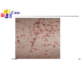
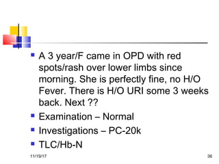
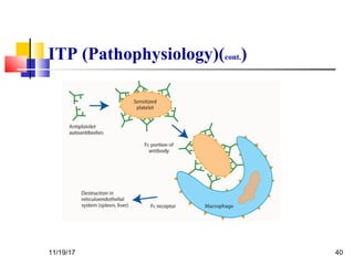
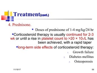
DR SHIV DUBEY
AIIMS Bhopal
11/19/17 1
 A 11 months/F child came to
emergency with severe right knee joint
pain and swelling. There is h/o easy
bruising and blue spots over body. She
has just started to stand with support
and fell.
 History ??
 D/D
 Investigations11/19/17 2
Differential Diagnosis
 Clinically impossible to differentiate from Hemophilia
B- FIX def- Christmas’ disease
 Type 2N vWD, transmitted as an autosomal
recessive trait, is characterized by mutations in VWF
within the factor VIII binding domain. Affected
patients present with low levels of factor VIII (usually
5 to 15 percent of normal), because of unimpeded
proteolytic cleavage of factor VIII, along with a clinical
pattern of bleeding similar to that seen in hemophilia
A, rather than that associated with classical vWD
Should be suspected in families in which an
autosomal recessive (rather than X-linked)
inheritance pattern is seen.
11/19/17 3
11/19/17 4
Definition
 Hemophilia- “love of bleeding”
 Hemophilia A: X linked recessive
hereditary disorder that is due to
defective or deficient factor VIII
11/19/17 5
Incidence
 It is the second most common inherited
clotting factor abnormality (after von
Willebrand disease)
 1 in 5000-10000 live male births
 No difference between racial groups
11/19/17 6
Pathophysiology
 Sequential activation of
a series of proenzymes
or inactive precursor
proteins (zymogens) to
active enzymes,
resulting in significant
stepwise response
amplification.
 Two pathways: intrinsic
and extrinsic measured
by two lab tests
11/19/17 7
Pathophysiology
 F VIII is a cofactor for
intrinsic Xa
 FvW is its carrier
 Activated by Xa and
thrombin
 Inactivated by activated
protein C in conjunction
with protein S
11/19/17 8
Genetics
 Transmitted by females, suffered by males
 The female carrier transmits the disorder to half their
sons and the carrier state to half her dtrs
 The affected male does not transmit the disease to his
sons (Y is nl) but all his dtrs are all carriers
(transmission of defected X)
11/19/17 9
Genetics
 Hemophilia in females

If a carrier female mates with an affected
male there’s the possibility that half their
daughters are homozygous for the
disease

Other possibility: Turner syndrome
(45,X0) with a defective X
11/19/17 10
Clinical manifestations
 Frequency and severity of bleeding are related to F VIII levels
Severity F VIII activity Clinical manifestations
Severe <1% Spontaneous hemorrhage from early
infancy
Freq sp hemarthrosis
Moderate 2-5% Hemorrhage sec to trauma or surgery
Occ sp hemarthrosis
Mild >5% Hemorrhage sec to trauma or surgery
Rare sp bleeding
 Coinheritance of prothrombotic mutations (i.e. Factor V Leiden) can decrease the risk of bleeding
11/19/17 11
Clinical Manifestations:
Hemarthrosis
 The most common, painful and most
physically, economically and
psychologically debilitating manifestation.
 Clinically:
 Aura: tingling warm sensation
 Excruciating pain
 Generally affects one joint at the time
 Most commonly: knee; but there are others
as elbows, wrists and ankles.
 Edema, erythema, warmth and LOM
 If treated early it can subside in 6 to 8 hs
and disappear in 12 to 24 hs.
11/19/17 12
Clinical Manifestations:
Hemarthrosis
 Pathophysiology:
 Bleeding probably starts from synovial vessels into
the synovial space.
 Reabsorption of this blood is often incomplete leading
to chronic proliferative synovitis, where the synovium
is more thickened and vascular, creating a “target
joint” with recurrence of bleeding.
 There is destruction of surrounding structures as
well-bone necrosis and cyst formations, osteophytes
 Terminal stage: Chronic Hemophiliac arthropathy:
fibrous or bony ankilosing of the joint.
11/19/17 13
Clinical Manifestations
Hematomas
 Subcutaneous bleeding
spreads in characteristic
manner- in the site of origin
the tissue is indurated
purplish black and when it
extends the origin starts to
fade
 Muscle hematomas:
1)calf,2)thigh,3)buttocks,4)fo
rearms
 Psoas hematoma- if right
sided may mimic acute
appendicitis
11/19/17 14
Clinical manifestations
Intracranial hemorrhage
 Leading cause of death
of hemophiliacs
 Spontaneous or
following trauma
 Suspect always in
hemophilic patient that
presents with unusual
headache
11/19/17 15
Clinical manifestations
Others
 Gastrointestinal Bleeding:
 Mucous Bleeding:
Epistaxis, gum bleeding.
 Genitourinary Bleeding:
Frequently severe hemophiliac can experience
hematuria and a structural lesion should be ruled out.
11/19/17 16
Laboratory diagnosis
Deficit can be quantitative or qualitative
General Lab: prolonged aPTT, nl PT and BT
Mixing studies: aPTT corrects with normal plasma –if
there are no factor VIII antibodies present
Clotting assays: F VIII activity, expressed in % of
normal DecreasedQUANTITATIVE
Immunoassays: “Cross Reactive Material” Positive-
there is an antigen similar to the F VIII protein-
QUALITATIVE
11/19/17 17
Carrier detection and
Antenatal diagnosis
 Family history: if we follow the inheritance
pattern a female is a carrier if she:
 Has an hemophilic father
 Has two hemophilic sons
 Has one hemophilic son and has a family
history
 Has a son but no family history, there is a
67% chance that she is.
11/19/17 18
Carrier detection and
Antenatal diagnosis
For prenatal diagnosis: DNA testing
on choronic villi samples obtained by
biopsy at week 12
11/19/17 19
Treatment
General Considerations
 Avoidance of aspirin and NSAIDs if at
all possible sometimes it is difficult
because of the painful hemarthrosis
 No IM injections
 Counseling for patient and family, both
genetic and psychosocial, encouraging
normal socialization
11/19/17 20
Treatment
Factor replacement
Replacement of F VIII is the cardinal step to prevent
or reverse acute bleeding episodes
 Dosing: Replacement products can be given on the
basis of body weight or plasma volume ( aprox 5% of
body weight)
 1 U/ml = 100% factor activity
 Practically 1 unit of F VIII/kg increases F VIII about
0.02 U/ml
 In a severe hemophiliac, to raise F VIII to 100%
activity or 1 U/ml, we need 50 U/kg
 Redosing is based on half life: 8-12 hs
 Monitoring of Factor activity is crucial during therapy
11/19/17 21
Treatment
Factor replacement
 Choice of treatment: is based on
 Purity of the factor (how concentrated
or “purified” the factor is)
 Safety
 Cost
 Nowadays most used therapies are
believed to be effective and relatively
safe
11/19/17 22
Treatment
Factor replacement
Sources of F VIII
 Plasma
• FFP was used as the only replacement therapy until
1960s.
• Not really much effective since it could only raise f
VIII to 20%, by giving the patient many liters
• Usually patients experienced severe volume overload
(luckily furosemide was introduced around this time)
• Patients used to have to spend most of their time in
the hospital
11/19/17 23
Treatment
Factor replacement
 Cryoprecipitate
• By mid 1960s Pool et al demonstrated that cold
insoluble material obtained from plasma contained
high levels of F VIII and fibrinogen, achieving a major
advance in hemophilia treatment
• 1 unit of FFP prepared by cryoprecipitate contains
50-120 U of VIII
 Plasma Derived f VIII prepared by monoclonal
antibodies.
11/19/17 24
Treatment
Factor replacement
 Before 1985 all plasma derived products were
highly contaminated by blood borne virus
such as HIV, HBV and HCV which is now
incredibly reduced by the introduction of
donor screening and viral inactivation
techniques such as pasteurization, solvent
detergent treatment and ultrafiltration.
 However, there is still some theoretical
concern about non lipid coated parvovirus,
HAV and prion disease such as Creutzfeld-
Jakob
11/19/17 25
Treatment
Factor replacement
 Recombinant F VIII
• First generation: derived from hamster cell culture.
Contains human albumin for stabilization (possible
source of viral contamination)
• Second Generation: Mutated F VIII, lacking B domain
(no role in clotting) that can be stabilized by
sucrose “albumin free”
11/19/17 26
Treatment
Factor replacement
 Target level and duration of treatment: depend of
severity and site of bleeding
Site of hemorrhage Desired F VIII
level
Duration of treatment
(days)
Hemarthrosis 30-50 1-2
Superficial intramuscular hematoma 30-50 1-2
GI tract 50 7-10
Epistaxis 30-50 Until resolved
Oral Mucosa 30-50 Until resolved
Hematuria 30-100 Until resolved
CNS 50-100 At least 7-10 days
Retropharyngeal 50-100 At least 7-10 days
Retroperitoneal 50-100 At least 7-10 days
11/19/17 27
Treatment
Others
 Fibrin Glue

Contains fibrinogen, thrombin and factor XIII

It’s placed in the site of injury and stabilizes clot

Used in dental procedures and after circumcision
 Antifibrinolyitic Agents

Epsilon aminocaproic acid

Inhibit fibrinolysis by inhibiting plasminogen activator

Adjuvant therapy for dental procedures

Contraindicated in hematuria
 Desmopressin

Transient increase in F VIII levels in pts with mild hemophilia(2-4 times
above baseline)

Mechanism: release from endothelial storage sites

Has spared many hemophiliacs of blood borne products in the 1970s

Repeated administration results in a diminished response-
tachyphylaxis

Side effects: hyponatremia, facial flushing and headache
11/19/17 28
Treatment
Gene Therapy
 Hemophilia is an ideal disease to target for gene
therapy since it is caused by mutations in a single
identified gene.
 A slight increase in factor activity can make a severe
hemophilic in mild.
 Tight regulation of gene expression is not essential.
 Many animal models trials have been studied, being
the main problems encountered: immunogenicity and
short gene expression.
 To date 3 hemophilia A trials in human (aprox 20
patients): transient increase of factor VIII activity and
good safety profile.
 Main issue remains: finding of a gene delivery system
which is nonimmunogenic so as to allow for long term
expression.11/19/17 29
Course and prognosis
 Replacement therapy has its
complications and includes:
 Development of F VIII antibodies
 Liver disease resulting from hepatitis B
and C
 Infection with HIV
11/19/17 30
So…
WHY IS IT CALLED
THE ROYAL
DISEASE?!!?
11/19/17 31
History
Why the Royal disease?
 This is because Queen Victoria, Queen of England from 1837 to
1901, was a carrier.
 Most likely a spontaneous mutation since the duke of Kent (her
father) was not affected and her mother did not have any
affected children from the previous marriage.
 Her eighth child, Leopold, had hemophilia and suffered from
frequent hemorrhages. These were reported in the British
Medical Journal in 1868.
 Leopold died of a brain hemorrhage at the age of 31, but not
before he had children. His daughter, Alice, was a carrier and
her son, Viscount Trematon, also died of a brain hemorrhage in
1928.
 The British family descends from Victoria’s first child Edward
who was not affected. Hence this house is disease free.
11/19/17 32
History
Why the Royal disease?
 Beatrice, Victoria’s youngest child had two
hemophilic sons and a daughter- Victoria
Eugene that was a carrier
 She introduced the hemophilia gene into the
Spanish royal family by marrying king Alfonso
XIII.
 By this time, Queen Victoria’s blood was
recognized as “defective” and the king may
have been warned about Eugene’s carrier
state. However, Royalty was more important
than X chromosomes.
11/19/17 33
History
Queen Victoria’s pedigree
Spanish
House
Russian House
British House
11/19/17 34
Case
11/19/17 35
 A 3 year/F came in OPD with red
spots/rash over lower limbs since
morning. She is perfectly fine, no H/O
Fever. There is H/O URI some 3 weeks
back. Next ??
 Examination – Normal
 Investigations – PC-20k
 TLC/Hb-N
11/19/17 36
Idiopathic (Autoimmune)
Thrombocytopenic Purpura(ITP)
 The most common cause of acute onset of
thrombocytopenia in an otherwise well child
 A recent history of viral illness is described in
50-65% of cases of childhood ITP
11/19/17 37
ITP (cont.)
 1 - 4 wk after exposure to a common viral
infection
 The peak age is 1-4 yr.
 ITP seems to occur more often in late winter
and spring after the peak season of viral
respiratory illness.
11/19/17 38
ITP (Pathophysiology)
 An autoantibody directed against the
platelet surface develops with resultant
sudden onset of thrombocytopenia
 After binding of the antibody to the platelet
surface, circulating antibody-coated platelets
are recognized by the Fc receptor on splenic
macrophages, ingested, and destroyed
11/19/17 39
ITP (Pathophysiology)(cont.)
11/19/17 40
ITP (Pathophysiology)(cont.)
 Most common viruses have been described in
association with ITP, including Epstein-Barr
virus
 In some patients ITP appears to arise in
children infected with Helicobacter pylori or
rarely following the measles, mumps, rubella
vaccine
11/19/17 41
Clinical Manifestations (Cont.)
 The classic presentation of ITP is a
previously healthy 1-4 yr old child who has
sudden onset of generalized petechiae and
purpura
 Often there is bleeding from the gums and
mucous membranes, particularly with
profound thrombocytopenia (platelet count
<10 × 109/L).
11/19/17 42
Clinical Manifestations (cont.)
11/19/17 43
Clinical Manifestations (cont.)
11/19/17 44
Clinical Manifestations (Cont .)
 The presence of abnormal findings such as
hepatosplenomegaly, bone or joint pain, or
remarkable lymphadenopathy suggests other
diagnoses
11/19/17 45
Prognosis
 Severe bleeding is rare (<3% of cases)
 In 70-80% of children who present with acute
ITP, spontaneous resolution occurs within 6
mo
 Fewer than 1% of patients develop an
intracranial hemorrhage.
 Approximately 20% of children who present
with acute ITP go on to have chronic ITP
11/19/17 46
Prognosis(cont.)
 The outcome/prognosis may be related more to
age, as:

ITP in younger children is more likely to
resolve
 The development of chronic ITP in
adolescents approaches 50%.
11/19/17 47
Laboratory Findings
 Severe thrombocytopenia (platelet count <20 ×
109/L) is common, and platelet size is normal
or increased, reflective of increased platelet
turnover
 In acute ITP, the hemoglobin value, white
blood cell (WBC) count, and differential count
should be normal.
11/19/17 48
Laboratory Findings(cont.)
 Bone marrow examination shows normal
granulocytic and erythrocytic series, with
characteristically normal or increased numbers
of megakaryocytes
11/19/17 49
Diagnosis/
Differential Diagnosis
 Autoimmune thrombocytopenia may be an
initial manifestation of :
1. SLE
2. HIV infection
3. Common variable immunodeficiency
4. Lymphoma(rarely)
11/19/17 50
Treatment
 Platelet transfusion in ITP is usually
contraindicated unless life-threatening
bleeding is present (Antiplatelet antibodies
bind to transfused platelets as well as they do
to autologous platelets)
11/19/17 51
Treatment (cont.)
 Initial approaches to the management of ITP
include the following:
1. No therapy other than education and
counseling of the family and patient for
patients with minimal, mild, and moderate
symptoms, as defined earlier.
 This approach is:
 Far less costly
 Side effects are minimal
11/19/17 52
Treatment(cont.)
2.Intravenous immunoglobulin (IVIG).

IVIG at a dose of 0.8- 1.0 g/kg/day for 1-2
days induces a rapid rise in platelet count
(usually >20 × 109/L) in 95% of patients
within 48 hr.
 IVIG appears to induce a response by
downregulating Fc-mediated phagocytosis
of antibody-coated platelets.
11/19/17 53
Treatment(cont.)
2.Intravenous immunoglobulin (IVIG).(cont.)
IVIG therapy is :
 Expensive
 Time-consuming to administer
 After infusion, there is a high frequency
of headaches and vomiting, suggestive of
IVIG-induced aseptic meningitis.
11/19/17 54
Treatment(cont.)
3.Intravenous anti-D therapy.
For Rh positive patients:
IV anti-D at a dose of 50-75 μg/kg causes a
rise in platelet count to >20 × 109
/L in
80-90% of patients within48-72 hr.
11/19/17 55
Treatment(cont.)
4. Prednisone.
 Doses of prednisone of 1-4 mg/kg/24 hr
Corticosteroid therapy is usually continued for 2-3
wk or until a rise in platelet count to >20 × 109/L has
been achieved, with a rapid taper
long-term side effects of corticosteroid therapy:
1. Growth failure
2. Diabetes mellitus
3. Osteoporosis
11/19/17 56
Treatment(Cont.)
 The role of splenectomy in ITP should be reserved for 1 of 2
circumstances.
1. The older child (≥4 yr) with severe ITP that has lasted >1
yr (chronic ITP)
2. Whose symptoms are not easily controlled with therapy
3. Life-threatening hemorrhage (intracranial hemorrhage)
complicates acute ITP
4. Platelet count cannot be corrected rapidly with
transfusion of platelets and administration of IVIG and
corticosteroids
11/19/17 57
All Options
 Steroids
 IVIg / anti-D
 Rituximab (anti-CD20)
 Cyclophosphamide
 Danazol
 Accessory splenectomy
 H. pylori eradication
11/19/17 58
Hematopoietic Malignancies
Lymphoma is a general term used for
proliferations that arise as discrete tissue
masses.
Leukemia is used for neoplasms that present
with widespread involvement of the bone
marrow and the peripheral blood(usually).
59
What is Lymphoma
Malignant lymphoma is a term given to tumors
of the lymphoid system and specifically of
lymphocytes and their precursor cells
i.e.
Cancer of the lymphatic system.
 Many lymphomas are known to be due to
specific genetic mutations.
60
61
 Associated with EBV infection mainly
with mixed cellularity type.
 High socio economic status.
 Prolonged use of of human growth
hormone.
11/19/17 62
 Most common presentation is asymptomatic lymph node
enlargement, typically in the neck.
 Cervical lymphnodes are involved in 80% cases.
 Mediastinal involvement is seen in about 50% cases. They
produce symptoms like chest pain, cough and dyspnoea.
 Other less common symptoms are :
Pruritis, alcohol induced pain over involved lymphnodes,
nephrotic syndrome, erythema nodosum, cerebellar
degeneration, immune hemolytic anaemia, thrombocytopenia,
hypercalcemia.
11/19/17 63
2008 WHO Classification of Hodgkin
Lymphoma
64
Reed-Sternberg cell
65
Ann Arbor Staging System
 Stage I: Single lymph node region (I) or single extralymphatic organ or
site (IE)
 Stage II: > 2 lymph node regions on same side of diaphragm (II) or with
limited, contiguous extra lymphatic tissue involvement (IIE)
 Stage III: both sides of diaphragm involved, may include spleen (IIIS) or
local tissue involvement (IIIE)
 Stage IV: multiple/disseminated foci involved with > 1 extralymphatic
organs (i.e. bone marrow)
 or (B) designates absence/presence of “B” symptoms
*(E) Localized, solitary involvement of extralymphatic tissue, excluding liver
and bone marrow 66
Stage I Stage II Stage III Stage IV
Staging of lymphoma
A: absence of B symptoms
B: fever, night sweats, weight loss
67
68
Chemotherapy
Regimen Medication Regimen Medication
1. ABVD
(US)
•ADRIAMYCIN
•BLEOMYCIN
•VINBLASTINE
•DACARBAZINE
2. STANFORD V
(NEW)
•ADRIAMYCIN
•BLEOMYCIN
•VINBLASTINE
•VINCRISTINE
•PREDNISONE
•MECHLORETHAMIN
E
ETOPOSIDE
3. MOPP •Mechlorethamine
• Vincristine
•Procarbazine
• Prednisone
4. BEACOPP
(EUROPE)
•BLEOMYCIN
•ETOPOSIDE
•ADRIAMYCIN
•CYCLOPHOSPHAMI
DE
•ONCOVIN
•PROCARBAZINE
•PREDNISONE 69
THANK YOU!
11/19/17 70

Hemophilia itp lymphoma

  • 1.
    DR SHIV DUBEY AIIMSBhopal 11/19/17 1
  • 2.
     A 11months/F child came to emergency with severe right knee joint pain and swelling. There is h/o easy bruising and blue spots over body. She has just started to stand with support and fell.  History ??  D/D  Investigations11/19/17 2
  • 3.
    Differential Diagnosis  Clinicallyimpossible to differentiate from Hemophilia B- FIX def- Christmas’ disease  Type 2N vWD, transmitted as an autosomal recessive trait, is characterized by mutations in VWF within the factor VIII binding domain. Affected patients present with low levels of factor VIII (usually 5 to 15 percent of normal), because of unimpeded proteolytic cleavage of factor VIII, along with a clinical pattern of bleeding similar to that seen in hemophilia A, rather than that associated with classical vWD Should be suspected in families in which an autosomal recessive (rather than X-linked) inheritance pattern is seen. 11/19/17 3
  • 4.
  • 5.
    Definition  Hemophilia- “loveof bleeding”  Hemophilia A: X linked recessive hereditary disorder that is due to defective or deficient factor VIII 11/19/17 5
  • 6.
    Incidence  It isthe second most common inherited clotting factor abnormality (after von Willebrand disease)  1 in 5000-10000 live male births  No difference between racial groups 11/19/17 6
  • 7.
    Pathophysiology  Sequential activationof a series of proenzymes or inactive precursor proteins (zymogens) to active enzymes, resulting in significant stepwise response amplification.  Two pathways: intrinsic and extrinsic measured by two lab tests 11/19/17 7
  • 8.
    Pathophysiology  F VIIIis a cofactor for intrinsic Xa  FvW is its carrier  Activated by Xa and thrombin  Inactivated by activated protein C in conjunction with protein S 11/19/17 8
  • 9.
    Genetics  Transmitted byfemales, suffered by males  The female carrier transmits the disorder to half their sons and the carrier state to half her dtrs  The affected male does not transmit the disease to his sons (Y is nl) but all his dtrs are all carriers (transmission of defected X) 11/19/17 9
  • 10.
    Genetics  Hemophilia infemales  If a carrier female mates with an affected male there’s the possibility that half their daughters are homozygous for the disease  Other possibility: Turner syndrome (45,X0) with a defective X 11/19/17 10
  • 11.
    Clinical manifestations  Frequencyand severity of bleeding are related to F VIII levels Severity F VIII activity Clinical manifestations Severe <1% Spontaneous hemorrhage from early infancy Freq sp hemarthrosis Moderate 2-5% Hemorrhage sec to trauma or surgery Occ sp hemarthrosis Mild >5% Hemorrhage sec to trauma or surgery Rare sp bleeding  Coinheritance of prothrombotic mutations (i.e. Factor V Leiden) can decrease the risk of bleeding 11/19/17 11
  • 12.
    Clinical Manifestations: Hemarthrosis  Themost common, painful and most physically, economically and psychologically debilitating manifestation.  Clinically:  Aura: tingling warm sensation  Excruciating pain  Generally affects one joint at the time  Most commonly: knee; but there are others as elbows, wrists and ankles.  Edema, erythema, warmth and LOM  If treated early it can subside in 6 to 8 hs and disappear in 12 to 24 hs. 11/19/17 12
  • 13.
    Clinical Manifestations: Hemarthrosis  Pathophysiology: Bleeding probably starts from synovial vessels into the synovial space.  Reabsorption of this blood is often incomplete leading to chronic proliferative synovitis, where the synovium is more thickened and vascular, creating a “target joint” with recurrence of bleeding.  There is destruction of surrounding structures as well-bone necrosis and cyst formations, osteophytes  Terminal stage: Chronic Hemophiliac arthropathy: fibrous or bony ankilosing of the joint. 11/19/17 13
  • 14.
    Clinical Manifestations Hematomas  Subcutaneousbleeding spreads in characteristic manner- in the site of origin the tissue is indurated purplish black and when it extends the origin starts to fade  Muscle hematomas: 1)calf,2)thigh,3)buttocks,4)fo rearms  Psoas hematoma- if right sided may mimic acute appendicitis 11/19/17 14
  • 15.
    Clinical manifestations Intracranial hemorrhage Leading cause of death of hemophiliacs  Spontaneous or following trauma  Suspect always in hemophilic patient that presents with unusual headache 11/19/17 15
  • 16.
    Clinical manifestations Others  GastrointestinalBleeding:  Mucous Bleeding: Epistaxis, gum bleeding.  Genitourinary Bleeding: Frequently severe hemophiliac can experience hematuria and a structural lesion should be ruled out. 11/19/17 16
  • 17.
    Laboratory diagnosis Deficit canbe quantitative or qualitative General Lab: prolonged aPTT, nl PT and BT Mixing studies: aPTT corrects with normal plasma –if there are no factor VIII antibodies present Clotting assays: F VIII activity, expressed in % of normal DecreasedQUANTITATIVE Immunoassays: “Cross Reactive Material” Positive- there is an antigen similar to the F VIII protein- QUALITATIVE 11/19/17 17
  • 18.
    Carrier detection and Antenataldiagnosis  Family history: if we follow the inheritance pattern a female is a carrier if she:  Has an hemophilic father  Has two hemophilic sons  Has one hemophilic son and has a family history  Has a son but no family history, there is a 67% chance that she is. 11/19/17 18
  • 19.
    Carrier detection and Antenataldiagnosis For prenatal diagnosis: DNA testing on choronic villi samples obtained by biopsy at week 12 11/19/17 19
  • 20.
    Treatment General Considerations  Avoidanceof aspirin and NSAIDs if at all possible sometimes it is difficult because of the painful hemarthrosis  No IM injections  Counseling for patient and family, both genetic and psychosocial, encouraging normal socialization 11/19/17 20
  • 21.
    Treatment Factor replacement Replacement ofF VIII is the cardinal step to prevent or reverse acute bleeding episodes  Dosing: Replacement products can be given on the basis of body weight or plasma volume ( aprox 5% of body weight)  1 U/ml = 100% factor activity  Practically 1 unit of F VIII/kg increases F VIII about 0.02 U/ml  In a severe hemophiliac, to raise F VIII to 100% activity or 1 U/ml, we need 50 U/kg  Redosing is based on half life: 8-12 hs  Monitoring of Factor activity is crucial during therapy 11/19/17 21
  • 22.
    Treatment Factor replacement  Choiceof treatment: is based on  Purity of the factor (how concentrated or “purified” the factor is)  Safety  Cost  Nowadays most used therapies are believed to be effective and relatively safe 11/19/17 22
  • 23.
    Treatment Factor replacement Sources ofF VIII  Plasma • FFP was used as the only replacement therapy until 1960s. • Not really much effective since it could only raise f VIII to 20%, by giving the patient many liters • Usually patients experienced severe volume overload (luckily furosemide was introduced around this time) • Patients used to have to spend most of their time in the hospital 11/19/17 23
  • 24.
    Treatment Factor replacement  Cryoprecipitate •By mid 1960s Pool et al demonstrated that cold insoluble material obtained from plasma contained high levels of F VIII and fibrinogen, achieving a major advance in hemophilia treatment • 1 unit of FFP prepared by cryoprecipitate contains 50-120 U of VIII  Plasma Derived f VIII prepared by monoclonal antibodies. 11/19/17 24
  • 25.
    Treatment Factor replacement  Before1985 all plasma derived products were highly contaminated by blood borne virus such as HIV, HBV and HCV which is now incredibly reduced by the introduction of donor screening and viral inactivation techniques such as pasteurization, solvent detergent treatment and ultrafiltration.  However, there is still some theoretical concern about non lipid coated parvovirus, HAV and prion disease such as Creutzfeld- Jakob 11/19/17 25
  • 26.
    Treatment Factor replacement  RecombinantF VIII • First generation: derived from hamster cell culture. Contains human albumin for stabilization (possible source of viral contamination) • Second Generation: Mutated F VIII, lacking B domain (no role in clotting) that can be stabilized by sucrose “albumin free” 11/19/17 26
  • 27.
    Treatment Factor replacement  Targetlevel and duration of treatment: depend of severity and site of bleeding Site of hemorrhage Desired F VIII level Duration of treatment (days) Hemarthrosis 30-50 1-2 Superficial intramuscular hematoma 30-50 1-2 GI tract 50 7-10 Epistaxis 30-50 Until resolved Oral Mucosa 30-50 Until resolved Hematuria 30-100 Until resolved CNS 50-100 At least 7-10 days Retropharyngeal 50-100 At least 7-10 days Retroperitoneal 50-100 At least 7-10 days 11/19/17 27
  • 28.
    Treatment Others  Fibrin Glue  Containsfibrinogen, thrombin and factor XIII  It’s placed in the site of injury and stabilizes clot  Used in dental procedures and after circumcision  Antifibrinolyitic Agents  Epsilon aminocaproic acid  Inhibit fibrinolysis by inhibiting plasminogen activator  Adjuvant therapy for dental procedures  Contraindicated in hematuria  Desmopressin  Transient increase in F VIII levels in pts with mild hemophilia(2-4 times above baseline)  Mechanism: release from endothelial storage sites  Has spared many hemophiliacs of blood borne products in the 1970s  Repeated administration results in a diminished response- tachyphylaxis  Side effects: hyponatremia, facial flushing and headache 11/19/17 28
  • 29.
    Treatment Gene Therapy  Hemophiliais an ideal disease to target for gene therapy since it is caused by mutations in a single identified gene.  A slight increase in factor activity can make a severe hemophilic in mild.  Tight regulation of gene expression is not essential.  Many animal models trials have been studied, being the main problems encountered: immunogenicity and short gene expression.  To date 3 hemophilia A trials in human (aprox 20 patients): transient increase of factor VIII activity and good safety profile.  Main issue remains: finding of a gene delivery system which is nonimmunogenic so as to allow for long term expression.11/19/17 29
  • 30.
    Course and prognosis Replacement therapy has its complications and includes:  Development of F VIII antibodies  Liver disease resulting from hepatitis B and C  Infection with HIV 11/19/17 30
  • 31.
    So… WHY IS ITCALLED THE ROYAL DISEASE?!!? 11/19/17 31
  • 32.
    History Why the Royaldisease?  This is because Queen Victoria, Queen of England from 1837 to 1901, was a carrier.  Most likely a spontaneous mutation since the duke of Kent (her father) was not affected and her mother did not have any affected children from the previous marriage.  Her eighth child, Leopold, had hemophilia and suffered from frequent hemorrhages. These were reported in the British Medical Journal in 1868.  Leopold died of a brain hemorrhage at the age of 31, but not before he had children. His daughter, Alice, was a carrier and her son, Viscount Trematon, also died of a brain hemorrhage in 1928.  The British family descends from Victoria’s first child Edward who was not affected. Hence this house is disease free. 11/19/17 32
  • 33.
    History Why the Royaldisease?  Beatrice, Victoria’s youngest child had two hemophilic sons and a daughter- Victoria Eugene that was a carrier  She introduced the hemophilia gene into the Spanish royal family by marrying king Alfonso XIII.  By this time, Queen Victoria’s blood was recognized as “defective” and the king may have been warned about Eugene’s carrier state. However, Royalty was more important than X chromosomes. 11/19/17 33
  • 34.
  • 35.
  • 36.
     A 3year/F came in OPD with red spots/rash over lower limbs since morning. She is perfectly fine, no H/O Fever. There is H/O URI some 3 weeks back. Next ??  Examination – Normal  Investigations – PC-20k  TLC/Hb-N 11/19/17 36
  • 37.
    Idiopathic (Autoimmune) Thrombocytopenic Purpura(ITP) The most common cause of acute onset of thrombocytopenia in an otherwise well child  A recent history of viral illness is described in 50-65% of cases of childhood ITP 11/19/17 37
  • 38.
    ITP (cont.)  1- 4 wk after exposure to a common viral infection  The peak age is 1-4 yr.  ITP seems to occur more often in late winter and spring after the peak season of viral respiratory illness. 11/19/17 38
  • 39.
    ITP (Pathophysiology)  Anautoantibody directed against the platelet surface develops with resultant sudden onset of thrombocytopenia  After binding of the antibody to the platelet surface, circulating antibody-coated platelets are recognized by the Fc receptor on splenic macrophages, ingested, and destroyed 11/19/17 39
  • 40.
  • 41.
    ITP (Pathophysiology)(cont.)  Mostcommon viruses have been described in association with ITP, including Epstein-Barr virus  In some patients ITP appears to arise in children infected with Helicobacter pylori or rarely following the measles, mumps, rubella vaccine 11/19/17 41
  • 42.
    Clinical Manifestations (Cont.) The classic presentation of ITP is a previously healthy 1-4 yr old child who has sudden onset of generalized petechiae and purpura  Often there is bleeding from the gums and mucous membranes, particularly with profound thrombocytopenia (platelet count <10 × 109/L). 11/19/17 42
  • 43.
  • 44.
  • 45.
    Clinical Manifestations (Cont.)  The presence of abnormal findings such as hepatosplenomegaly, bone or joint pain, or remarkable lymphadenopathy suggests other diagnoses 11/19/17 45
  • 46.
    Prognosis  Severe bleedingis rare (<3% of cases)  In 70-80% of children who present with acute ITP, spontaneous resolution occurs within 6 mo  Fewer than 1% of patients develop an intracranial hemorrhage.  Approximately 20% of children who present with acute ITP go on to have chronic ITP 11/19/17 46
  • 47.
    Prognosis(cont.)  The outcome/prognosismay be related more to age, as:  ITP in younger children is more likely to resolve  The development of chronic ITP in adolescents approaches 50%. 11/19/17 47
  • 48.
    Laboratory Findings  Severethrombocytopenia (platelet count <20 × 109/L) is common, and platelet size is normal or increased, reflective of increased platelet turnover  In acute ITP, the hemoglobin value, white blood cell (WBC) count, and differential count should be normal. 11/19/17 48
  • 49.
    Laboratory Findings(cont.)  Bonemarrow examination shows normal granulocytic and erythrocytic series, with characteristically normal or increased numbers of megakaryocytes 11/19/17 49
  • 50.
    Diagnosis/ Differential Diagnosis  Autoimmunethrombocytopenia may be an initial manifestation of : 1. SLE 2. HIV infection 3. Common variable immunodeficiency 4. Lymphoma(rarely) 11/19/17 50
  • 51.
    Treatment  Platelet transfusionin ITP is usually contraindicated unless life-threatening bleeding is present (Antiplatelet antibodies bind to transfused platelets as well as they do to autologous platelets) 11/19/17 51
  • 52.
    Treatment (cont.)  Initialapproaches to the management of ITP include the following: 1. No therapy other than education and counseling of the family and patient for patients with minimal, mild, and moderate symptoms, as defined earlier.  This approach is:  Far less costly  Side effects are minimal 11/19/17 52
  • 53.
    Treatment(cont.) 2.Intravenous immunoglobulin (IVIG).  IVIGat a dose of 0.8- 1.0 g/kg/day for 1-2 days induces a rapid rise in platelet count (usually >20 × 109/L) in 95% of patients within 48 hr.  IVIG appears to induce a response by downregulating Fc-mediated phagocytosis of antibody-coated platelets. 11/19/17 53
  • 54.
    Treatment(cont.) 2.Intravenous immunoglobulin (IVIG).(cont.) IVIGtherapy is :  Expensive  Time-consuming to administer  After infusion, there is a high frequency of headaches and vomiting, suggestive of IVIG-induced aseptic meningitis. 11/19/17 54
  • 55.
    Treatment(cont.) 3.Intravenous anti-D therapy. ForRh positive patients: IV anti-D at a dose of 50-75 μg/kg causes a rise in platelet count to >20 × 109 /L in 80-90% of patients within48-72 hr. 11/19/17 55
  • 56.
    Treatment(cont.) 4. Prednisone.  Dosesof prednisone of 1-4 mg/kg/24 hr Corticosteroid therapy is usually continued for 2-3 wk or until a rise in platelet count to >20 × 109/L has been achieved, with a rapid taper long-term side effects of corticosteroid therapy: 1. Growth failure 2. Diabetes mellitus 3. Osteoporosis 11/19/17 56
  • 57.
    Treatment(Cont.)  The roleof splenectomy in ITP should be reserved for 1 of 2 circumstances. 1. The older child (≥4 yr) with severe ITP that has lasted >1 yr (chronic ITP) 2. Whose symptoms are not easily controlled with therapy 3. Life-threatening hemorrhage (intracranial hemorrhage) complicates acute ITP 4. Platelet count cannot be corrected rapidly with transfusion of platelets and administration of IVIG and corticosteroids 11/19/17 57
  • 58.
    All Options  Steroids IVIg / anti-D  Rituximab (anti-CD20)  Cyclophosphamide  Danazol  Accessory splenectomy  H. pylori eradication 11/19/17 58
  • 59.
    Hematopoietic Malignancies Lymphoma isa general term used for proliferations that arise as discrete tissue masses. Leukemia is used for neoplasms that present with widespread involvement of the bone marrow and the peripheral blood(usually). 59
  • 60.
    What is Lymphoma Malignantlymphoma is a term given to tumors of the lymphoid system and specifically of lymphocytes and their precursor cells i.e. Cancer of the lymphatic system.  Many lymphomas are known to be due to specific genetic mutations. 60
  • 61.
  • 62.
     Associated withEBV infection mainly with mixed cellularity type.  High socio economic status.  Prolonged use of of human growth hormone. 11/19/17 62
  • 63.
     Most commonpresentation is asymptomatic lymph node enlargement, typically in the neck.  Cervical lymphnodes are involved in 80% cases.  Mediastinal involvement is seen in about 50% cases. They produce symptoms like chest pain, cough and dyspnoea.  Other less common symptoms are : Pruritis, alcohol induced pain over involved lymphnodes, nephrotic syndrome, erythema nodosum, cerebellar degeneration, immune hemolytic anaemia, thrombocytopenia, hypercalcemia. 11/19/17 63
  • 64.
    2008 WHO Classificationof Hodgkin Lymphoma 64
  • 65.
  • 66.
    Ann Arbor StagingSystem  Stage I: Single lymph node region (I) or single extralymphatic organ or site (IE)  Stage II: > 2 lymph node regions on same side of diaphragm (II) or with limited, contiguous extra lymphatic tissue involvement (IIE)  Stage III: both sides of diaphragm involved, may include spleen (IIIS) or local tissue involvement (IIIE)  Stage IV: multiple/disseminated foci involved with > 1 extralymphatic organs (i.e. bone marrow)  or (B) designates absence/presence of “B” symptoms *(E) Localized, solitary involvement of extralymphatic tissue, excluding liver and bone marrow 66
  • 67.
    Stage I StageII Stage III Stage IV Staging of lymphoma A: absence of B symptoms B: fever, night sweats, weight loss 67
  • 68.
  • 69.
    Chemotherapy Regimen Medication RegimenMedication 1. ABVD (US) •ADRIAMYCIN •BLEOMYCIN •VINBLASTINE •DACARBAZINE 2. STANFORD V (NEW) •ADRIAMYCIN •BLEOMYCIN •VINBLASTINE •VINCRISTINE •PREDNISONE •MECHLORETHAMIN E ETOPOSIDE 3. MOPP •Mechlorethamine • Vincristine •Procarbazine • Prednisone 4. BEACOPP (EUROPE) •BLEOMYCIN •ETOPOSIDE •ADRIAMYCIN •CYCLOPHOSPHAMI DE •ONCOVIN •PROCARBAZINE •PREDNISONE 69
  • 70.