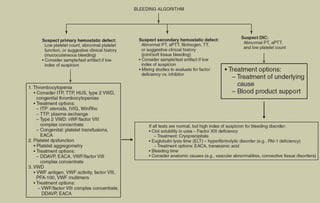
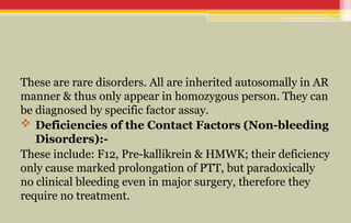
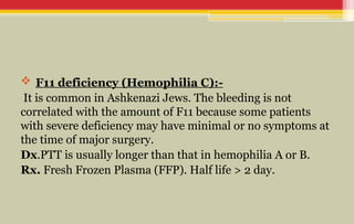
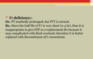
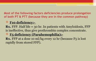
Bleeding tendency
Supervisor: DR. Haifa Ali Bin Dohman
Prepared by: Mohsen Alsolaimani
• 3month baby develop bleeding after circumcision…
• Other thing you want to ask
GENERAL APPROACH
TO PATIENT WITH
BLEEDING TENDENCY
Tests of Platelet disorders
-:
1. Platelet Count: Thrombocytopenia is the most common
acquired cause of bleeding disorder in children. N.R.: 150-
450 Ɨ 103/mm3.
2. Bleeding Time: It assesses platelets and VWF disorders
& their interaction with the vascular wall (in vivo). N.R.: 4-8
min.
3. Platelet Aggregation: It assess platelets function
through their aggregation (in vitro) by; ristocetin, ADP,
epinephrine, collagen, or thrombin. Some platelet
aggregometers measure specific ADP release from platelets.
4. Platelet Function Analyzer (PFA-100): It also assess
platelet adhesion-aggregation in whole blood at high shear
when exposed to collagen-epinephrine or collagen-ADP. It
also can measure some variants of VWD.
Tests of Coagulation factors disorders
-:
1. ProthrombinTime (PT): It measures the factors of extrinsic
pathway by activation of F7 after addition of the Tissue Factor
(TF). PT measures only the following factors: 1, 2, 5, 7, & 10.N.R.:
ā‰ˆ20-13 sec (to validity, it should be standardized by INR).
↑
2. Activated Partial Thromboplastin Time (aPTT or PTT):
It measures the factors of intrinsic pathway by activation of F12
with a surface e.g. silica, glass or a contact activator e.g.kaolin,
ellagic acid.
PTT measures the following factors: 1, 2, 5, 8, 9, 10, 11, 12 & VWF.
N.R.: ā‰ˆ 15-40 sec (N.R. is much more variable with age than PT).
3. Thrombin Time (TT): It measures the final step in the
clotting cascade, in which fibrinogen is converted to fibrin. It
is prolonged when there are disorders of fibrinogen, but may
be falsely prolonged in the presence of heparin or FDP
(fibrin degradation product). N.R.: 11–15 sec.
4. Reptilase Time: It uses snake venom to clot fibrinogen & used
mainly when there is suspicion of heparin contamination (because it not
sensitive to heparin). prolonged in dysfunctional fibrinogen
5. Mixing Studies: It is used when there is unexplained prolongation
of PT or PTT by mixing patient's plasma with normal plasma (1:1) &
repeats the tests. If PT & PTT are corrected, it means a clotting factor
deficiency is present because the presence of 50% level of individual
clotting proteins is sufficient to produce normal PT or PTT; whereas if
there is no or partial correction of PT & PTT, it means an inhibitor is
present (which affect the normal plasma as well). The inhibitor is
mainly either antibodies or heparin, less commonly due to phospholipid
used in the clotting tests.
Note: Bethesda assay measures the amount of inhibitor (which mainly
antibodies against F8 & F9). One Bethesda unit means the amount that
inhibits 50% of the clotting factor in normal plasma.
7. Clotting Factor Assays: Each of the clotting factors can
be measured in the laboratory using individual factor-
deficient plasma. Activity of 100% is expressed as 100
IU/dL. One IU for each factor means the amount of the
factor in 1 ml of normal plasma (referenced against a
standard). For most clotting factors, N.R. is 50–150 IU/dL
(i.e. 50–150% of activity).
HISTORY
It is the most valuable, take details about the site, severity,
previous surgery, menstrual, and family hx.
Ex. Generally, mucocutaneous bleeding is caused by
platelets disorders or VWD, whereas deep bleeding into
muscles and joints is caused by clotting factors deficiency.
Inv. Do 1st Platelet count, PT, & PTT. If abnormal, do
specific factor work-up, whereas if normal, do VWF testing &
Thrombin time.
HEMOPHILIA A & B
These are the most common severe inherited bleeding
disorders that due to deficiencies of F8 (85%) & F9 (15%)
respectively.
These are XL disorders, but female can also be affected if
she is homozygous or by Lyon hypothesis.
Spontaneous mutation is common.
Note: Neither F8 nor F9 can cross placenta, although F8
may in newborn during the birth process (as acute phase
↑
response), whereas F9 do not.
Clinical manifestation
.
It depends on the degree of deficiency as follows:-
- Mild deficiency; factor level >5% of normal, it may be asymptomatic for many years or
require significant trauma to bleed.
- Moderate deficiency; 1-5%, it require a minor trauma.
- Severe deficiency; <1%, there may be spontaneous bleeding.
Bleeding, including intracranial hemorrhage, can occur before (in fetus) or at birth, but more
commonly after circumcision in ā‰ˆ 30% of affected males; however, the most common
manifestations of hemophilia is bleeding into joints or muscles.
Hemarthrosis is mainly occur in large joints, especially ankles, knees and elbows; when
bleeding is repeated in only 1 joint, this will be the "target joint" which eventually result in
erosion of that joint.
Hematoma of muscles e.g. iliopsoas hematoma may cause hypovolemic shock, vague area of
referred pain in groin; hip is held in flexed, internally rotated position; It can be diagnosed US
or CT scan of iliopsoas muscle. Life-threatening hemorrhage & shock may also occur in any
organ or system e.g. CNS, GIT, RT...etc.
Investigation
.
PTT is prolonged 2-3 times above the normal range, whereas
other screening tests are usually normal (including PT).
• Mixing study is positive.
• Specific F8 or F9 assay is used to confirm the Dx.
• Hemophilia carriers (e.g. other family members) can be
screened by Genetic studies or F8:VWF ratio.
Management
Early appropriate therapy is the hallmark of excellent hemophilia care.
• Desmopressin Acetate (DDAVP) Nasal Spray can be used only to treat
patient with mild hemophilia A (not B) in dose, 150 μg (1 puff) for patients
<50 kg & 300 μg (2 puffs) if >50 kg.
• Specific Factor (F8 or F9) Concentrate: The dose of each as follows:-
1. F8 = % of activity desired Ɨ body weight Ɨ 0.5
2. F9 = % of activity desired Ɨ body weight Ɨ 1.4
For mild to moderate bleeding, their levels of must be raised to hemostatic
range (35–50% of activity); whereas for major or life-threatening
hemorrhages, the dose should be raised up to 100% of activity.
However, there are specific situations that require specific doses of F8 in
hemophilia A; where as in hemophilia B, F9 dose should be up to 50- 100%
↑
of below doses:-
• Major surgery or life-threatening hemorrhage; 50–75
IU/kg, then give continuous infusion of 2–4 IU/kg/hr in the
1st day to maintain F8 > 100 IU/dL , then progressively ↓
dose in the subsequent days.
• Iliopsoas hemorrhage; 50 IU/kg, then half the dose every
12 hr until resolution.
• Hemarthrosis; 40 IU/kg on day 1, then half the dose until
resolution. Bleeding into the hip joint may require aspiration.
• Muscle or significant SC hematoma; 20 IU/kg, may need
every-other-day Rx until resolved.
• Mouth bleeding, tooth extraction or epistaxis; 20 IU/kg, antifibrinolytic
Rx (e.g. aminocaproic acid); apply pressure.
• Hematuria; 20 IU/kg, Bed rest, Prednisone!.
• Supportive Care; include:-
• Hemophilia may require comprehensive Hemophilia Care Center which involves
a team of many specialists.
• Education of the patient & his family about the disease, its Rx & Cxs; with
psychological support.
• Avoidance of trauma (including IM injection) or any violent contact sports with
anticipatory guidance.
• Avoidance of Aspirin & other NSAI drugs.
• Vaccination against HBV with periodic screening for blood-borne infections.
• Gene therapy is a promising therapy (in the future) for hemophilia.
Prophylaxis
.
Bleeding can be prevented by giving the specific factor every
other day (or every 3 days) to maintain a measurable clotting
factor (>1% trough level). It used in 2 conditions:-
1. Severe Hemophilia A or B to prevent spontaneous
bleeding
2. Target joint development.
Complication
.
Chronic arthropathy, Transfusion-transmitted infectious
diseases, and response to treatment due to development
↓
of Inhibitors against F8 & F9.
INHIBITORS
These are antibodies against F8 or F9 after repeated infusions that
block their activities which manifested as failure of bleeding to stop
after appropriate replacement therapy. They develop in ā‰ˆ 25–35% in
patient with hemophilia A & to lesser extent in hemophilia B, although
most patients lose their inhibitors after continued regular infusions.
Note: Hemophilia due to mutation that cause inactive dysfunctional
protein factor (which occur in minority of hemophilia A & upto half of
patient with hemophilia B) are less susceptible to the inhibitors.
Dx of inhibitors is by Mixing studies; it can be measured in
Bethesda unit.
Management
1. Desensitization programs, in which high doses of F8 or F9
are infused in an attempt to saturate the antibody and permit the
body to develop tolerance (although this had result in nephrotic
syndrome in some patients with hemophilia B).
2. If desensitization fails, patient can be given rituximab (to Ab
↓
production) or factors that can bypass the inhibitor effect e.g.
recombinant aF7 (Novo7) or activated prothrombin complex
concentrates (which carry a risk of thrombosis).
OTHER CLOTTING FACTORS
DEFICIENCY
These are rare disorders. All are inherited autosomally in AR
manner & thus only appear in homozygous person. They can
be diagnosed by specific factor assay.
 Deficiencies of the Contact Factors (Non-bleeding
Disorders):-
These include: F12, Pre-kallikrein & HMWK; their deficiency
only cause marked prolongation of PTT, but paradoxically
no clinical bleeding even in major surgery, therefore they
require no treatment.
 F11 deficiency (Hemophilia C):-
It is common in Ashkenazi Jews. The bleeding is not
correlated with the amount of F11 because some patients
with severe deficiency may have minimal or no symptoms at
the time of major surgery.
Dx.PTT is usually longer than that in hemophilia A or B.
Rx. Fresh Frozen Plasma (FFP). Half life > 2 day.
 F7 deficiency:-
Dx. PT markedly prolonged, but PTT is normal.
Rx. Since the half life of F7 is very short (2-4 hr), thus it is
inappropriate to give FFP as a replacement Rx because it
may complicated with fluid overload; therefore it is better
replaced with Recombinant aF7 concentrate.
Most of the following factors deficiencies produce prolongation
of both PT & PTT (because they are in the common pathway)
 F10 deficiency:-
Rx. FFP. Half life ā‰ˆ 30 hr. In patients with Amyloidosis, FFP
is ineffective, thus give prothrombin complex concentrate.
 F5 deficiency (Parahemophilia):-
Rx. FFP at a dose 10 ml/kg every 12 hr (because F5 is lost
rapidly from stored FFP).
 F2 (Prothrombin) deficiency:-
It is due to either hypoprothrombinemia or
dysprothrombinemia.
Rx. FFP or Prothrombin complex concentrate. Half life ā‰ˆ 3.5
days.
 F1 (Fibrinogen)deficiency:-
It is due to afibrinogenemia, hypofibrinogenemia, or
dysfibrinogenemia. It may be manifested in the newborn as
hematomas or GIT bleeding.
Dx. Prolonged PT, PTT & TT.
Rx. FFP or Cryoprecipitate. Half life ā‰ˆ 3 days.
 F13 (Transglutaminase or Fibrin-Stabilizing Factor) deficiency:-
Typically, patient has a bruise or hematoma in the next day of
trauma. Other manifestations include: delayed separation of the
umbilical stump beyond 4 wk, poor wound healing, mild bruising &
recurrent spontaneous abortions in women.
Dx. Screening tests are normal, but clot solubility in the presence of 5
↑
M urea.
Rx. FFP, Cryoprecipitate, or F13 concentrate. Half life ā‰ˆ 6 days.
 Antiplasmin & Plasminogen Activator Inhibitor Deficiency:-
These are antifibrinolytic proteins & their deficiency plasmin
→↑
generation and premature lysis of the fibrin clot.
Dx. Screening tests are normal, but euglobulin clot lysis time .
↓
Rx. FFP or Aminocaproic acid (for oral bleeding).
VON WILLEBRAND
DISEASE
VWD is the most common hereditary bleeding disorder that affect ā‰ˆ 1-
2% of population. It inherited as AD (homozygous is affected >
heterozygous); its gene is located on chromosome 12. VWF is an acute-
phase reactant that may be during stressful conditions & pregnancy;
↑
whereas certain diseases (e.g. hypothyroidism) and medications (e.g.
valproic acid) can VWF levels.Level of VWF is also vary according to
↓
blood group, O < A < B < AB!.
Pathophysiology. VWF is a large multimeric glycoprotein that is
synthesized in the endothelial cells and megakaryocytes then stored in
platelets. VWF has 2 functions; 1st it adheres to subendothelial matrix
after vascular damage & then it causes platelets to adhere to it through
their glycoprotein IB (GPIb) receptor; the 2nd function it is a carrier
protein for F8 in plasma.
 Types of VWD:-
ļ‚· Type 1 is the most common form and accounts ā‰ˆ70%. It is due to heterozygous
mutation that causes partial reduction in VWF.
Type 1C is a subtype due to rapid "Clearance" of VWF rather than reduction its
production.
ļ‚· Type 3 is less common & due to homozygous (or compound heterozygous) mutation
which cause a complete reduction in VWF (but measurable F8).
ļ‚· Type 2 is due to abnormal structure which results in dysfunctional protein.
It is divided into the following subtypes:-
2A. Small VWF multimers VWF antigen & activity.
→↓
2B. Hyperactive VWF that binds spontaneously to platelets rapid clearance of both.
→
Platelet-type (or pseudo-) VWD is in contrast to type 2B because the defect is in
the platelets (not VWF) due to hyperactive GPIb receptor that binds spontaneously to
VWF rapid clearance of both.
→
2M. Hypoactive VWF with reduced binding to platelets.
2N. Autosomal Hemophilia due to reduced binding of VWF to F8 rapid clearance
→
of F8.
Clinical manifestation
.
VWD usually cause mucocutaneous hemorrhage e.g.
excessive bruising, epistaxis, postoperative hemorrhage, and
menorrhagia.
Note: Menorrhagia may not be recognized as abnormal
because other females in the family may also be affected
with VWD, thus patient may present later on with iron
deficiency anemia.
Investigation
ļ‚· Platelet function analysis is considered as a screening test for VWD.
ļ‚· PTT & Bleeding Time are usually prolonged (although it may be
normal in type 1 VWD).F8 activity measurement is also reduced in some
types(2N).
ļ‚· VWF antigen.
ļ‚· VWF structure (VWF multimers distribution).
ļ‚· VWF activity (ristocetin cofactor activity).
Type 2B can be diagnosed in vitro by VWF binding to platelets and agglutinates
them at low concentrations of ristocetin.
ļ‚· Platelet count; thrombocytopenia occur in both Type 2B & Pseudo-
VWD.
ļ‚· Genetic studies.
management
ļ‚· Desmopressin acetate (DDAVP) nasal spray can VWF 3- to 5-folds;
↑
however, it is mainly effective in Type 1VWD, whereas in other types of
VWD, it is less effective (or ineffective). Dose is the same as that in mild
hemophilia A.
ļ‚· Cryoprecipitate or VWF concentrate are effective in treatment of all types
of VWD. Half life of both F8 & VWF is 12 hr.
ļ‚· Aminocaproic acid can be used in epistaxis & dental extractions in
dose 100 mg/kg loading dose orally followed by 50 mg/kg every 6 hrs;
or by Tranexamic acid, 1300 mg orally Ɨ3 daily for 5 days.
ļ‚· In severe bleeding, patient should be given platelets (to replace the
VWF in platelets), and F8 (because endogenous correction of F8 require
12-24 hr).
THANK YOU

Bleeding tendency in pediatrics age .pptx

  • 1.
    Bleeding tendency Supervisor: DR.Haifa Ali Bin Dohman Prepared by: Mohsen Alsolaimani
  • 2.
    • 3month babydevelop bleeding after circumcision… • Other thing you want to ask
  • 3.
    GENERAL APPROACH TO PATIENTWITH BLEEDING TENDENCY
  • 6.
    Tests of Plateletdisorders -: 1. Platelet Count: Thrombocytopenia is the most common acquired cause of bleeding disorder in children. N.R.: 150- 450 Ɨ 103/mm3. 2. Bleeding Time: It assesses platelets and VWF disorders & their interaction with the vascular wall (in vivo). N.R.: 4-8 min. 3. Platelet Aggregation: It assess platelets function through their aggregation (in vitro) by; ristocetin, ADP, epinephrine, collagen, or thrombin. Some platelet aggregometers measure specific ADP release from platelets.
  • 7.
    4. Platelet FunctionAnalyzer (PFA-100): It also assess platelet adhesion-aggregation in whole blood at high shear when exposed to collagen-epinephrine or collagen-ADP. It also can measure some variants of VWD.
  • 8.
    Tests of Coagulationfactors disorders -: 1. ProthrombinTime (PT): It measures the factors of extrinsic pathway by activation of F7 after addition of the Tissue Factor (TF). PT measures only the following factors: 1, 2, 5, 7, & 10.N.R.: ā‰ˆ20-13 sec (to validity, it should be standardized by INR). ↑ 2. Activated Partial Thromboplastin Time (aPTT or PTT): It measures the factors of intrinsic pathway by activation of F12 with a surface e.g. silica, glass or a contact activator e.g.kaolin, ellagic acid. PTT measures the following factors: 1, 2, 5, 8, 9, 10, 11, 12 & VWF. N.R.: ā‰ˆ 15-40 sec (N.R. is much more variable with age than PT).
  • 9.
    3. Thrombin Time(TT): It measures the final step in the clotting cascade, in which fibrinogen is converted to fibrin. It is prolonged when there are disorders of fibrinogen, but may be falsely prolonged in the presence of heparin or FDP (fibrin degradation product). N.R.: 11–15 sec.
  • 10.
    4. Reptilase Time:It uses snake venom to clot fibrinogen & used mainly when there is suspicion of heparin contamination (because it not sensitive to heparin). prolonged in dysfunctional fibrinogen 5. Mixing Studies: It is used when there is unexplained prolongation of PT or PTT by mixing patient's plasma with normal plasma (1:1) & repeats the tests. If PT & PTT are corrected, it means a clotting factor deficiency is present because the presence of 50% level of individual clotting proteins is sufficient to produce normal PT or PTT; whereas if there is no or partial correction of PT & PTT, it means an inhibitor is present (which affect the normal plasma as well). The inhibitor is mainly either antibodies or heparin, less commonly due to phospholipid used in the clotting tests. Note: Bethesda assay measures the amount of inhibitor (which mainly antibodies against F8 & F9). One Bethesda unit means the amount that inhibits 50% of the clotting factor in normal plasma.
  • 11.
    7. Clotting FactorAssays: Each of the clotting factors can be measured in the laboratory using individual factor- deficient plasma. Activity of 100% is expressed as 100 IU/dL. One IU for each factor means the amount of the factor in 1 ml of normal plasma (referenced against a standard). For most clotting factors, N.R. is 50–150 IU/dL (i.e. 50–150% of activity).
  • 12.
    HISTORY It is themost valuable, take details about the site, severity, previous surgery, menstrual, and family hx. Ex. Generally, mucocutaneous bleeding is caused by platelets disorders or VWD, whereas deep bleeding into muscles and joints is caused by clotting factors deficiency. Inv. Do 1st Platelet count, PT, & PTT. If abnormal, do specific factor work-up, whereas if normal, do VWF testing & Thrombin time.
  • 13.
  • 14.
    These are themost common severe inherited bleeding disorders that due to deficiencies of F8 (85%) & F9 (15%) respectively. These are XL disorders, but female can also be affected if she is homozygous or by Lyon hypothesis. Spontaneous mutation is common. Note: Neither F8 nor F9 can cross placenta, although F8 may in newborn during the birth process (as acute phase ↑ response), whereas F9 do not.
  • 15.
    Clinical manifestation . It dependson the degree of deficiency as follows:- - Mild deficiency; factor level >5% of normal, it may be asymptomatic for many years or require significant trauma to bleed. - Moderate deficiency; 1-5%, it require a minor trauma. - Severe deficiency; <1%, there may be spontaneous bleeding. Bleeding, including intracranial hemorrhage, can occur before (in fetus) or at birth, but more commonly after circumcision in ā‰ˆ 30% of affected males; however, the most common manifestations of hemophilia is bleeding into joints or muscles. Hemarthrosis is mainly occur in large joints, especially ankles, knees and elbows; when bleeding is repeated in only 1 joint, this will be the "target joint" which eventually result in erosion of that joint. Hematoma of muscles e.g. iliopsoas hematoma may cause hypovolemic shock, vague area of referred pain in groin; hip is held in flexed, internally rotated position; It can be diagnosed US or CT scan of iliopsoas muscle. Life-threatening hemorrhage & shock may also occur in any organ or system e.g. CNS, GIT, RT...etc.
  • 16.
    Investigation . PTT is prolonged2-3 times above the normal range, whereas other screening tests are usually normal (including PT). • Mixing study is positive. • Specific F8 or F9 assay is used to confirm the Dx. • Hemophilia carriers (e.g. other family members) can be screened by Genetic studies or F8:VWF ratio.
  • 17.
    Management Early appropriate therapyis the hallmark of excellent hemophilia care. • Desmopressin Acetate (DDAVP) Nasal Spray can be used only to treat patient with mild hemophilia A (not B) in dose, 150 μg (1 puff) for patients <50 kg & 300 μg (2 puffs) if >50 kg. • Specific Factor (F8 or F9) Concentrate: The dose of each as follows:- 1. F8 = % of activity desired Ɨ body weight Ɨ 0.5 2. F9 = % of activity desired Ɨ body weight Ɨ 1.4 For mild to moderate bleeding, their levels of must be raised to hemostatic range (35–50% of activity); whereas for major or life-threatening hemorrhages, the dose should be raised up to 100% of activity. However, there are specific situations that require specific doses of F8 in hemophilia A; where as in hemophilia B, F9 dose should be up to 50- 100% ↑ of below doses:-
  • 18.
    • Major surgeryor life-threatening hemorrhage; 50–75 IU/kg, then give continuous infusion of 2–4 IU/kg/hr in the 1st day to maintain F8 > 100 IU/dL , then progressively ↓ dose in the subsequent days. • Iliopsoas hemorrhage; 50 IU/kg, then half the dose every 12 hr until resolution. • Hemarthrosis; 40 IU/kg on day 1, then half the dose until resolution. Bleeding into the hip joint may require aspiration. • Muscle or significant SC hematoma; 20 IU/kg, may need every-other-day Rx until resolved.
  • 19.
    • Mouth bleeding,tooth extraction or epistaxis; 20 IU/kg, antifibrinolytic Rx (e.g. aminocaproic acid); apply pressure. • Hematuria; 20 IU/kg, Bed rest, Prednisone!. • Supportive Care; include:- • Hemophilia may require comprehensive Hemophilia Care Center which involves a team of many specialists. • Education of the patient & his family about the disease, its Rx & Cxs; with psychological support. • Avoidance of trauma (including IM injection) or any violent contact sports with anticipatory guidance. • Avoidance of Aspirin & other NSAI drugs. • Vaccination against HBV with periodic screening for blood-borne infections. • Gene therapy is a promising therapy (in the future) for hemophilia.
  • 20.
    Prophylaxis . Bleeding can beprevented by giving the specific factor every other day (or every 3 days) to maintain a measurable clotting factor (>1% trough level). It used in 2 conditions:- 1. Severe Hemophilia A or B to prevent spontaneous bleeding 2. Target joint development.
  • 21.
    Complication . Chronic arthropathy, Transfusion-transmittedinfectious diseases, and response to treatment due to development ↓ of Inhibitors against F8 & F9.
  • 22.
    INHIBITORS These are antibodiesagainst F8 or F9 after repeated infusions that block their activities which manifested as failure of bleeding to stop after appropriate replacement therapy. They develop in ā‰ˆ 25–35% in patient with hemophilia A & to lesser extent in hemophilia B, although most patients lose their inhibitors after continued regular infusions. Note: Hemophilia due to mutation that cause inactive dysfunctional protein factor (which occur in minority of hemophilia A & upto half of patient with hemophilia B) are less susceptible to the inhibitors. Dx of inhibitors is by Mixing studies; it can be measured in Bethesda unit.
  • 23.
    Management 1. Desensitization programs,in which high doses of F8 or F9 are infused in an attempt to saturate the antibody and permit the body to develop tolerance (although this had result in nephrotic syndrome in some patients with hemophilia B). 2. If desensitization fails, patient can be given rituximab (to Ab ↓ production) or factors that can bypass the inhibitor effect e.g. recombinant aF7 (Novo7) or activated prothrombin complex concentrates (which carry a risk of thrombosis).
  • 24.
  • 25.
    These are raredisorders. All are inherited autosomally in AR manner & thus only appear in homozygous person. They can be diagnosed by specific factor assay.  Deficiencies of the Contact Factors (Non-bleeding Disorders):- These include: F12, Pre-kallikrein & HMWK; their deficiency only cause marked prolongation of PTT, but paradoxically no clinical bleeding even in major surgery, therefore they require no treatment.
  • 26.
     F11 deficiency(Hemophilia C):- It is common in Ashkenazi Jews. The bleeding is not correlated with the amount of F11 because some patients with severe deficiency may have minimal or no symptoms at the time of major surgery. Dx.PTT is usually longer than that in hemophilia A or B. Rx. Fresh Frozen Plasma (FFP). Half life > 2 day.
  • 27.
     F7 deficiency:- Dx.PT markedly prolonged, but PTT is normal. Rx. Since the half life of F7 is very short (2-4 hr), thus it is inappropriate to give FFP as a replacement Rx because it may complicated with fluid overload; therefore it is better replaced with Recombinant aF7 concentrate.
  • 28.
    Most of thefollowing factors deficiencies produce prolongation of both PT & PTT (because they are in the common pathway)  F10 deficiency:- Rx. FFP. Half life ā‰ˆ 30 hr. In patients with Amyloidosis, FFP is ineffective, thus give prothrombin complex concentrate.  F5 deficiency (Parahemophilia):- Rx. FFP at a dose 10 ml/kg every 12 hr (because F5 is lost rapidly from stored FFP).
  • 29.
     F2 (Prothrombin)deficiency:- It is due to either hypoprothrombinemia or dysprothrombinemia. Rx. FFP or Prothrombin complex concentrate. Half life ā‰ˆ 3.5 days.  F1 (Fibrinogen)deficiency:- It is due to afibrinogenemia, hypofibrinogenemia, or dysfibrinogenemia. It may be manifested in the newborn as hematomas or GIT bleeding. Dx. Prolonged PT, PTT & TT. Rx. FFP or Cryoprecipitate. Half life ā‰ˆ 3 days.
  • 30.
     F13 (Transglutaminaseor Fibrin-Stabilizing Factor) deficiency:- Typically, patient has a bruise or hematoma in the next day of trauma. Other manifestations include: delayed separation of the umbilical stump beyond 4 wk, poor wound healing, mild bruising & recurrent spontaneous abortions in women. Dx. Screening tests are normal, but clot solubility in the presence of 5 ↑ M urea. Rx. FFP, Cryoprecipitate, or F13 concentrate. Half life ā‰ˆ 6 days.  Antiplasmin & Plasminogen Activator Inhibitor Deficiency:- These are antifibrinolytic proteins & their deficiency plasmin →↑ generation and premature lysis of the fibrin clot. Dx. Screening tests are normal, but euglobulin clot lysis time . ↓ Rx. FFP or Aminocaproic acid (for oral bleeding).
  • 31.
  • 32.
    VWD is themost common hereditary bleeding disorder that affect ā‰ˆ 1- 2% of population. It inherited as AD (homozygous is affected > heterozygous); its gene is located on chromosome 12. VWF is an acute- phase reactant that may be during stressful conditions & pregnancy; ↑ whereas certain diseases (e.g. hypothyroidism) and medications (e.g. valproic acid) can VWF levels.Level of VWF is also vary according to ↓ blood group, O < A < B < AB!. Pathophysiology. VWF is a large multimeric glycoprotein that is synthesized in the endothelial cells and megakaryocytes then stored in platelets. VWF has 2 functions; 1st it adheres to subendothelial matrix after vascular damage & then it causes platelets to adhere to it through their glycoprotein IB (GPIb) receptor; the 2nd function it is a carrier protein for F8 in plasma.
  • 33.
     Types ofVWD:- ļ‚· Type 1 is the most common form and accounts ā‰ˆ70%. It is due to heterozygous mutation that causes partial reduction in VWF. Type 1C is a subtype due to rapid "Clearance" of VWF rather than reduction its production. ļ‚· Type 3 is less common & due to homozygous (or compound heterozygous) mutation which cause a complete reduction in VWF (but measurable F8). ļ‚· Type 2 is due to abnormal structure which results in dysfunctional protein. It is divided into the following subtypes:- 2A. Small VWF multimers VWF antigen & activity. →↓ 2B. Hyperactive VWF that binds spontaneously to platelets rapid clearance of both. → Platelet-type (or pseudo-) VWD is in contrast to type 2B because the defect is in the platelets (not VWF) due to hyperactive GPIb receptor that binds spontaneously to VWF rapid clearance of both. → 2M. Hypoactive VWF with reduced binding to platelets. 2N. Autosomal Hemophilia due to reduced binding of VWF to F8 rapid clearance → of F8.
  • 34.
    Clinical manifestation . VWD usuallycause mucocutaneous hemorrhage e.g. excessive bruising, epistaxis, postoperative hemorrhage, and menorrhagia. Note: Menorrhagia may not be recognized as abnormal because other females in the family may also be affected with VWD, thus patient may present later on with iron deficiency anemia.
  • 35.
    Investigation ļ‚· Platelet functionanalysis is considered as a screening test for VWD. ļ‚· PTT & Bleeding Time are usually prolonged (although it may be normal in type 1 VWD).F8 activity measurement is also reduced in some types(2N). ļ‚· VWF antigen. ļ‚· VWF structure (VWF multimers distribution). ļ‚· VWF activity (ristocetin cofactor activity). Type 2B can be diagnosed in vitro by VWF binding to platelets and agglutinates them at low concentrations of ristocetin. ļ‚· Platelet count; thrombocytopenia occur in both Type 2B & Pseudo- VWD. ļ‚· Genetic studies.
  • 36.
    management ļ‚· Desmopressin acetate(DDAVP) nasal spray can VWF 3- to 5-folds; ↑ however, it is mainly effective in Type 1VWD, whereas in other types of VWD, it is less effective (or ineffective). Dose is the same as that in mild hemophilia A. ļ‚· Cryoprecipitate or VWF concentrate are effective in treatment of all types of VWD. Half life of both F8 & VWF is 12 hr. ļ‚· Aminocaproic acid can be used in epistaxis & dental extractions in dose 100 mg/kg loading dose orally followed by 50 mg/kg every 6 hrs; or by Tranexamic acid, 1300 mg orally Ɨ3 daily for 5 days. ļ‚· In severe bleeding, patient should be given platelets (to replace the VWF in platelets), and F8 (because endogenous correction of F8 require 12-24 hr).
  • 37.