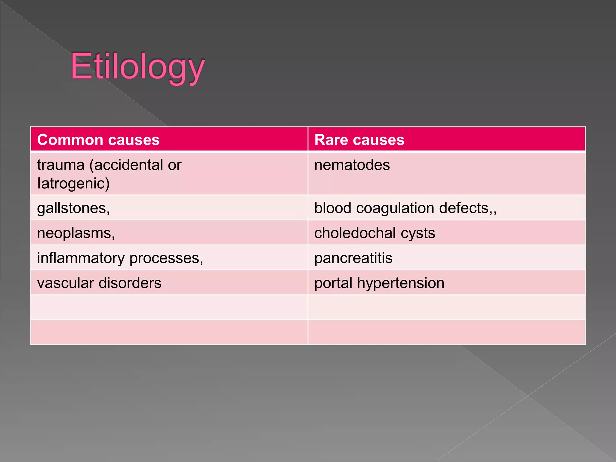
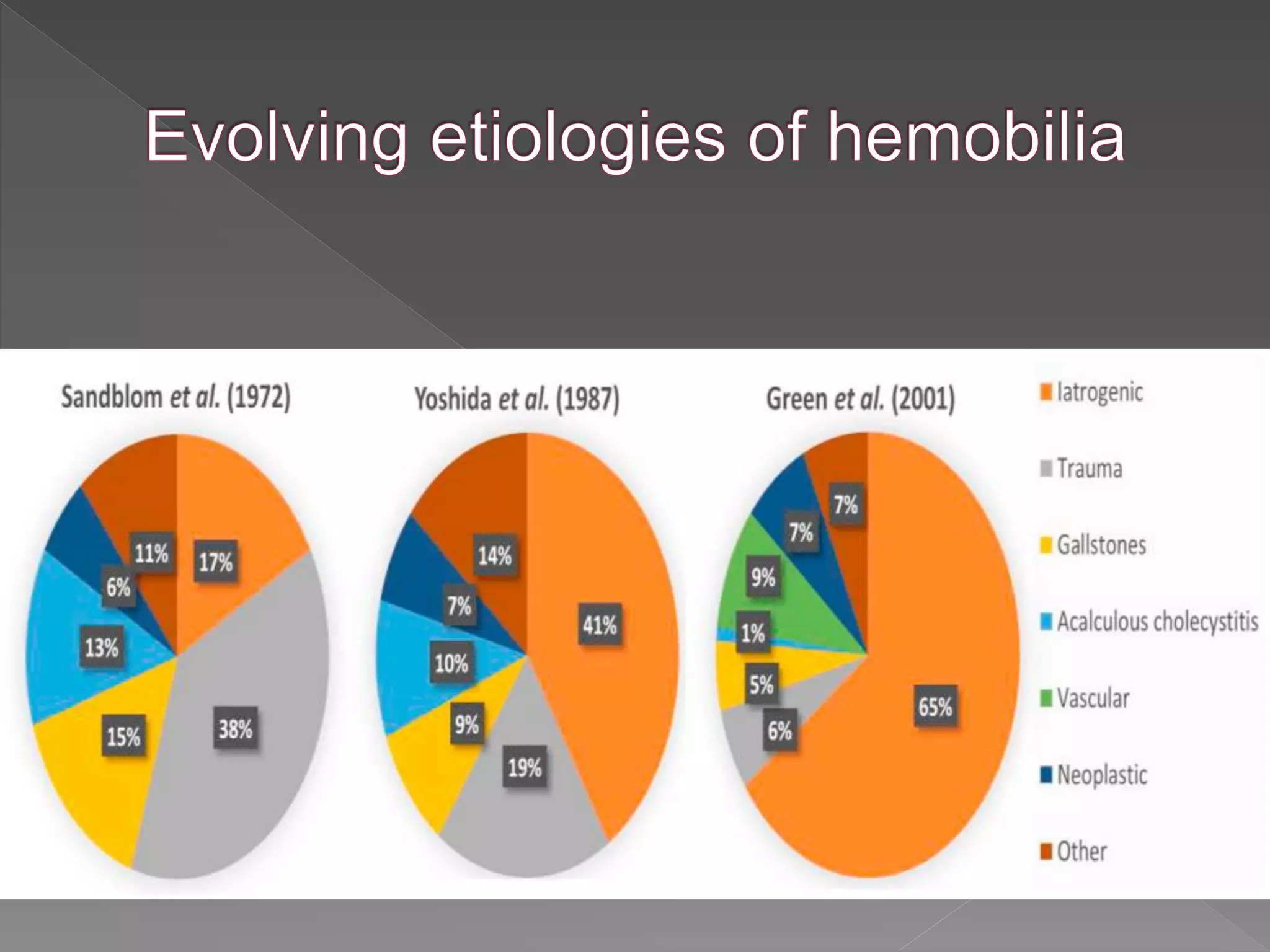
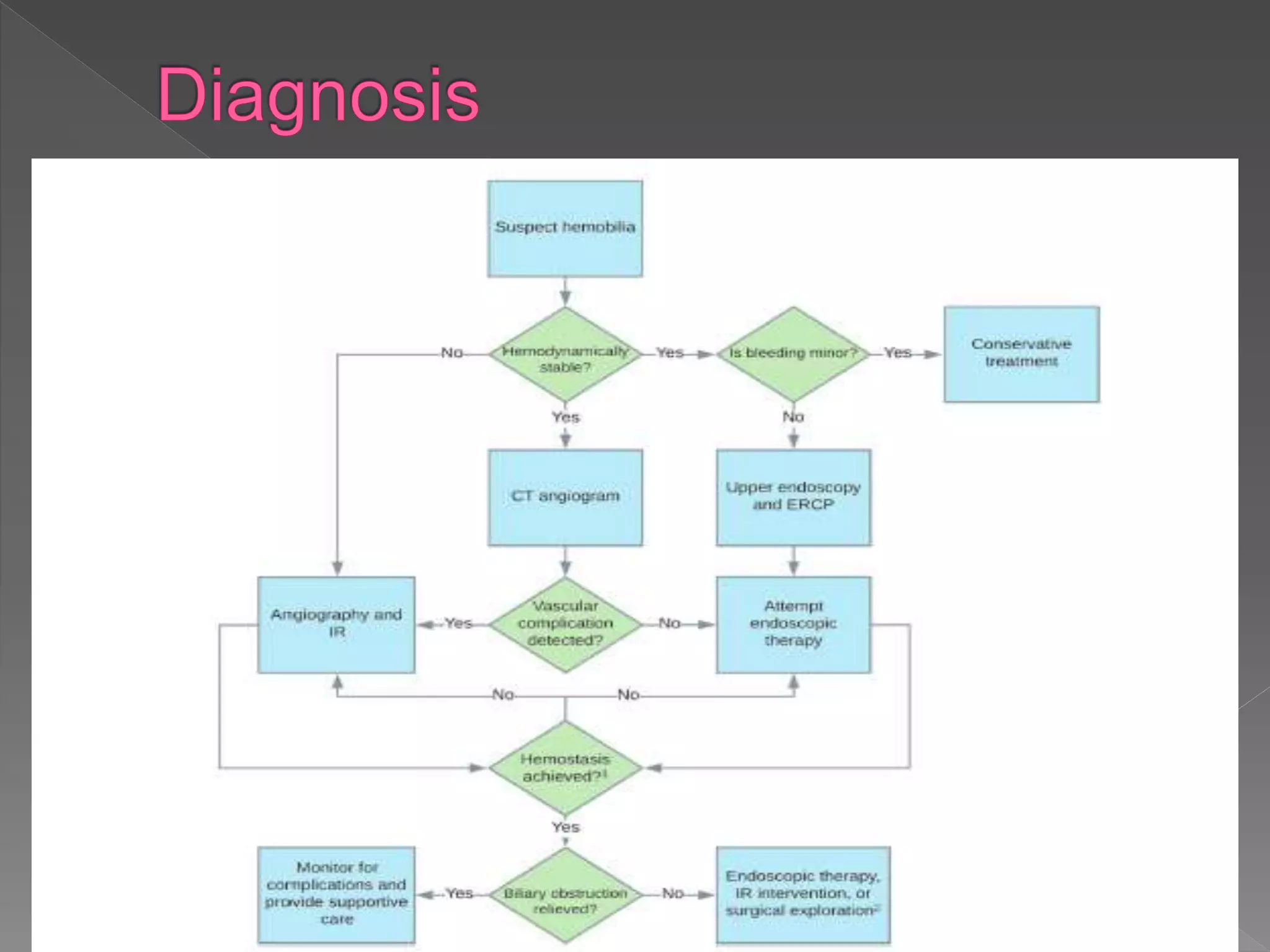
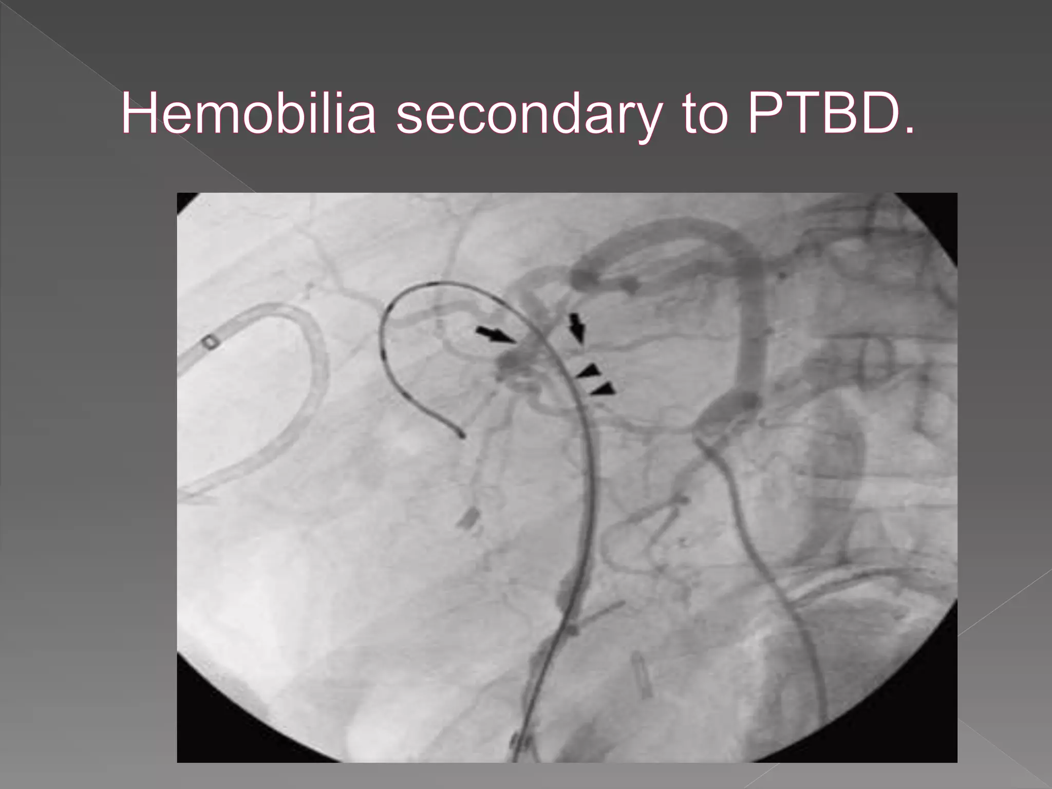
3. Refers to hemorrhage
into the biliary tract
occurs when disease or
trauma produces an
abnormal
communication
between blood vessels
and bile duct
4. The first published case in North
America was by a Boston
surgeon, Jackson (1834), who
reported on the clinical and
pathologic consequences of an
“aneurysm of the hepatic artery
bursting into the hepatic duct,”
the first direct observation of an
abnormal communication
between the blood vessels and
biliary ducts
In 1871, the German surgeon
Quincke (1871) further
characterized biliary tract
hemorrhage by establishing three
cardinal symptoms: GI
hemorrhage, biliary colic, and
jaundice, commonly called
“Quincke’s triad.”
Francis Glisson
7. The incidence of clinically significant hemobilia after
percutaneous liver biopsy is between 0.01% and
0.06% (Howlett et al, 2013)
The risk of hemobilia
chronic liver disease because of the presence of
ascites,
coagulopathy,
platelet dysfunction
liver transplant .
elevated INR >1.3
platelet count <60,000
8. PTBD and PTC are associated with a significantly
higher rate of hemobilia than percutaneous liver
biopsy because of the proximity of hepatic
vasculature to the biliary tree
Delayed presentation of hemobilia
Large bore needle gauge size was associated
with hemobilia
left-sided
PTBD was associated with hemobilia
9. risk of hemobilia is higher with PTBD (2.2%)
than with PTC alone (0.7%). (Rivera et al)
This three-fold increase in hemobilia with
PTBD compared to PTC alone may be
due to the greater size of the aperture made
in the bile duct wall with PTBD
presence of a foreign material remaining in
the duct which can serve as a cause of
inflammation or erosion.
11. endoscopic sphincterotomy associated bleeding typically occurs at
the level of cut papillary sphincter, but blood can occasionally reflux
from the duodenum into the biliary tree.
Risk of hemobilia
stricturoplasty,
extraction of large stones)
Coagulopathy
presence of diseased tissue (e.g. hypervascular tumor)
presence of variant anatomy
, aggressive biliary balloon dilation
intraductal biopsy acquisition,
12. Surgeries performed near the cystic and right
hepatic artery
Cholecystectomy, liver transplantation, and
pancreaticoduodenectomy
Mode of injury of the hepatic artery-suture,
dissection, diathermy injury, clip migration, or
erosion and subsequent arteriobiliary fistula or
pseudoaneurysm
The majority of pseudoaneurysms occur in the
right hepatic artery, though they can occur in
any artery near the hepatobiliary system.
13. the most common spontaneous cause of hemobilia is
primary or metastatic hepatobiliary malignancy.
Eg. cholangiocarcinoma, pancreatic cancer, gallbladder
cancer, liver metastasis, and hepatocellular carcinoma
(HCC)
due to more friable tissue and
vasculaturespontaneous hemorrhage.
Cases have been reported hemobilia as an initial
presentation of cholangiocarcinoma (Manolakis AC et al
2008 Ryu DK et al 2010)
HCC also can cause hemobilia with biliary ductal
invasion- spontaneous rupturing (very rare)
14. Chronic obstruction of the pancreatobiliary tract
can potentially lead to inflammation, erosion,
and fistulization with adjacent vascular
structures and resultant hemobilia. Even when
gallstones are present, however, there can still
be concurrent hemobilia, especially in cases
wherein the stone erodes through the cystic
artery or other vascular, analogous to how a
stone can erode and fistulize into the duodenum
and cause duodenal outlet obstruction in
Bouveret's syndrome.
15. Cause of infectious hemobilia is “tropical
hemobilia”, a result of parasitic infestation of
the biliary tract.
Commonly implicated organisms include
roundworms (e.g. Ascaris lumbricoides), the
Chinese liver fluke (Clonorchis sinensis), and
the sheep liver fluke (Fasciola hepatica).
Echinococcal infections can also cause
hemobilia indirectly
16. Quincke's triad: jaundice, right upper quadrant abdominal
pain, and upper gastrointestinal hemorrhage
Typically, hemobilia can present as hematemesis, melena,
or hematochezia, with or without choluria
ERCP-related hemobilia tends to present immediately or
within a few days after the inciting biliary duct injury (e.g.
sphincterotomy or biliary stricturoplasty).
Hemobilia due to venous sources tends to be lower volume
or selflimited compared to the arterial sources.
Because of their similar echogenicity, it is important to
recognize that clots can masquerade as biliary stones on
imaging studies thus requiring a high index of suspicion
congruent with the clinical presentation.
19. CT of the abdomen with
angiography protocol has become
a first choice diagnostic test for
hemobilia.
The advantages include
non-invasive nature
and low radiation exposure
compared to conventional
angiography,
rapid results
excellent diagnostic
performance characteristics
Hemobilia in a child due to right hepatic artery pseudoaneurysm: Multidetector-row
computed tomography demonstration(Wani Nisar A et al. 2011)
20. Up to 60% of hemobilia cases can be diagnosed by
upper endoscopy.
ERCP can be used to further visualize the biliary
tree and may offer therapeutic options in patients
with hemobilia and associated biliary obstruction.
Characteristic ERCP findings that suggest the
presence of blood clots include amorphous, tubular,
or cast-like filling defects with otherwise
unexplained common bile duct or peri-hilar ductal
dilation.
21. Although formal angiography
is no longer used as a first-line
study, it remains the gold-
standard for both diagnosis
and treatment of hemobilia in
most settings.
If the bleeding vessel has not
already been identified on non-
invasive imaging, the first
angiographic study should be
a celiac arteriogram with
delayed phase imaging to
visualize both the hepatic
arteries as well as the portal
vein.
22. Is the procedure really indicated?
Procedure
Liver biopsy Tract embolization
PTBD Avoid insertion near liver hilum
Cholecystectomy Avoid injury to hepatic arteries by
e.g. diathermy, suture, clips
Minimize infection, biloma
Patient’s own risk
Ascites Drainage
Coagulopathy Replacement of clotting factors, vitamin K1, withhold
anti-platelet agents
23. Main objectives:
achieving hemostasis
maintaining bile flow
The approach depends on several factors:
including the suspected source of bleeding
(arterial vs. venous bleeding),
degree of hemodynamic instability
etiology/cause
24. Indication :Minor hemobilia
Often due to injury related to PTBD catheters
Exchanging a PTBD catheter with a larger sized one
Adjusting its position
Minor hemobilia will often resolve with maturation of the
surgically created tract.
A tractogram or “tubogram”, can be performed
If hemobilia persists, options such as embolization of the
existing percutaneous tract and creation of a new tract can
be considered
25.
26. ERCP are typically the initial therapeutic procedure
of choice
postsphincterotomy hemobiliaduring
sphincterotomy, can be treated
-by spraying diluted epinephrine (1:10,000) over the
area of hemorrhage
-injection of epinephrine into the adjacent tissue
-monopolar or bipolar coagulation
-fibrin sealant injection
-hemoclipping
-balloon tamponading
-stent placement
27. (e.g. peri-hilar) bleeding
source, other
accessories and
methods to treat the
hemobilia tend to be
needed, including
devices to extract
intraductal clots, e.g.
extraction balloon
catheters and retrieval
baskets, followed by
stent placement, among
other options
28.
29. Indication
if non-invasive imaging shows significant
arterial extravasation
presence of large arterial aneurysms or
pseudoaneurysms
presence of arterio-biliary fistulae
intrahepatic or extrahepatic vascular lesions
The success rate of TAE has been reported to be as high as
80% to 100%.
TAE should be avoided, in patients
-liver allografts
-cirrhosis with concurrent shock
-portal vein
30. Once the bleeding site has been identified angiographically,
superselection of the injured artery via threading of a microcatheter to
the target area is performed, followed by TAE using coils.
Coiling should be performed in a distal-to-proximal fashion to avoid
back bleeding via intrahepatic arterial collaterals.
Pseudoaneurysms should be embolized with coils from the two ends
to reduce the risk of enlarging the aneurysm
Complications of TAE include
- hepatic abscesses
-postembolization syndrome
-hyperaminotransaminasemia
- hepatic ischemia
-hepatic infarction or rarely failure
31. An alternative to embolization, as alluded to earlier,
is the placement of a covered stent across the site
of vascular injury.
Advantage of preserving flow through the artery,
which may be beneficial.
The diameter of most hepatic vessels is similar to
the size of coronary vessels, making coronary
stents ideal for this application.
Stent diameter should be slightly oversized by
about 10%e20% of the diameter of the target
vessel and extend approximately 10mm to either
side/end of the site of injury to ensure proper
tamponade.
32. Surgical intervention is rarely necessary and usually reserved
for failed endoscopic, endovascular, and/or percutaneous
therapies.
However, it is first-line if pseudoaneurysms are infected or if
they are compressing other vascular structures. Surgery may
also be indicated if cholecystitis is present, among other
uncommon scenarios.
Options for surgery
hepatic artery ligation,
Pseudoaneurysm excision
hepatic segmentectomy/lobectomy with the potential for
concurrent cholecystectomy if cholecystitis is present or the
gallbladder neck is involved.
Although surgery has a high success rate of above 90%, it is
also associated with a high mortality of up to 10%.
33. Hemobilia is an unusual but important cause of GI bleeding and most
commonly due to hepatopancreatobiliary tract procedures, regional trauma,
and malignancy.
The CT angiography and endoscopy/ERCP have become common initial
diagnostic testing modalities.
Most cases of minor hemobilia can be treated conservatively or with minimally-
invasive endoscopic management.
While TAE is a mainstay, vascular stenting has gained traction as an alternative
to embolization due to the preservation of hepatic arterial blood supply.
Surgery is typically reserved as a last resort due to its high mortality rate and
invasive nature relative to alternative approaches.
Editor's Notes
Francis Glisson (Fig. 105.1) discussed the possibility of hemorrhage into the biliary tract when describing the case of a man who died from presumed GI hemorrhage following a stab wound to the liver suffered in a duel
The increase in hemobilia caused by iatrogenic trauma parallels the advent and increasing use of both diagnostic and therapeutic instrumentation
of the biliary tract.
Evolving etiologies of hemobilia over time. A transition can be seen from traumatogenic to iatrogenic cases of hemobilia over time
It was on repeat ERCP that the biliary ductal systemwas noted to have been infiltrated with tumor and lined by friable tissue with blood clots evident in the papilla of Vater, consistent with hemobilia. This case highlights the rare but important consideration of hemobilia and underlying malignancy in a patient presenting with classic symptoms despite a lack of previous trauma or instrumentation
,as hydatid cysts may cause inflammation of perivascular tissue, weakening of vessel walls, and/or pseudoaneurysm formation with resultant bleeding into the biliary tract
, depending on the rate of bleeding and anatomical factors (e.g. post-bilioenteric surgical anatomy).
However, the clinical presentation of hemobilia often depends on the etiology. For example, patients with a percutaneous transhepatic biliary drain (PTBD) may present with bloody output from the biliary drain . The timing of presentation can also vary and possibly aid in diagnosis. arterial (except in cases of portal hypertension, in which case it may be larger volume and/or persistent).
Hemobilia should be suspected in any patient with an unclear source of gastro-intestinal (GI) bleed, recent blunt force or penetrating trauma to the upper abdomen, or biliary instrumentation or manipulation, particularly in the context of contemporaneous signs or symptoms of biliary obstruction Vascular complications include hepatic artery aneurysms, pseudoaneurysms, and cholangiovenous or arterio-ductal fistulae. (ii) If endoscopic therapy is not successful in achieving hemostasis, IR techniques should be attempted. The converse may also apply, depending on the clinical scenario. (iii) Surgical exploration is indicated for biliary obstruction (or hemostasis) when endoscopic and IR techniques fail or are not applicable. Abbreviations: CT, computed tomography; IR, interventional radiology; ERCP, endoscopic retrograde cholangiopancreatography.
:Axial plane thick multiplanar reformation CT angiography image showing pseudoaneurysm of the right branch of the hepatic artery (upward arrow)
Coronal plane thick multiplanar reformation CT angiography image showing right hepatic artery pseudoaneurysm with normal common hepatic artery (upward arrow), left hepatic artery (leftward arrow), and gastroduodenal artery (rightward arrow); proximal right hepatic artery is normal (small downward arrow) with pseudoaneurysm arising further distally (large downward arrow)
If EUS is available, it may be used as an adjunctive non-invasive method to evaluate for vascular aneurysms and blood clots within the biliary tree when ERCP findings are equivocal.
It is necessary to ensure that the portal vein is patent prior to hepatic artery embolization because the liver is supplied by both the hepatic artery and the portal vein; performing hepatic artery embolization when the portal vein is thrombosed or otherwise obstructed could potentially cause significant hepatic ischemia
Hepatic artery angiography shows contrast medium extravasating from the cystic artery to the gallbladder
Selective arteriogram shows a pseudoaneurysm of the hepatic artery located at the liver hilum.
The latter is important because the formation of blood clots within the biliary tract can cause complications such as obstructive jaundice, acute cholangitis, acute cholecystitis, and pancreatitis
Minor hemobilia, which typically presents as blood-tinged output from a biliary drainage catheter, is often due to injury related to PTBD catheters and can often be treated conservatively. Adjusting its position such that the side-holes of the tube are not in the same location as potential portal vein transgression sites can also help tamponade blood by increasing pressure on the walls of the bile ducts
”, an imaging study where contrast is injected into the tract to visualize its course and patency, can be performed if bleeding persists or if there is impaired drainage (e.g. due to obstruction from clot material).
Hemobilia secondary to percutaneous transhepatic biliary drainage (PTBD). Angiogram obtained following placement of PTBD demonstrates a connection between the left hepatic artery (arrows) and biliary tree (arrowheads).
because of their utility in concurrently managing both bleeding and biliary obstruction
, which typically is a result of injury to the posterior branch of the superior pancreaticoduodenal artery (itself a branch of the gastroduodenal artery)
Cholangiographic images of hemobilia. (A) Intraductal lucencies (arrows) suggestive of occlusive clot formation are seen fluoroscopically. A self-expanding metallic stent is also noted in the biliary tree. (B) A flower basket is used to extract clot from the biliary tree. Resultant air cholangiograms are seen, with arrows denoting pneumobilia.
The success rate of TAE has been reported to be as high as 80% to 100%.69,70 TAE should be avoided, however, in patients with liver allografts, cirrhosis with concurrent shock, and portal vein thrombosis given these patients have compromised collateral blood flow from the portal vein, as a result of which TAE can lead to ischemic liver injury.46 Such patients may benefit from arterial stenting (as a tamponading measure) instead.