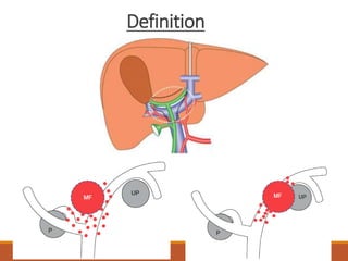
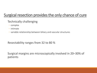
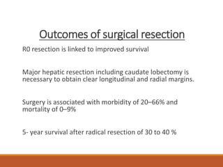
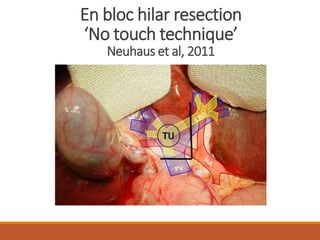
Surgery for Hilar
Cholangiocarcinoma
Decision Making & Technical
Aspects
DR HARSH SHAH
MS, FMAS, DNB, MCH(GI)
KAIZEN HOSPITAL
Abbreviations
RH – Right hepatectomy
LH – Left hepatectomy
ERH – Extended right hepatectomy
ELH – Extended left hepatectomy
Objective
Preparing patient for surgery
How to decide type of hepatectomy
Few surgical tips
Definition
Bismuth-Corlette classification
Surgical resection provides the only chance of cure
Technically challenging
◦ complex
◦ intimate
◦ variable relationship between biliary and vascular structures
Resectability ranges from 32 to 80 %
Surgical margins are microscopically involved in 20–30% of
patients
Outcomes of surgical resection
R0 resection is linked to improved survival
Major hepatic resection including caudate lobectomy is
necessary to obtain clear longitudinal and radial margins.
Surgery is associated with morbidity of 20–66% and
mortality of 0–9%
5- year survival after radical resection of 30 to 40 %
Pre-operative Investigations
CECT scan with 3DVR with volumetry
◦ PV bifurcation
◦ RHA
MRCP
MDCT scan with 3DVR
Pre-operative preparations
PTBD
◦ Vascular injury
◦ Infection
◦ Tumour seeding
ENBD (Kawashima et al, Ann Surg 2013)
◦ 80% success, 7F single catheter in remnant lobe
◦ Internal bile drainage
◦ Biopsy possible
◦ Cholangitis, Pancreatitis
Future Liver Remnant
Normal liver >20%
Cholestatic liver >30%
Cirrhotic >40%
RH - 31% (Seg 2,3,4)
ERH - 19% (Seg 2,3)
LH - 66% (Seg 5,6,7,8)
ELH - 30% (Seg 6,7)
Small liver remnant
RPVE + Seg 4 Embolization
◦ 2-4 weeks
◦ 50% hypertrophy of seg 2,3
◦ Prevents resection in 10-15%
ALPPS
◦ Rapid(1 week)
◦ Significant(Median 70%)
◦ High morbidity & upto 15% mortality
◦ Bile leak
Oral synbiotic preparations
Enhance immune responses
Attenuate systemic postoperative inflammatory responses
Improve intestinal microbial environment
Reduce postoperative infectious complications
Sugawara et al, Ann Surg 2006
Blood Donation
For Autologous blood transfusion
Surgical Anatomy
Liver resecion
Extent of resection
Always Segment 1 & 4
Extent of bile duct involvement (Bismuth-Corlette Type)
Vascular involvement
Lobar atrophy
Right sided resection
Short RHD, wider margin on LHD
Bile duct confluence lies on the PV confluence & RPV
RHA is usually involved, LHA remains uninvolved
No need for vascular reconstruction
Small remnant
Right Sided resections
RH/ERH + Caudate
Rule out peritoneal metastasis
Right lobe & caudate mobilized
Bile duct division in retroduodenal part
RHA ligation, MHA ligation
Ligation of RPV, caudate branches, seg 4 branches in umbilical
fissure
ERH – 4,5,6,7,8
RH – 5,6,7,8
Parenchymal transection
RHV, MHV ligation
Cholangiogram
Right hepatectomy + Caudate resection
Extended Right hepatectomy +
Caudate resection
En bloc hilar resection
‘No touch technique’
Neuhaus et al, 2011
Rex recess approach
(Rela et al, 2014)
Rex recess approach
Left sided resection
Adequate remnant size – seg 6,7
Need for vascular reconstruction – RHA, RPV
RPSD Anatomy
Hjortsjo’s Crook
Higher margin positivity
Difficult anastomosis
◦ Depth
◦ Behind portal vein
Higher leak rate
Supra portal
LH vs ELH
ELH preferred when
Not possible to get negative RPSD margin (supraportal type)
Right anterior sector atrophy
Right anterior sector vascular involvement
Type 4 tumours
How to select between ELH vs ERH
ELH ERH
FLR adequate Not adequate
PVE/PBD Not required Required
AR Required Not required
PVR Usually Required Usually required
Post-op
sepsis/liver failure
No Yes
Post-op vascular
complications
Yes Usually No
Portal Vein Resection
No added morbidity if performed in high volume centers
Wu XS (2013) Combined portal vein resection for hilar cholangiocarcinoma: a
meta-analysis of comparative studies. J Gastrointest Surg 17:1107–1115
Left Portal vein resection
Long extrahepatic length and easy access to the vein within the
umbilical fissure beyond the limit of the tumour
Almost always possible to anastomose the ends of the vein directly
Not much reduction in diameter of the vein along its length because its
main branches are given off within the umbilical fissure
Interposition grafts are necessary only when more than 5–6 cm of vein
is resected
5-0 prolene continuous
Right Portal vein resection
short and bifurcates early
Limits of RPV resection are the point where the first order branches of the
RPV can be safely controlled with a vascular clamp
A Y-graft from External iliac vein harvested through an extraperitoneal
approach is necessary
There is considerable discrepancy in diameter between the MPV and the
sectional branches.
Anastomosis with interrupted sutures.
Arterial resection
RHA has intimate relationship with biliary confluence
Should be performed after parenchymal transection
End to End anastomosis with RHA/RPSA
Interposition graft of IMV/Gonadal vein/Great saphenous
vein/radial artery
Post-operative care after AR
Nagino et al, 2010, Ann Surg
Doppler on POD-1
Routine DVT prophylaxis only
If interposition graft used – Aspirin 150mg may be added
Triphasic CT scan before discharge
Extent of surgery based on Bismuth-Corlette type
Type 1 – EHBDE
Type 2 – Seg 1 & 4b Excision, Median Hepatectomy
Type 3a – ERH, Right Hepatectomy
Type 3b – Left Hepatectomy, ERH
Type 4 – ERH, ELH
Thank You

Hilar Cholangiocarcinoma

  • 1.
    Surgery for Hilar Cholangiocarcinoma DecisionMaking & Technical Aspects DR HARSH SHAH MS, FMAS, DNB, MCH(GI) KAIZEN HOSPITAL
  • 2.
    Abbreviations RH – Righthepatectomy LH – Left hepatectomy ERH – Extended right hepatectomy ELH – Extended left hepatectomy
  • 3.
    Objective Preparing patient forsurgery How to decide type of hepatectomy Few surgical tips
  • 4.
  • 6.
  • 7.
    Surgical resection providesthe only chance of cure Technically challenging ◦ complex ◦ intimate ◦ variable relationship between biliary and vascular structures Resectability ranges from 32 to 80 % Surgical margins are microscopically involved in 20–30% of patients
  • 8.
    Outcomes of surgicalresection R0 resection is linked to improved survival Major hepatic resection including caudate lobectomy is necessary to obtain clear longitudinal and radial margins. Surgery is associated with morbidity of 20–66% and mortality of 0–9% 5- year survival after radical resection of 30 to 40 %
  • 9.
    Pre-operative Investigations CECT scanwith 3DVR with volumetry ◦ PV bifurcation ◦ RHA MRCP
  • 10.
  • 11.
    Pre-operative preparations PTBD ◦ Vascularinjury ◦ Infection ◦ Tumour seeding ENBD (Kawashima et al, Ann Surg 2013) ◦ 80% success, 7F single catheter in remnant lobe ◦ Internal bile drainage ◦ Biopsy possible ◦ Cholangitis, Pancreatitis
  • 12.
    Future Liver Remnant Normalliver >20% Cholestatic liver >30% Cirrhotic >40% RH - 31% (Seg 2,3,4) ERH - 19% (Seg 2,3) LH - 66% (Seg 5,6,7,8) ELH - 30% (Seg 6,7)
  • 13.
    Small liver remnant RPVE+ Seg 4 Embolization ◦ 2-4 weeks ◦ 50% hypertrophy of seg 2,3 ◦ Prevents resection in 10-15% ALPPS ◦ Rapid(1 week) ◦ Significant(Median 70%) ◦ High morbidity & upto 15% mortality ◦ Bile leak
  • 14.
    Oral synbiotic preparations Enhanceimmune responses Attenuate systemic postoperative inflammatory responses Improve intestinal microbial environment Reduce postoperative infectious complications Sugawara et al, Ann Surg 2006
  • 15.
  • 16.
  • 18.
  • 19.
    Extent of resection AlwaysSegment 1 & 4 Extent of bile duct involvement (Bismuth-Corlette Type) Vascular involvement Lobar atrophy
  • 20.
    Right sided resection ShortRHD, wider margin on LHD Bile duct confluence lies on the PV confluence & RPV RHA is usually involved, LHA remains uninvolved No need for vascular reconstruction Small remnant
  • 21.
    Right Sided resections RH/ERH+ Caudate Rule out peritoneal metastasis Right lobe & caudate mobilized Bile duct division in retroduodenal part RHA ligation, MHA ligation Ligation of RPV, caudate branches, seg 4 branches in umbilical fissure ERH – 4,5,6,7,8 RH – 5,6,7,8 Parenchymal transection RHV, MHV ligation
  • 22.
  • 23.
    Extended Right hepatectomy+ Caudate resection
  • 25.
    En bloc hilarresection ‘No touch technique’ Neuhaus et al, 2011
  • 26.
  • 27.
  • 28.
    Left sided resection Adequateremnant size – seg 6,7 Need for vascular reconstruction – RHA, RPV
  • 29.
  • 31.
  • 33.
    Higher margin positivity Difficultanastomosis ◦ Depth ◦ Behind portal vein Higher leak rate Supra portal
  • 34.
    LH vs ELH ELHpreferred when Not possible to get negative RPSD margin (supraportal type) Right anterior sector atrophy Right anterior sector vascular involvement
  • 35.
    Type 4 tumours Howto select between ELH vs ERH ELH ERH FLR adequate Not adequate PVE/PBD Not required Required AR Required Not required PVR Usually Required Usually required Post-op sepsis/liver failure No Yes Post-op vascular complications Yes Usually No
  • 36.
    Portal Vein Resection Noadded morbidity if performed in high volume centers Wu XS (2013) Combined portal vein resection for hilar cholangiocarcinoma: a meta-analysis of comparative studies. J Gastrointest Surg 17:1107–1115
  • 37.
    Left Portal veinresection Long extrahepatic length and easy access to the vein within the umbilical fissure beyond the limit of the tumour Almost always possible to anastomose the ends of the vein directly Not much reduction in diameter of the vein along its length because its main branches are given off within the umbilical fissure Interposition grafts are necessary only when more than 5–6 cm of vein is resected 5-0 prolene continuous
  • 38.
    Right Portal veinresection short and bifurcates early Limits of RPV resection are the point where the first order branches of the RPV can be safely controlled with a vascular clamp A Y-graft from External iliac vein harvested through an extraperitoneal approach is necessary There is considerable discrepancy in diameter between the MPV and the sectional branches. Anastomosis with interrupted sutures.
  • 39.
    Arterial resection RHA hasintimate relationship with biliary confluence Should be performed after parenchymal transection End to End anastomosis with RHA/RPSA Interposition graft of IMV/Gonadal vein/Great saphenous vein/radial artery
  • 41.
    Post-operative care afterAR Nagino et al, 2010, Ann Surg Doppler on POD-1 Routine DVT prophylaxis only If interposition graft used – Aspirin 150mg may be added Triphasic CT scan before discharge
  • 42.
    Extent of surgerybased on Bismuth-Corlette type Type 1 – EHBDE Type 2 – Seg 1 & 4b Excision, Median Hepatectomy Type 3a – ERH, Right Hepatectomy Type 3b – Left Hepatectomy, ERH Type 4 – ERH, ELH
  • 43.

Editor's Notes

  • #5 These tumours are defined by the Japanese Study Group on Perihilar Cholangiocarcinomas (JSGPH) to involve hilar bile ducts between the right side of the umbilical portion of the left portal vein and the left side of the origin of the right posterior portal vein. For tumours with a significant intrahepatic component, the epicenter of the mass should lie between the above-stated landmarks
  • #11 Three-dimensional volume-rendered images. A, Arterial phase; B, Portal phase. Encasement of the proper and right hepatic artery was found (‚). The left portal vein was completely occluded and the right anterior portal vein was markedly stenotic (arrow). This patient underwent left trisectionectomy and caudate lobectomy with vascular resection.
  • #18 Direct infiltration - Caudate ducts & segment 4 Superior part of portal bifurcation usually right PV Right hepatic artery – Normal course gets involved, anomalous origin from SMA may be spared Left hepatic artery courses away Right hepatic duct short, Left hepatic duct longer
  • #24 Preoperative cholangiograms of patients who underwent a right hepatic lobectomy and caudate lobectomy and b right trisegmentectomy and caudate lobectomy. The solid arrowed lines indicate the beginning of tapering of the bile duct, which means the margin of the cancer. The dotted arrowed lines show the resection limits in each patient. The circles are located on the bottom of the umbilical portion of the left portal vein which is presumed from the image of the cranial curve of the anterior lateral branch (B3) and the landmark of the middle segment branch (B4). B2, left posterior duct
  • #25 Resection limits of three types of hepatectomy. A The straight double-arrowed line on the left indicates the limit of ductal resection for caudate lobectomy alone, which corresponds to the right margin of the umbilical portion of the left portal vein. The double-arrowed lines in the middle and on the right show the limits of ductal resection for right lobectomy and right trisegmentectomy, respectively. b In right hepatic lobectomy, division of the right portal vein and the Arantius ligament causes the bottom of the umbilical portion of the left portal vein to be mobilized completely to the left. The bile duct is exposed to the center of the umbilical portion of the left portal vein where the limit of ductal resection for right hepatic lobectomy is located (double-arrowed line). c In right trisegmentectomy, as the middle segmental branch of the portal vein (P4) is divided and the umbilical portion, the left portal vein is completely mobilized to the left side. The resection limit of the ducts is the left margin of the umbilical portion (double-arrowed line). The caudate branches and inferior lateral branch of the portal vein are omitted from the schema. P3, left lateral anterior branch of the portal vein; B4, middle segmental duct; B3, left anterior duct; B2, left lateral posterior duct
  • #26 Local recurrence is most common Perineural & perilymphatic invasion, tumour dissected off the vascular structures 58% 5 year survival rate with no touch technique
  • #27 CT scan demonstrating the location of the umbilical segment of the LPV between segments 3 and 4 at the Rex recess. A: Segments 2 and 3 branches to the left and segment 4 branches to the right; B: Reconstructed portal venous system shows the horizontal and the vertical segment (red arrows) of LPV with segmental branches. The black line shows the transection line on the LPV
  • #28 Intra-operative images of the Rex recess approach. A: Exposure of the LPV in the Rex recess with isolated segmental veins. The black line indicates the point of transection on the LPV; B: Application of Satinsky vascular clamp behind the segmental veins for facilitating anastomosis.The lateral position of the skeletonized left HA; C: A longitudinal venotomy was done on the exposed LPV for an end-side anastomosis with the interposition graft (*); D: The completed anastomosis after restoring portal blood flow to the remnant liver.
  • #30 Confluence patterns of the right posterior sectional bile duct (RPSBD). (A); Supraportal type, the RPSBD runs cranially around the RPV to form a confluence with the right anterior sectional bile duct (RASBD) duct at cranial side of the RPV. (B); Infraportal type, the RPSBD runs caudal to the RPV and joins to the RASBD at caudal side of the RPV. (C); Combined type, there is no RPSBD. The ducts from segments 6 and 7 have separate confluences with the remaining biliary tree; one segmental duct runs in a supraportal position and the other in an infraportal position. Bl, left hepatic duct; Br, right hepatic duct; B6, segment 6 duct; B7, segment 7 duct; PV portal vein.
  • #31 Diagnosis of the confluence pattern of the right posterior sectional bile duct (RPSBD) using contrast enhanced computed tomography. (A); Supraportal type, the RPSBD (arrow) joins to the distal bile duct at cranial side of the right portal vein (RPV) (arrowhead). (B); Infraportal type, the RPBD (arrow) joins to the distal bile duct at caudal side of the RPV (arrowhead)
  • #32 Cholangiography. The typical configuration of the right posterior sectional bile duct (RPSBD), which is called Hjortsjo’s Crook (arrow), can be observed in the supraportal type
  • #34 Stump of the right posterior sectional bile duct (RPSBD) after left hemihepatectomy extended to the hilar part of segment 5 and the entire segment 1. (A); Supraportal type, the stump of the RPSBD is posterior (dorsal) to the right portal vein (RPV). (B); Infraportal type, the stump of the RPSBD is anterior (ventral) side to the RPV.