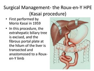
Biliary Atresia
Anup Shrestha
CMC
Introduction
• Biliary atresia (BA) is a progressive, idiopathic,
fibro-obliterative disease of the extrahepatic
biliary tree that presents with biliary obstruction
exclusively in the neonatal period.
• Overall incidence is low (approximately 1 in
10,000 to 20,000 live births )
• BA is the most common indication for liver
transplantation in children.
Japanese Association of Pediatric
Surgeons classification
• In type I, or “distal” BA,
atresia affects only the CBD,
with the GB and hepatic
ducts remaining patent.
• type IIa, in which the
gallbladder and CBD are both
present and patent
• Type IIb, in which the GB,
CBD, and hepatic ducts are all
obliterated
• type III or “complete,” :
obliteration of both
intrahepatic bile ducts as well
as the entire extrahepatic
biliary
Distinct clinical forms
• Fetal-embryonic (or syndromic): is characterized by
early cholestasis, appears in the first 2 weeks of life.
- In this form, the bile ducts are discontinuous at birth,
and affected neonates have associated congenital
defects: situs inversus, polysplenia, malrotation, intestinal
atresia, and cardiac anomalies.
• Perinatal (or acquired): This form is typically found in
neonates and infants aged 2-8 weeks.
- Progressive inflammation and obliteration of the
extrahepatic bile ducts occurs after birth.
- -This form is not associated with congenital anomalies,
and infants may have a short jaundice –free interval.
Etiology
• Genetic: Recent work has identified mutations in jagged 1, a
Notch signaling ligand, and the left right patterning gene
Nodal Cofactor Cryptic (CFC1).
• Immunologic: Overexpression of adhesion molecules in
biliary epithelium.
-Aberrant expression of class I and II HLAs
-Expression of Fas ligand and increased apoptosis of bile duct
epithelial cells
• Vascular Abnormalities : impaired arterial flow may lead to
necrosis and fibrous obliteration of extrahepatic bile ducts
• Viral :Reovirus type3, Rotavirus, Cytomegalovirus,
Papillomavirus
• Environmental/miscellaneous- Gestational use of drugs
(amphetamines, alcohol), phytotoxins, mycotoxins, Industrial
toxins, gestational diabetes, maternal age
Epidemiology
• It is most commonly observed in East Asian
countries, with a reported incidence as high as
1 in 5,000 to 10,000 live births
• More common in females than in males
• Chinese infants seem to be particularly at risk
Clinical Features
• Jaundice lasting longer than the first 2 weeks
of life
• Acholic stools
• Dark urine
• Hepatomegaly
• Vitamin K deficiency
• Coagulopathy
• Cardiac Murmurs
Workup
• Labs- LFT
• infants show moderate elevations in total
bilirubin, which is commonly 6-12 mg/dL
• . Elevated Alkaline phosphatase (ALP), gamma-
glutamyl transpeptidase(GGTP), serum
aminotransferases, serum bile acids
• albumin, total protein are in normal range.
USG
• A shrunken gallbladder
despite fasting and lack
of intrahepatic bile
ducts.
• The presence of a
hyperechogenic hilum
on ultrasound, or
“triangular cord sign
Hepatobiliary Iminodiacetic
Acid (HIDA) Scan
• Lack of bile excretion into the
intestine on HIDA scan is consistent
with BA.
• The specificity is 93% specific, and
94.6% accurate in diagnosing BA.
• Limitation-
-infants with severe hepatitis may
exhibit impaired radiotracer uptake and
thus have limited excretion into the
bowel.
- Additionally, given the progressive
nature of BA, some infants may initially
demonstrate some flow of radiotracer
into the duodenum with the gradual
obliteration of this tract later in the
disease
MRCP
• Provide more detailed definition of the
biliary tree.
• Findings- include incomplete visualization
of the extrahepatic biliary system and
periportal high-signal intensity on T2-
weighted MRI scans
• Sensitivity and specificity of greater than
90%
ERCP
• ERCP allows direct
visualization of the
extrahepatic biliary tree.
• It requires a general
anesthetic, substantial
expertise, and the
availability of sufficiently
small endoscopes.
Preoperative liver biopsy
• Used method to help
exclude other causes
of neonatal jaundice .
• Findings suggestive of
BA include portal or
bridging fibrosis, bile
duct proliferation,
portal inflammation
and bile duct plugs
Surgical Management- the Roux-en-Y HPE
(Kasai procedure)
• First performed by
Morio Kasai in 1959
• In this procedure, the
extrahepatic biliary tree
is excised, and the
fibrous portal plate at
the hilum of the liver is
transected and
anastomosed to a Roux-
en-Y limb
Procedure
• Incision – rooftop or Chevron
Procedure
• the abdomen is grossly inspected to identify any
evidence of associated anomalies, such as
intestinal malrotation or abnormalities of the
spleen and portal vein
• the liver often appears cholestatic or fibrotic with
a fibrotic and shrunken-appearing gallbladder.
• If the gallbladder is normal in appearance, is
patent, the contents of the gallbladder may be
aspirated to assess.
• If clear (“white”) bile is aspirated, no additional
maneuvers are required. If the fluid is darker
appearing, it is recommended to proceed with a
cholangiogram
Intra-op Cholangiogram
• Is considered gold standard
for diagnosis of BA
• If contrast appears to freely
flow into both the
intrahepatic ducts as well as
the duodenum, BA may be
safely excluded.
• If a complete biliary tree is
not visualized, biliary-enteric
continuity must be restored
• The peritoneum overlying the
hepatoduodenal ligament is opened to
allow identification of the structures in
this area.
• Fibrous remnant of the distal common
bile duct is often present here.
• As dissection continues proximally, the
biliary remnant develops into a cone of
fibrotic tissue that is located at the
bifurcation of the main portal vein into
its left and right branches.
• –most important landmark during the
dissection of the portal plate and should
be the goal of every dissection
Roux Limb Construction
• Proximal jejunum is identified and
transected about 10 centimeters distal
to the ligament of Treitz
• The distal end, destined for the right
upper quadrant, is oversewn and the
Roux limb is measured to 40 to 50
centimeters. At this location, an end-
to-side jejunojejunostomy is created
with interrupted absorbable sutures.
• The oversewn end of the Roux limb is
carefully brought into the right upper
quadrant via a small defect created in
the avascular portion of the transverse
mesocolon.
Portoenterostomy
• An end-to-end HPE is created rather than an
end-to-side to avoid creating a long unused
blind end as the bowel continues to grow over
time.
Post-op care
• Drainage of gastric secretions with a nasogastric tube
should continue for the first 48 hours, then removed
• NPO can be broken at 2nd or 3rd day
• High calorie diet – 3 to 4 gm/pg/ day
• Drain removed at 5th P.O.D
• Ursodeoxycholic Acid for choleretic effect
• Antibiotics
• Vitamin A, D ,E ,K supplements
• Methylprednisolone should be given for it’s anti-
inflammatory and choleretic effects
Outcome
• 30-day postoperative mortality following HPE is
low, and is estimated to be 0% to 5%
• Patients in whom biliary drainage is adequate
develop pigmented stools with an associated
downtrend in serum bilirubin levels, typically
within the first 10 to 14 days after surgery.
• Half of patients will continue to experience liver
inflammation, fibrosis, and eventual liver failure
despite an initial good response following HPE.
• Ultimately need liver transplantation by a mean
age of 5.4 years
Long-term native-liver survival by age at
hepatoportoenterostomy
groups 1 and 2 are operative age < 6 days; group 3, 61 to 90
days; group 4, 91 to 120 days; group 5, 121 to 150 days;
group 6, >151 days
Complications
• Cholangitis- MC Complication
- Mechanism- reflux of intestinal content
- Bacterial Translocation
- Impaired portal lymphatic drainage
Prevention :- Initial construction of an adequate
Roux limb is fundamental
-Prophylactic antibiotics
- Intractable cholangitis : Liver Transplant should be
considered
Portal Hypertension
• It is estimated that as many as 30% to 70% of
patients will continue to have elevated portal
pressures following HPE
• Results in esophageal varices and ascites
• Half of the patient will have atleast one
episode of variceal bleeding
• β-blockers and endoscopic interventions
• (sclerotherapy or banding) as primary
prophylaxis in children
Hepatopulmonary Syndrome
• characterized by hypoxia associated with chronic
liver disease with evidence of intrapulmonary
shunting
• inability of the liver to metabolize vasoactive
substances may lead to abnormal shunting within
the lungs, ultimately resulting in symptoms of
dyspnea, platypnea, and orthodoexia
• No effective medical therapy
• Liver transplant – definitive treatment
Thank You

Extrahepatic biliary atresia

  • 1.
  • 2.
    Introduction • Biliary atresia(BA) is a progressive, idiopathic, fibro-obliterative disease of the extrahepatic biliary tree that presents with biliary obstruction exclusively in the neonatal period. • Overall incidence is low (approximately 1 in 10,000 to 20,000 live births ) • BA is the most common indication for liver transplantation in children.
  • 3.
    Japanese Association ofPediatric Surgeons classification • In type I, or “distal” BA, atresia affects only the CBD, with the GB and hepatic ducts remaining patent. • type IIa, in which the gallbladder and CBD are both present and patent • Type IIb, in which the GB, CBD, and hepatic ducts are all obliterated • type III or “complete,” : obliteration of both intrahepatic bile ducts as well as the entire extrahepatic biliary
  • 4.
    Distinct clinical forms •Fetal-embryonic (or syndromic): is characterized by early cholestasis, appears in the first 2 weeks of life. - In this form, the bile ducts are discontinuous at birth, and affected neonates have associated congenital defects: situs inversus, polysplenia, malrotation, intestinal atresia, and cardiac anomalies. • Perinatal (or acquired): This form is typically found in neonates and infants aged 2-8 weeks. - Progressive inflammation and obliteration of the extrahepatic bile ducts occurs after birth. - -This form is not associated with congenital anomalies, and infants may have a short jaundice –free interval.
  • 5.
    Etiology • Genetic: Recentwork has identified mutations in jagged 1, a Notch signaling ligand, and the left right patterning gene Nodal Cofactor Cryptic (CFC1). • Immunologic: Overexpression of adhesion molecules in biliary epithelium. -Aberrant expression of class I and II HLAs -Expression of Fas ligand and increased apoptosis of bile duct epithelial cells • Vascular Abnormalities : impaired arterial flow may lead to necrosis and fibrous obliteration of extrahepatic bile ducts • Viral :Reovirus type3, Rotavirus, Cytomegalovirus, Papillomavirus • Environmental/miscellaneous- Gestational use of drugs (amphetamines, alcohol), phytotoxins, mycotoxins, Industrial toxins, gestational diabetes, maternal age
  • 6.
    Epidemiology • It ismost commonly observed in East Asian countries, with a reported incidence as high as 1 in 5,000 to 10,000 live births • More common in females than in males • Chinese infants seem to be particularly at risk
  • 7.
    Clinical Features • Jaundicelasting longer than the first 2 weeks of life • Acholic stools • Dark urine • Hepatomegaly • Vitamin K deficiency • Coagulopathy • Cardiac Murmurs
  • 8.
    Workup • Labs- LFT •infants show moderate elevations in total bilirubin, which is commonly 6-12 mg/dL • . Elevated Alkaline phosphatase (ALP), gamma- glutamyl transpeptidase(GGTP), serum aminotransferases, serum bile acids • albumin, total protein are in normal range.
  • 9.
    USG • A shrunkengallbladder despite fasting and lack of intrahepatic bile ducts. • The presence of a hyperechogenic hilum on ultrasound, or “triangular cord sign
  • 10.
    Hepatobiliary Iminodiacetic Acid (HIDA)Scan • Lack of bile excretion into the intestine on HIDA scan is consistent with BA. • The specificity is 93% specific, and 94.6% accurate in diagnosing BA. • Limitation- -infants with severe hepatitis may exhibit impaired radiotracer uptake and thus have limited excretion into the bowel. - Additionally, given the progressive nature of BA, some infants may initially demonstrate some flow of radiotracer into the duodenum with the gradual obliteration of this tract later in the disease
  • 11.
    MRCP • Provide moredetailed definition of the biliary tree. • Findings- include incomplete visualization of the extrahepatic biliary system and periportal high-signal intensity on T2- weighted MRI scans • Sensitivity and specificity of greater than 90%
  • 12.
    ERCP • ERCP allowsdirect visualization of the extrahepatic biliary tree. • It requires a general anesthetic, substantial expertise, and the availability of sufficiently small endoscopes.
  • 13.
    Preoperative liver biopsy •Used method to help exclude other causes of neonatal jaundice . • Findings suggestive of BA include portal or bridging fibrosis, bile duct proliferation, portal inflammation and bile duct plugs
  • 14.
    Surgical Management- theRoux-en-Y HPE (Kasai procedure) • First performed by Morio Kasai in 1959 • In this procedure, the extrahepatic biliary tree is excised, and the fibrous portal plate at the hilum of the liver is transected and anastomosed to a Roux- en-Y limb
  • 15.
    Procedure • Incision –rooftop or Chevron
  • 16.
    Procedure • the abdomenis grossly inspected to identify any evidence of associated anomalies, such as intestinal malrotation or abnormalities of the spleen and portal vein • the liver often appears cholestatic or fibrotic with a fibrotic and shrunken-appearing gallbladder. • If the gallbladder is normal in appearance, is patent, the contents of the gallbladder may be aspirated to assess. • If clear (“white”) bile is aspirated, no additional maneuvers are required. If the fluid is darker appearing, it is recommended to proceed with a cholangiogram
  • 17.
    Intra-op Cholangiogram • Isconsidered gold standard for diagnosis of BA • If contrast appears to freely flow into both the intrahepatic ducts as well as the duodenum, BA may be safely excluded. • If a complete biliary tree is not visualized, biliary-enteric continuity must be restored
  • 18.
    • The peritoneumoverlying the hepatoduodenal ligament is opened to allow identification of the structures in this area. • Fibrous remnant of the distal common bile duct is often present here. • As dissection continues proximally, the biliary remnant develops into a cone of fibrotic tissue that is located at the bifurcation of the main portal vein into its left and right branches. • –most important landmark during the dissection of the portal plate and should be the goal of every dissection
  • 19.
    Roux Limb Construction •Proximal jejunum is identified and transected about 10 centimeters distal to the ligament of Treitz • The distal end, destined for the right upper quadrant, is oversewn and the Roux limb is measured to 40 to 50 centimeters. At this location, an end- to-side jejunojejunostomy is created with interrupted absorbable sutures. • The oversewn end of the Roux limb is carefully brought into the right upper quadrant via a small defect created in the avascular portion of the transverse mesocolon.
  • 20.
    Portoenterostomy • An end-to-endHPE is created rather than an end-to-side to avoid creating a long unused blind end as the bowel continues to grow over time.
  • 21.
    Post-op care • Drainageof gastric secretions with a nasogastric tube should continue for the first 48 hours, then removed • NPO can be broken at 2nd or 3rd day • High calorie diet – 3 to 4 gm/pg/ day • Drain removed at 5th P.O.D • Ursodeoxycholic Acid for choleretic effect • Antibiotics • Vitamin A, D ,E ,K supplements • Methylprednisolone should be given for it’s anti- inflammatory and choleretic effects
  • 22.
    Outcome • 30-day postoperativemortality following HPE is low, and is estimated to be 0% to 5% • Patients in whom biliary drainage is adequate develop pigmented stools with an associated downtrend in serum bilirubin levels, typically within the first 10 to 14 days after surgery. • Half of patients will continue to experience liver inflammation, fibrosis, and eventual liver failure despite an initial good response following HPE. • Ultimately need liver transplantation by a mean age of 5.4 years
  • 23.
    Long-term native-liver survivalby age at hepatoportoenterostomy groups 1 and 2 are operative age < 6 days; group 3, 61 to 90 days; group 4, 91 to 120 days; group 5, 121 to 150 days; group 6, >151 days
  • 24.
    Complications • Cholangitis- MCComplication - Mechanism- reflux of intestinal content - Bacterial Translocation - Impaired portal lymphatic drainage Prevention :- Initial construction of an adequate Roux limb is fundamental -Prophylactic antibiotics - Intractable cholangitis : Liver Transplant should be considered
  • 25.
    Portal Hypertension • Itis estimated that as many as 30% to 70% of patients will continue to have elevated portal pressures following HPE • Results in esophageal varices and ascites • Half of the patient will have atleast one episode of variceal bleeding • β-blockers and endoscopic interventions • (sclerotherapy or banding) as primary prophylaxis in children
  • 26.
    Hepatopulmonary Syndrome • characterizedby hypoxia associated with chronic liver disease with evidence of intrapulmonary shunting • inability of the liver to metabolize vasoactive substances may lead to abnormal shunting within the lungs, ultimately resulting in symptoms of dyspnea, platypnea, and orthodoexia • No effective medical therapy • Liver transplant – definitive treatment
  • 27.

Editor's Notes

  • #3 leading to cholestasis, cirrhosis, and, ultimately, death within the first several years of life if left untreated.
  • #4 Type 3 most common
  • #11 Technetium Tc 99m lidofenin
  • #26 Although the Kasai procedure may improve portal hypertension in some infants and children with biliary atresia,