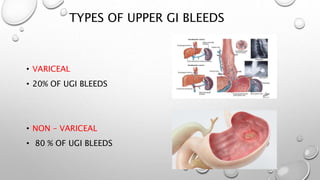
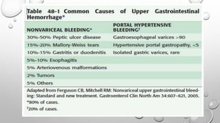
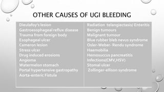
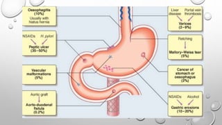
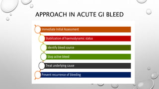
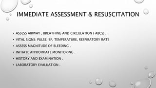
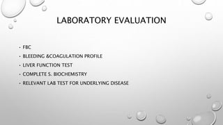
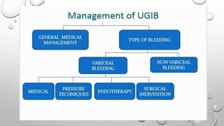
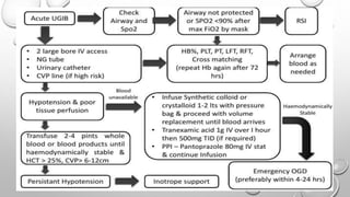
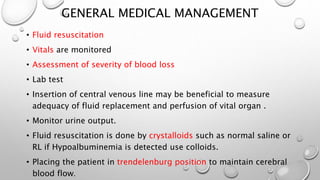
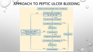
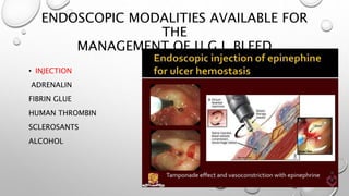
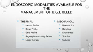
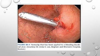
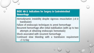
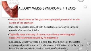
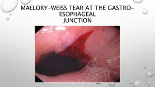
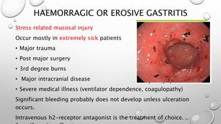
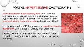
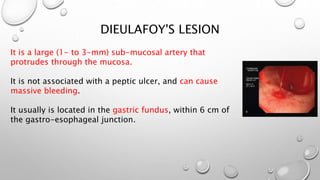
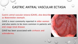
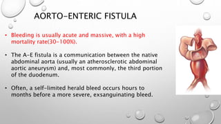
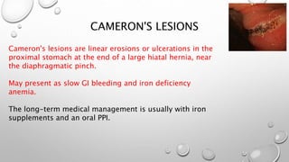
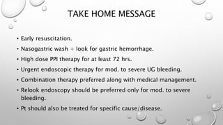
MANAGEMENT OF UPPER GI
BLEEDING
ANNIA RAMOS PEREZ
CONSULTANT GENERAL SURGEON
DEFINITION
• Loss of blood anywhere into the gastrointestinal tract.
• Depending on location of ligament of treitz
• UGIB
• LGIB
Sources of GI bleeding
• Upper GI tract
• Proximal to the ligament of Treitz
• 80% of acute GI bleeds
• Lower GI tract
• Distal to the ligament of Treitz
• 20% of acute GI bleeds
LIGAMENT OF TREITZ
UPPER GI BLEEDING
• Upper GI bleed : lower GI bleed = 4:1
• Incidence: 170 patients/ 100,000 population /year( USA data).
• 40% due to peptic ulcer(most common).
• 80% are self-limited.
TYPES OF UPPER GI BLEEDS
• VARICEAL
• 20% OF UGI BLEEDS
• NON – VARICEAL
• 80 % OF UGI BLEEDS
OTHER CAUSES OF UGI BLEEDING
CLINICAL FEATURES
ON EXAMINATION
APPROACH IN ACUTE GI BLEED
IMMEDIATE ASSESSMENT & RESUSCITATION
• ASSESS AIRWAY , BREATHING AND CIRCULATION ( ABCS) .
• VITAL SIGNS: PULSE, BP, TEMPERATURE, RESPIRATORY RATE
• ASSESS MAGNITUDE OF BLEEDING .
• INITIATE APPROPRIATE MONITORING .
• HISTORY AND EXAMINATION .
• LABORATORY EVALUATION .
ESTIMATED FLUID AND BLOOD LOSSES IN
SHOCK
Differential diagnosis
LABORATORY EVALUATION
• FBC
• BLEEDING &COAGULATION PROFILE
• LIVER FUNCTION TEST
• COMPLETE S. BIOCHEMISTRY
• RELEVANT LAB TEST FOR UNDERLYING DISEASE
GENERAL MEDICAL MANAGEMENT
• Fluid resuscitation
• Vitals are monitored
• Assessment of severity of blood loss
• Lab test
• Insertion of central venous line may be beneficial to measure
adequacy of fluid replacement and perfusion of vital organ .
• Monitor urine output.
• Fluid resuscitation is done by crystalloids such as normal saline or
RL if Hypoalbuminemia is detected use colloids.
• Placing the patient in trendelenburg position to maintain cerebral
blood flow.
GENERAL MEDICAL MANAGEMENT
1.Oxygen support to prevent hypoxia of tissues.
2.IV route - crystalloid solution/colloids/ blood.
3. Blood transfusion:
• Maintain Htc at 30% in the elderly, esp. With comorbid diseases
e.g.. CHF, CRF
• 20-25% in younger pt.
• 25-28% in portal HTN
• Administration of Vit k
4.In symptomatic thrombocytopenia (<50000 )infused platelets.
5.FFP-the transfusion of plasma should not be based solely on the
patient’s abnormal INR and/or PTT.
The decision to transfuse should be based on the patient’s clinical
APPROACH TO VARICEAL BLEEDING
VARICEAL BLEEDING
Patients with Variceal hemorrhage have poorer outcomes than patients
with other sources of UGIB
Ligation is the endoscopic therapy of choice for esophageal varices
Primary prophylaxis: non-selective beta blockers
Chronic therapy with beta blockers plus endoscopic ligation is
recommended for prevention of recurrent esophageal Variceal bleeding.
Endoscopic management
EVL, sclerotherapy( cyanoacrylate , Na morrhuate , ethanolamine ,etc.)
Surgical management
TIPS, esophageal transection, Suguira procedure
Liver transplantation
VARICEAL BLEEDING
BALLOON TAMPONADE -SENGSTAKEN
BLAKEMORE TUBE
SURGICAL ALTERNATIVE - SUGIURA
PROCEDURE
• A transthoracic-abdominal esophageal transection
• • Para-esophageal Devascularization, Esophageal Transection and
Anastomosis, splenectomy, and Pyloroplasty.
TRANS-JUGULAR INTRAHEPATIC PORTO-
SYSTEMIC
SHUNT (TIPS)
TIPS
MEDICAL MANAGEMENT OF VARICEAL
BLEEDING
APPROACH TO PEPTIC ULCER BLEEDING
ENDOSCOPIC MODALITIES AVAILABLE FOR
THE
MANAGEMENT OF U.G.I. BLEED
• INJECTION
ADRENALIN
FIBRIN GLUE
HUMAN THROMBIN
SCLEROSANTS
ALCOHOL
ENDOSCOPIC MODALITIES AVAILABLE FOR
THE
MANAGEMENT OF U.G.I. BLEED
MALLORY WEISS SYNDROME / TEARS
Mucosal lacerations at the gastro-esophageal junction or in the
cardia of the stomach
Patients generally present with hematemesis or coffee-ground
emesis after alcohol intake
Typically have a history of recent non-bloody vomiting with
excessive retching followed by hematemesis
Endoscopy usually reveals a single tear that begins at the gastro-
esophageal junction and extends several millimeters distally into a
hiatal hernia sac/within cardiac portion of stomach.
MALLORY-WEISS TEAR AT THE GASTRO-
ESOPHAGEAL
JUNCTION
HAEMORRAGIC OR EROSIVE GASTRITIS
Stress related mucosal injury
Occur mostly in extremely sick patients
• Major trauma
• Post major surgery
• 3rd degree burns
• Major intracranial disease
• Severe medical illness (ventilator dependence, coagulopathy)
Significant bleeding probably does not develop unless ulceration
occurs.
Intravenous h2-receptor antagonist is the treatment of choice.
PORTAL HYPERTENSIVE GASTROPATHY
Portal hypertensive gastropathy (PHG) is caused by
increased portal venous pressure and severe mucosal
hyperemia that results in ecstatic blood vessels in the
proximal gastric body and cardia and oozing of blood.
Less severe grades of PHG appear as a mosaic or snake skin
appearance and are not associated with bleeding.
Usually, patients with severe PHG present with chronic
blood loss, but they occasionally can present with acute
bleeding.
DIEULAFOY'S LESION
It is a large (1- to 3-mm) sub-mucosal artery that
protrudes through the mucosa.
It is not associated with a peptic ulcer, and can cause
massive bleeding.
It usually is located in the gastric fundus, within 6 cm of
the gastro-esophageal junction.
GASTRIC ANTRAL VASCULAR ECTASIA
Gastric antral vascular ectasia (GAVE), also described
as Watermelon stomach.
GAVE is most commonly reported in older women
and also seems to be more common in patients with
end-stage renal disease
GAVE has been associated with cirrhosis and
scleroderma.
AORTO-ENTERIC FISTULA
• Bleeding is usually acute and massive, with a high
mortality rate(30-100%).
• The A-E fistula is a communication between the native
abdominal aorta (usually an atherosclerotic abdominal
aortic aneurysm) and, most commonly, the third portion
of the duodenum.
• Often, a self-limited herald bleed occurs hours to
months before a more severe, exsanguinating bleed.
CAMERON'S LESIONS
Cameron's lesions are linear erosions or ulcerations in the
proximal stomach at the end of a large hiatal hernia, near
the diaphragmatic pinch.
May present as slow GI bleeding and iron deficiency
anemia.
The long-term medical management is usually with iron
supplements and an oral PPI.
RISK FACTORS AND RISK STRATIFICATION
• To identify patients with Non-variceal UGI bleeding at greatest risk
for mortality and rebleeding.
• Pts. may be categorized as low, intermediate and high risk .
MANAGEMENT AS PER RISK
1- Low risk(0-2)-usually 80 % of the pt. Recovers spontaneously
with medical
Tt( PPI)+ hospitalization for 24 hrs and may be discharge if
uneventful.
2-Intermediate risk(3-5)- same Tt. + hospitalization for at least 72
hrs.
ADVERSE PROGNOSTIC FACTOR IN UGIB
TAKE HOME MESSAGE
• Early resuscitation.
• Nasogastric wash + look for gastric hemorrhage.
• High dose PPI therapy for at least 72 hrs.
• Urgent endoscopic therapy for mod. to severe UG bleeding.
• Combination therapy preferred along with medical management.
• Relook endoscopy should be preferred only for mod. to severe
bleeding.
• Pt should also be treated for specific cause/disease.
HOMEWORK
• DEFINITION AND CLINICAL MANIFESTATIONS OF:
• Cushing ulcers
• Curling ulcers
• Hemobilia
• Hemosuccus pancreaticus
BIBLIOGRAPHY
• SABISTON. TEXTBOOK OF SURGERY.
• SCHWARTZ. TEXTBOOK OF SURGERY.
• SHACKELFORS. SURGERY OF THE ALIMENTARY TRACT
Management of upper gi bleeding

Management of upper gi bleeding

  • 1.
    MANAGEMENT OF UPPERGI BLEEDING ANNIA RAMOS PEREZ CONSULTANT GENERAL SURGEON
  • 2.
    DEFINITION • Loss ofblood anywhere into the gastrointestinal tract. • Depending on location of ligament of treitz • UGIB • LGIB
  • 3.
    Sources of GIbleeding • Upper GI tract • Proximal to the ligament of Treitz • 80% of acute GI bleeds • Lower GI tract • Distal to the ligament of Treitz • 20% of acute GI bleeds
  • 4.
  • 5.
    UPPER GI BLEEDING •Upper GI bleed : lower GI bleed = 4:1 • Incidence: 170 patients/ 100,000 population /year( USA data). • 40% due to peptic ulcer(most common). • 80% are self-limited.
  • 6.
    TYPES OF UPPERGI BLEEDS • VARICEAL • 20% OF UGI BLEEDS • NON – VARICEAL • 80 % OF UGI BLEEDS
  • 8.
    OTHER CAUSES OFUGI BLEEDING
  • 10.
  • 12.
  • 13.
  • 14.
    IMMEDIATE ASSESSMENT &RESUSCITATION • ASSESS AIRWAY , BREATHING AND CIRCULATION ( ABCS) . • VITAL SIGNS: PULSE, BP, TEMPERATURE, RESPIRATORY RATE • ASSESS MAGNITUDE OF BLEEDING . • INITIATE APPROPRIATE MONITORING . • HISTORY AND EXAMINATION . • LABORATORY EVALUATION .
  • 15.
    ESTIMATED FLUID ANDBLOOD LOSSES IN SHOCK
  • 16.
  • 17.
    LABORATORY EVALUATION • FBC •BLEEDING &COAGULATION PROFILE • LIVER FUNCTION TEST • COMPLETE S. BIOCHEMISTRY • RELEVANT LAB TEST FOR UNDERLYING DISEASE
  • 20.
    GENERAL MEDICAL MANAGEMENT •Fluid resuscitation • Vitals are monitored • Assessment of severity of blood loss • Lab test • Insertion of central venous line may be beneficial to measure adequacy of fluid replacement and perfusion of vital organ . • Monitor urine output. • Fluid resuscitation is done by crystalloids such as normal saline or RL if Hypoalbuminemia is detected use colloids. • Placing the patient in trendelenburg position to maintain cerebral blood flow.
  • 21.
    GENERAL MEDICAL MANAGEMENT 1.Oxygensupport to prevent hypoxia of tissues. 2.IV route - crystalloid solution/colloids/ blood. 3. Blood transfusion: • Maintain Htc at 30% in the elderly, esp. With comorbid diseases e.g.. CHF, CRF • 20-25% in younger pt. • 25-28% in portal HTN • Administration of Vit k 4.In symptomatic thrombocytopenia (<50000 )infused platelets. 5.FFP-the transfusion of plasma should not be based solely on the patient’s abnormal INR and/or PTT. The decision to transfuse should be based on the patient’s clinical
  • 22.
  • 26.
    VARICEAL BLEEDING Patients withVariceal hemorrhage have poorer outcomes than patients with other sources of UGIB Ligation is the endoscopic therapy of choice for esophageal varices Primary prophylaxis: non-selective beta blockers Chronic therapy with beta blockers plus endoscopic ligation is recommended for prevention of recurrent esophageal Variceal bleeding. Endoscopic management EVL, sclerotherapy( cyanoacrylate , Na morrhuate , ethanolamine ,etc.) Surgical management TIPS, esophageal transection, Suguira procedure Liver transplantation
  • 27.
  • 28.
  • 31.
    SURGICAL ALTERNATIVE -SUGIURA PROCEDURE • A transthoracic-abdominal esophageal transection • • Para-esophageal Devascularization, Esophageal Transection and Anastomosis, splenectomy, and Pyloroplasty.
  • 32.
  • 33.
  • 34.
    MEDICAL MANAGEMENT OFVARICEAL BLEEDING
  • 35.
    APPROACH TO PEPTICULCER BLEEDING
  • 39.
    ENDOSCOPIC MODALITIES AVAILABLEFOR THE MANAGEMENT OF U.G.I. BLEED • INJECTION ADRENALIN FIBRIN GLUE HUMAN THROMBIN SCLEROSANTS ALCOHOL
  • 40.
    ENDOSCOPIC MODALITIES AVAILABLEFOR THE MANAGEMENT OF U.G.I. BLEED
  • 47.
    MALLORY WEISS SYNDROME/ TEARS Mucosal lacerations at the gastro-esophageal junction or in the cardia of the stomach Patients generally present with hematemesis or coffee-ground emesis after alcohol intake Typically have a history of recent non-bloody vomiting with excessive retching followed by hematemesis Endoscopy usually reveals a single tear that begins at the gastro- esophageal junction and extends several millimeters distally into a hiatal hernia sac/within cardiac portion of stomach.
  • 48.
    MALLORY-WEISS TEAR ATTHE GASTRO- ESOPHAGEAL JUNCTION
  • 49.
    HAEMORRAGIC OR EROSIVEGASTRITIS Stress related mucosal injury Occur mostly in extremely sick patients • Major trauma • Post major surgery • 3rd degree burns • Major intracranial disease • Severe medical illness (ventilator dependence, coagulopathy) Significant bleeding probably does not develop unless ulceration occurs. Intravenous h2-receptor antagonist is the treatment of choice.
  • 50.
    PORTAL HYPERTENSIVE GASTROPATHY Portalhypertensive gastropathy (PHG) is caused by increased portal venous pressure and severe mucosal hyperemia that results in ecstatic blood vessels in the proximal gastric body and cardia and oozing of blood. Less severe grades of PHG appear as a mosaic or snake skin appearance and are not associated with bleeding. Usually, patients with severe PHG present with chronic blood loss, but they occasionally can present with acute bleeding.
  • 51.
    DIEULAFOY'S LESION It isa large (1- to 3-mm) sub-mucosal artery that protrudes through the mucosa. It is not associated with a peptic ulcer, and can cause massive bleeding. It usually is located in the gastric fundus, within 6 cm of the gastro-esophageal junction.
  • 52.
    GASTRIC ANTRAL VASCULARECTASIA Gastric antral vascular ectasia (GAVE), also described as Watermelon stomach. GAVE is most commonly reported in older women and also seems to be more common in patients with end-stage renal disease GAVE has been associated with cirrhosis and scleroderma.
  • 53.
    AORTO-ENTERIC FISTULA • Bleedingis usually acute and massive, with a high mortality rate(30-100%). • The A-E fistula is a communication between the native abdominal aorta (usually an atherosclerotic abdominal aortic aneurysm) and, most commonly, the third portion of the duodenum. • Often, a self-limited herald bleed occurs hours to months before a more severe, exsanguinating bleed.
  • 54.
    CAMERON'S LESIONS Cameron's lesionsare linear erosions or ulcerations in the proximal stomach at the end of a large hiatal hernia, near the diaphragmatic pinch. May present as slow GI bleeding and iron deficiency anemia. The long-term medical management is usually with iron supplements and an oral PPI.
  • 55.
    RISK FACTORS ANDRISK STRATIFICATION • To identify patients with Non-variceal UGI bleeding at greatest risk for mortality and rebleeding. • Pts. may be categorized as low, intermediate and high risk .
  • 57.
    MANAGEMENT AS PERRISK 1- Low risk(0-2)-usually 80 % of the pt. Recovers spontaneously with medical Tt( PPI)+ hospitalization for 24 hrs and may be discharge if uneventful. 2-Intermediate risk(3-5)- same Tt. + hospitalization for at least 72 hrs.
  • 58.
  • 59.
    TAKE HOME MESSAGE •Early resuscitation. • Nasogastric wash + look for gastric hemorrhage. • High dose PPI therapy for at least 72 hrs. • Urgent endoscopic therapy for mod. to severe UG bleeding. • Combination therapy preferred along with medical management. • Relook endoscopy should be preferred only for mod. to severe bleeding. • Pt should also be treated for specific cause/disease.
  • 60.
    HOMEWORK • DEFINITION ANDCLINICAL MANIFESTATIONS OF: • Cushing ulcers • Curling ulcers • Hemobilia • Hemosuccus pancreaticus
  • 61.
    BIBLIOGRAPHY • SABISTON. TEXTBOOKOF SURGERY. • SCHWARTZ. TEXTBOOK OF SURGERY. • SHACKELFORS. SURGERY OF THE ALIMENTARY TRACT