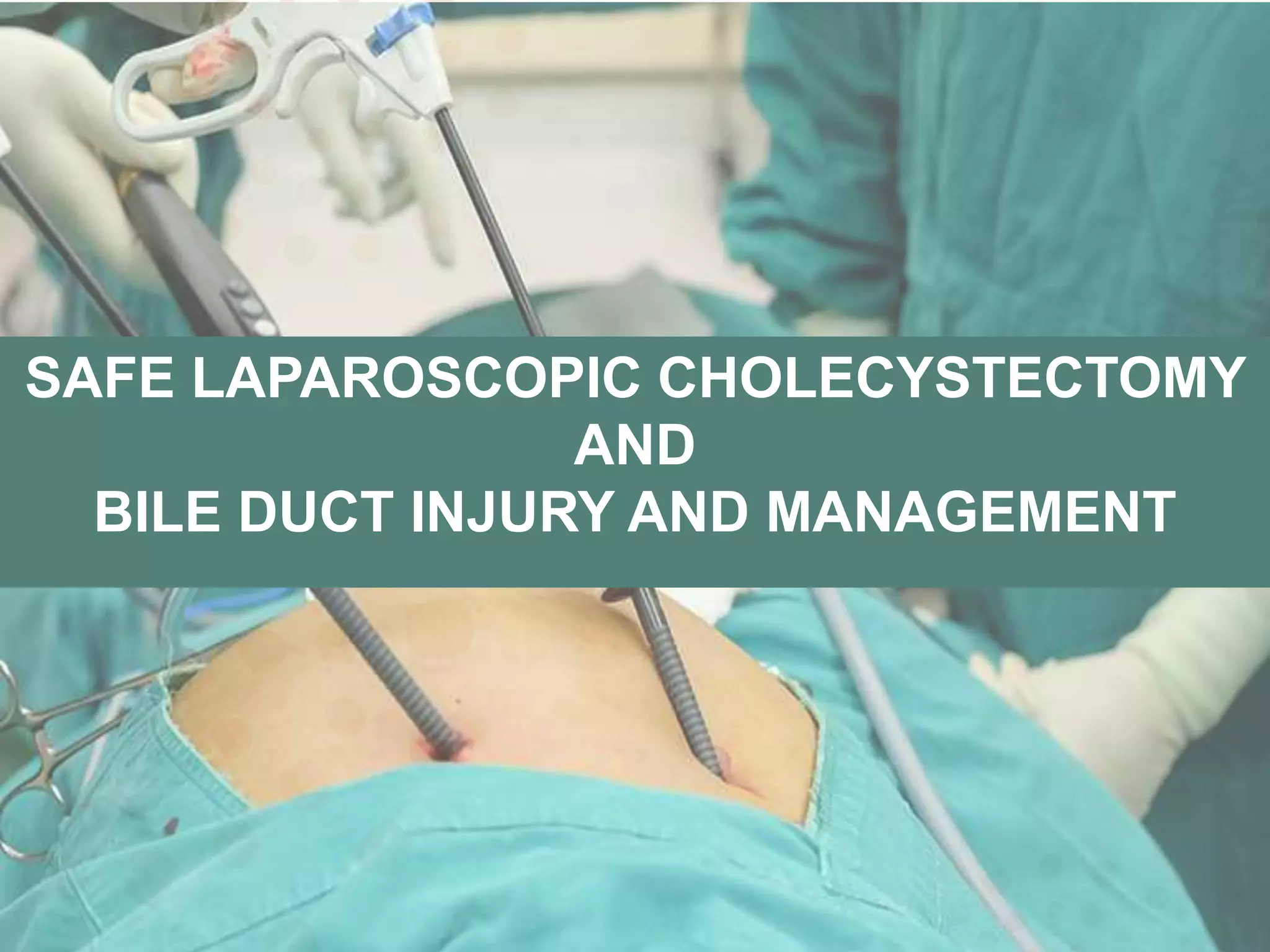
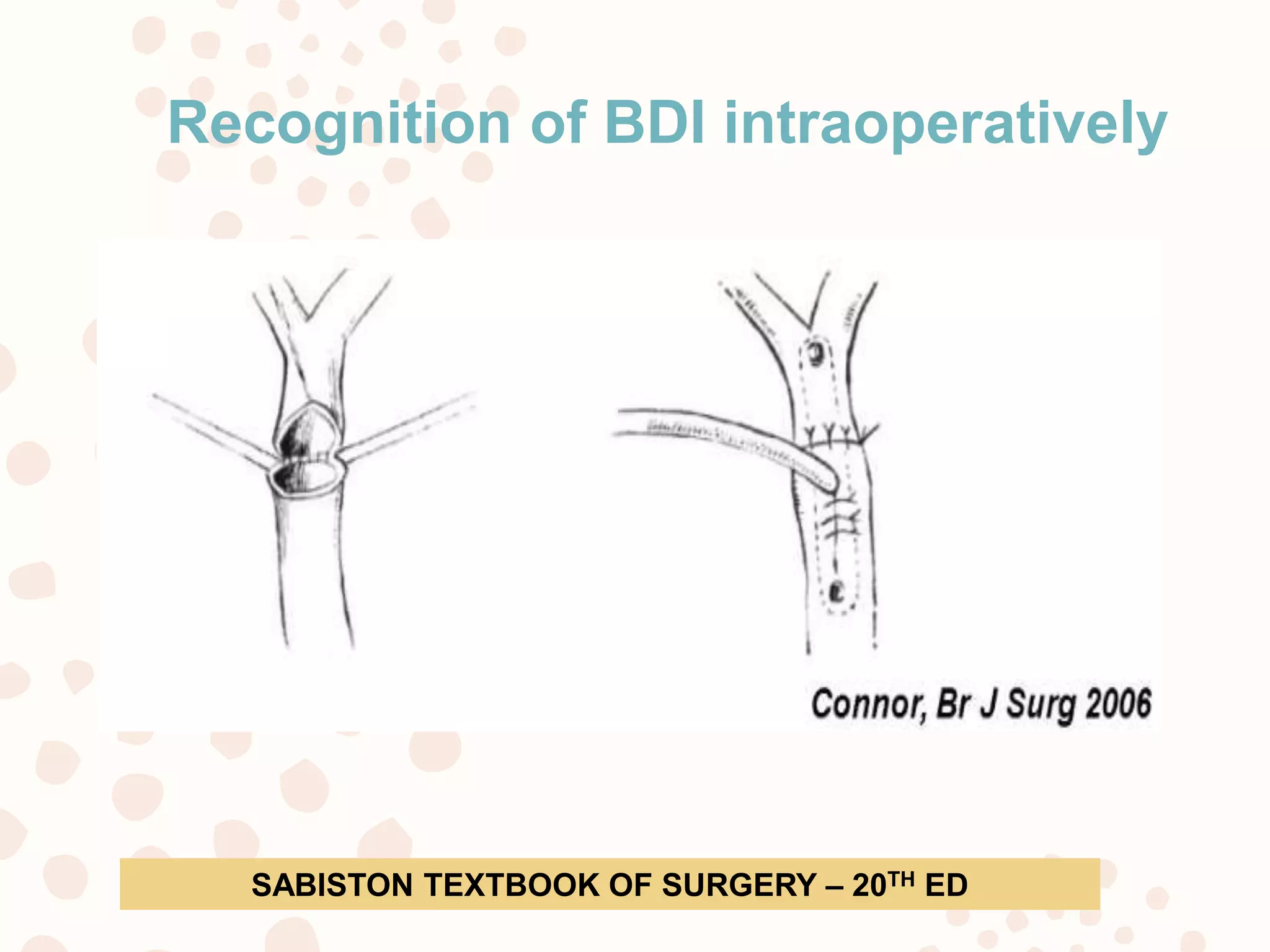
SAFE LAPAROSCOPIC CHOLECYSTECTOMY
AND
BILE DUCT INJURY AND MANAGEMENT
SEQUENCE OF FLOW
 HISTORY AND BACKGROUND
 OVERVIEW OF LC AND BILE DUCT INJURY
 BILIARY INJURY MECHANISM & CLASSIFICATIONS
 PREVENTION
 MANAGEMENT OF BILE DUCT INJURY
 CONCLUSION
HISTORY AND BACKGROUND
• Most common surgical procedures
performed- with over 750,000 *
• Open cholecystectomy- Carl
Langenbuch in 1882
• In 1985, the first Laparoscopic
cholecystectomy (LC)- Erich Mühe
of Bšblingen, Germany.
* Stinton LM, Myers RP, Shaffer EA. Epidemiology of gallstones.
Gastroenterol Clin North Am 2010;39:157–169
OVERVIEW
• Over 80% of -laparoscopic approach *
• Advantages of laparoscopic over open
cholecystectomy
* Keus F, et al. Laparoscopic versus open cholecystectomy for patients
with symptomatic cholecystolithiasis. Cochrane Database Syst Rev
2010;CD006231.
OVERVIEW…
• LC is the gold standard for management of
gallstones.
• Iatrogenic Bile Duct Injury- Two to three times
greater than the open procedure
• 3 per 1,000 procedures performed *
* Buddingh KT, Weersma RK, Savenije RA, van Dam GM, Nieuwenhuijs VB. Lower rate of major bile duct
injury and increased intraoperative management of common bile duct stones after implementation of
routine intraoperative cholangiography. Journal of the American College of Surgeons 2011; 213:267-74.
ANATOMY
a. Right hepatic duct.
b. Left hepatic duct.
c. Common hepatic duct.
d. Portal vein.
e. Hepatic artery.
f. Gastroduodenal artery.
g. Right gastroepiploic artery.
h. Common bile duct.
i. Fundus of the gallbladder.
j. Body of the gallbladder.
k. Infundibulum.
l. Cystic duct.
m. Cystic artery.
n. Superior
pancreaticoduodenal artery.
Schwartz‟s Principles of Surgery, 8th Ed.,McGraw-Hill Companies, 2005.
BLOOD SUPPLY TO CBD
The cranial segments :
cystic artery especially the
right hepatic artery.
The caudal segment:
pancreaticoduodenal artery
through the retroduodenal
artery.
The middle segment :
arterial anastomoses between
the cranial and caudal
supplies.
BLOOD SUPPLY
IATROGENIC BILIARY INJURY -
ETIOLOGY
ETIOLOGY
PATHOLOGICAL FACTOR
• Acute cholecystitis
• Acute biliary pancreatitis
• Bleeding in calot’s triangle
• Severely scarred or
shrunken gall bladder
• Difficult GB
TECHNICAL FACTOR
• 02 Dimensional
operative field
• Classic bile duct
injury
• Cognitive factors of
surgeons
• Skill of surgeon
Maingot’s abdominal operation- 12th ed
ETIOLOGY
ANATOMIC FACTOR
Abnormal biliary anatomy
• Short cystic duct, cystic duct
entering in the right duct
• Accessory right hepatic duct
Arterial anomalies
• Right hepatic artery running
parallel to the cystic duct
• Anomalous or accessory
right hepatic artery
PHYSIOLOGIC FACTOR
• Ischemia of bile duct
from excessive
periductal dissection
BILE DUCT INJURY- CLINICAL
PRESENTATION, INVESTIGATIONS
AND CLASSIFICATION
• Almost 85% of IBDI are not recognized- surgical
procedure*
• Only 15%-30% of IBDI are recognized during the initial
operation**
• 70% of IBDI are diagnosed within 6 months and 80%
within 12 months after the initial operation***
*De Wit LT, Rauws EA, Gouma DJ. Surgical management of iatrogenic bile duct injury.
Scand J Gastroenterol Suppl 1999; 230: 89-94
**Gouma DJ, Obertop H. Management of bile duct injuries: treatment and long-term
results. Dig Surg 2002; 19: 117-122
***Hall JG, Pappas TN. Current management of biliary strictures. J Gastrointest Surg
2004; 8: 1098-1110
FACTS
CLINICAL PRESENTATION
• Depends on type of injury and divided into groups:
 leak
 Stricture
Radiological investigations
• Ultrasonagraphy and CT – Ductal
dilatation intra-abdominal collection
and dilatation of biliary tree.
• Cholangiogram
• ERCP—biliary anatomy and assess
the injury
• PTC—define biliary anatomy proximal
to injury
• MRCP—noninvasive (can miss minor
leaks)
• HIDA scan -- If doubt exists, HIDA
scan can confirm leak but not the
specific leak site
• MR angiography—vascular injuries
Classification of bile duct injury(BDI)
 Bismuth-Corlette classification
 Strasberg classification
 Stewart-Way classification
 Lau classification
 Hannover classification
 Mattox classification
Bismuth classification
Strasberg classification
HOW TO AVOID IBDI ??
HOW TO AVOID BDI ??..
1. Correct exposure and identification
• “ Critical View Of Safety”
• Rouviere’s sulcus (RS)
2. Avoid error trap
3. Thermal injuries
4. Blind hemostasis
5. IOC
6. Surgeons characterstics
HOW TO AVOID BDI ??..
Strasberg in 1995- “Critical View of Safety” (CVS) *
1. Meticulous dissection of the Calot’s triangle from all
fatty and fibrous tissue.
2. Lowest part of gallbladder should be separated from
the cystic plate, which allows the visualization of
posterior liver bed.
3. Dissection and identification of only two structures
(cystic duct, cystic artery) entering the gallbladder.
* Strasberg SM, Hertl M, Soper NJ. An analysis of the problem of biliary injury during laparoscopic
cholecystectomy. J Am Coll Surg. 1995;180(1):101–25.
Critical View of Safety(CVS)
Critical view of safety anterior view Critical view of safety posterior view
SAGES 6 STRATEGIES
1. Critical View of Safety (CVS)
2. Intra-operative time-out prior to clipping, cutting or
transecting any ductal structures is advised.
3. Variations in anatomy should be considered in all cases.
4. Surgeon should use cholangiography or other instrument
for demonstrating biliary anatomy.
5. In case of difficulty to expose biliary anatomy alternatives
surgical techniques such as partial cholecystectomy,
cholecystostomy tube placement or conversion to an open
procedure
6. Consultation with an another surgeon in difficult cases may
be helpful.
SAGES (Society of American Gastrointestinal and Endoscopic Surgeons), 2008.
Rouviere’s sulcus (RS)
HOW TO AVOID BDI ??...
Error traps in laparoscopic
• Infundibular technique error trap
• Fundus down error trap
• Failure to perceive the presence of an aberrant right
hepatic duct on cholangiography
• “parallel union” cystic duct
STEVEN M. STRASBERG. Error traps and vasculo-biliary injury in laparoscopic and open
cholecystectomy. J Hepatobiliary Pancreat Surg (2008) 15:284–292
Infundibular technique error trap
Fundus down error trap
Failure to perceive the presence of an aberrant right
hepatic duct on cholangiography
“Parallel union” cystic duct
HOW TO AVOID BDI ??...
THERMAL INJURIES
• Misuse of cautery in dissecting the Calot’s triangle
• Not to use cautery to cut the cystic duct
AVOID BLIND HAEMOSTASIS
INTRAOPERATIVE CHOLANGIOGRAM (IOC)
SURGEONS CHARACTERISTICS OF RISK TAKING
CLASSIC BILIARY INJURY
• CBD is mistaken for cystic duct often includes injury
to RHA as it enters either above or below hepatic
duct
Maingot’s abdominal operation- 12th ed
Management of Strategy When Faced with
IBDI
1. INTRA OP MANAGEMENT
2. DELAYED MANAGEMENT
Treatment of BDI
1. Endoscopic and radiological treatment
2. Surgical treatment
 End-to-end Ductal Biliary Anastomosis (EE)
 jejunal interposition hepaticoduodenostomy
 Roux-en-Y HJ
 Blumgart (Hepp) anastomosis
 Heinecke-Mikulicz -biliary plastic reconstruction
 Smith mucosal graft
Recognition of BDI intraoperatively
• Immediate cholangiography and conversion to
an open procedure in order to define the extent of
the injury are required.
• Bile ducts of diameter < 3 mm -should be ligated
• Bile ducts of diameter > 3 mm should be repaired
• Interruption of common hepatic duct or
common bile duct continuity can be repaired by
immediate tension free EE with or without a T tube
SABISTON TEXTBOOK OF SURGERY – 20TH ED
• If the bile duct loss is too long and near the
bifurcation, EE is not possible without tension and
bilio-enteric anastomosis (Roux-en-Y HJ vs
choledochoduodenostomy) is recommended.
If detected in Post-Operative period
1. Control of infection limiting inflammation
 Parenteral antibiotics
 Percutaneous drainage of periportal fluid
2. Clear and thorough delineation of entire biliary
anatomy
 PTC
 ERCP/MRCP
3. Re-establishing biliary enteric continuity
 Tension free , mucosa to mucosa
SABISTON TEXTBOOK OF SURGERY – 20TH ED
Conclusion
• LC which is a gold standard therapeutic option
• Using well-described anatomical landmarks and fixed
extra biliary reference points, combined with other well
documented strategies should be followed
TAKE HOME MESSAGE
• The primary goal of LC is ‘‘safety first, total
cholecystectomy second.’’
• “Culture of safety”
• Failure - increase health care expenses, impaired
quality of life, and may even lead to death.
1. Critical View of Safety (CVS)
2. Variations in anatomy
3. Alternatives surgical techniques
4. Consultation with an another surgeon in difficult
cases may be helpful
5. Reporting of cases
THANK YOU !!!

Bile duct injury

  • 1.
  • 2.
    SEQUENCE OF FLOW HISTORY AND BACKGROUND  OVERVIEW OF LC AND BILE DUCT INJURY  BILIARY INJURY MECHANISM & CLASSIFICATIONS  PREVENTION  MANAGEMENT OF BILE DUCT INJURY  CONCLUSION
  • 3.
    HISTORY AND BACKGROUND •Most common surgical procedures performed- with over 750,000 * • Open cholecystectomy- Carl Langenbuch in 1882 • In 1985, the first Laparoscopic cholecystectomy (LC)- Erich Mühe of Bšblingen, Germany. * Stinton LM, Myers RP, Shaffer EA. Epidemiology of gallstones. Gastroenterol Clin North Am 2010;39:157–169
  • 4.
    OVERVIEW • Over 80%of -laparoscopic approach * • Advantages of laparoscopic over open cholecystectomy * Keus F, et al. Laparoscopic versus open cholecystectomy for patients with symptomatic cholecystolithiasis. Cochrane Database Syst Rev 2010;CD006231.
  • 5.
    OVERVIEW… • LC isthe gold standard for management of gallstones. • Iatrogenic Bile Duct Injury- Two to three times greater than the open procedure • 3 per 1,000 procedures performed * * Buddingh KT, Weersma RK, Savenije RA, van Dam GM, Nieuwenhuijs VB. Lower rate of major bile duct injury and increased intraoperative management of common bile duct stones after implementation of routine intraoperative cholangiography. Journal of the American College of Surgeons 2011; 213:267-74.
  • 7.
    ANATOMY a. Right hepaticduct. b. Left hepatic duct. c. Common hepatic duct. d. Portal vein. e. Hepatic artery. f. Gastroduodenal artery. g. Right gastroepiploic artery. h. Common bile duct. i. Fundus of the gallbladder. j. Body of the gallbladder. k. Infundibulum. l. Cystic duct. m. Cystic artery. n. Superior pancreaticoduodenal artery. Schwartz‟s Principles of Surgery, 8th Ed.,McGraw-Hill Companies, 2005.
  • 8.
    BLOOD SUPPLY TOCBD The cranial segments : cystic artery especially the right hepatic artery. The caudal segment: pancreaticoduodenal artery through the retroduodenal artery. The middle segment : arterial anastomoses between the cranial and caudal supplies.
  • 9.
  • 10.
  • 11.
    ETIOLOGY PATHOLOGICAL FACTOR • Acutecholecystitis • Acute biliary pancreatitis • Bleeding in calot’s triangle • Severely scarred or shrunken gall bladder • Difficult GB TECHNICAL FACTOR • 02 Dimensional operative field • Classic bile duct injury • Cognitive factors of surgeons • Skill of surgeon Maingot’s abdominal operation- 12th ed
  • 12.
    ETIOLOGY ANATOMIC FACTOR Abnormal biliaryanatomy • Short cystic duct, cystic duct entering in the right duct • Accessory right hepatic duct Arterial anomalies • Right hepatic artery running parallel to the cystic duct • Anomalous or accessory right hepatic artery PHYSIOLOGIC FACTOR • Ischemia of bile duct from excessive periductal dissection
  • 13.
    BILE DUCT INJURY-CLINICAL PRESENTATION, INVESTIGATIONS AND CLASSIFICATION
  • 14.
    • Almost 85%of IBDI are not recognized- surgical procedure* • Only 15%-30% of IBDI are recognized during the initial operation** • 70% of IBDI are diagnosed within 6 months and 80% within 12 months after the initial operation*** *De Wit LT, Rauws EA, Gouma DJ. Surgical management of iatrogenic bile duct injury. Scand J Gastroenterol Suppl 1999; 230: 89-94 **Gouma DJ, Obertop H. Management of bile duct injuries: treatment and long-term results. Dig Surg 2002; 19: 117-122 ***Hall JG, Pappas TN. Current management of biliary strictures. J Gastrointest Surg 2004; 8: 1098-1110 FACTS
  • 15.
    CLINICAL PRESENTATION • Dependson type of injury and divided into groups:  leak  Stricture
  • 16.
    Radiological investigations • Ultrasonagraphyand CT – Ductal dilatation intra-abdominal collection and dilatation of biliary tree. • Cholangiogram • ERCP—biliary anatomy and assess the injury • PTC—define biliary anatomy proximal to injury • MRCP—noninvasive (can miss minor leaks) • HIDA scan -- If doubt exists, HIDA scan can confirm leak but not the specific leak site • MR angiography—vascular injuries
  • 17.
    Classification of bileduct injury(BDI)  Bismuth-Corlette classification  Strasberg classification  Stewart-Way classification  Lau classification  Hannover classification  Mattox classification
  • 18.
  • 19.
  • 20.
    HOW TO AVOIDIBDI ??
  • 21.
    HOW TO AVOIDBDI ??.. 1. Correct exposure and identification • “ Critical View Of Safety” • Rouviere’s sulcus (RS) 2. Avoid error trap 3. Thermal injuries 4. Blind hemostasis 5. IOC 6. Surgeons characterstics
  • 22.
    HOW TO AVOIDBDI ??.. Strasberg in 1995- “Critical View of Safety” (CVS) * 1. Meticulous dissection of the Calot’s triangle from all fatty and fibrous tissue. 2. Lowest part of gallbladder should be separated from the cystic plate, which allows the visualization of posterior liver bed. 3. Dissection and identification of only two structures (cystic duct, cystic artery) entering the gallbladder. * Strasberg SM, Hertl M, Soper NJ. An analysis of the problem of biliary injury during laparoscopic cholecystectomy. J Am Coll Surg. 1995;180(1):101–25.
  • 23.
    Critical View ofSafety(CVS)
  • 24.
    Critical view ofsafety anterior view Critical view of safety posterior view
  • 25.
    SAGES 6 STRATEGIES 1.Critical View of Safety (CVS) 2. Intra-operative time-out prior to clipping, cutting or transecting any ductal structures is advised. 3. Variations in anatomy should be considered in all cases. 4. Surgeon should use cholangiography or other instrument for demonstrating biliary anatomy. 5. In case of difficulty to expose biliary anatomy alternatives surgical techniques such as partial cholecystectomy, cholecystostomy tube placement or conversion to an open procedure 6. Consultation with an another surgeon in difficult cases may be helpful. SAGES (Society of American Gastrointestinal and Endoscopic Surgeons), 2008.
  • 26.
  • 27.
    HOW TO AVOIDBDI ??... Error traps in laparoscopic • Infundibular technique error trap • Fundus down error trap • Failure to perceive the presence of an aberrant right hepatic duct on cholangiography • “parallel union” cystic duct STEVEN M. STRASBERG. Error traps and vasculo-biliary injury in laparoscopic and open cholecystectomy. J Hepatobiliary Pancreat Surg (2008) 15:284–292
  • 28.
  • 29.
  • 30.
    Failure to perceivethe presence of an aberrant right hepatic duct on cholangiography
  • 31.
  • 32.
    HOW TO AVOIDBDI ??... THERMAL INJURIES • Misuse of cautery in dissecting the Calot’s triangle • Not to use cautery to cut the cystic duct AVOID BLIND HAEMOSTASIS INTRAOPERATIVE CHOLANGIOGRAM (IOC) SURGEONS CHARACTERISTICS OF RISK TAKING
  • 33.
    CLASSIC BILIARY INJURY •CBD is mistaken for cystic duct often includes injury to RHA as it enters either above or below hepatic duct Maingot’s abdominal operation- 12th ed
  • 34.
    Management of StrategyWhen Faced with IBDI
  • 35.
    1. INTRA OPMANAGEMENT 2. DELAYED MANAGEMENT
  • 36.
    Treatment of BDI 1.Endoscopic and radiological treatment 2. Surgical treatment  End-to-end Ductal Biliary Anastomosis (EE)  jejunal interposition hepaticoduodenostomy  Roux-en-Y HJ  Blumgart (Hepp) anastomosis  Heinecke-Mikulicz -biliary plastic reconstruction  Smith mucosal graft
  • 37.
    Recognition of BDIintraoperatively • Immediate cholangiography and conversion to an open procedure in order to define the extent of the injury are required. • Bile ducts of diameter < 3 mm -should be ligated • Bile ducts of diameter > 3 mm should be repaired • Interruption of common hepatic duct or common bile duct continuity can be repaired by immediate tension free EE with or without a T tube SABISTON TEXTBOOK OF SURGERY – 20TH ED
  • 38.
    • If thebile duct loss is too long and near the bifurcation, EE is not possible without tension and bilio-enteric anastomosis (Roux-en-Y HJ vs choledochoduodenostomy) is recommended.
  • 39.
    If detected inPost-Operative period 1. Control of infection limiting inflammation  Parenteral antibiotics  Percutaneous drainage of periportal fluid 2. Clear and thorough delineation of entire biliary anatomy  PTC  ERCP/MRCP 3. Re-establishing biliary enteric continuity  Tension free , mucosa to mucosa SABISTON TEXTBOOK OF SURGERY – 20TH ED
  • 40.
    Conclusion • LC whichis a gold standard therapeutic option • Using well-described anatomical landmarks and fixed extra biliary reference points, combined with other well documented strategies should be followed
  • 41.
    TAKE HOME MESSAGE •The primary goal of LC is ‘‘safety first, total cholecystectomy second.’’ • “Culture of safety” • Failure - increase health care expenses, impaired quality of life, and may even lead to death. 1. Critical View of Safety (CVS) 2. Variations in anatomy 3. Alternatives surgical techniques 4. Consultation with an another surgeon in difficult cases may be helpful 5. Reporting of cases
  • 42.

Editor's Notes

  • #4 Calculous biliary disease is a common condition in the United States that affects more than 30 million Americans. Over 750,000 cholecystectomies are performed annually, making gallstone disease one of the most common digestive health problems.1 The treatment of calculous biliary disease has evolved over the last 2 decades. With the development of laparoscopic technology in the late 1980s, new techniques for cholecystectomy were introduced.2–4 By the early 1990s, laparoscopic cholecystectomy (LC) had supplanted open cholecystectomy in the operative management of gallbladder stone disease. Unfortunately, the widespreadCholecystectomy is one of the most common surgical procedures performed in the United States with over 600,000 procedures performed each year. Open cholecystectomy, first performed by Carl Langenbuch in 1882, In 1985, the first endoscopic cholecystectomy was performed by Erich Mühe of Bšblingen, Germany. Since then,laparoscopic cholecystectomy has been adopted around the world, and subsequently been recognized as the gold standard for the treatment of gallstone disease. 2– Stinton LM, Myers RP, Shaffer EA. Epidemiology of gallstones. Gastroenterol Clin North Am 2010;39:157–169, vii.
  • #5 Currently it is estimated that over 80% of cholecystectomies are performed using the laparoscopic approach. advantages of laparoscopic over open cholecystectomy have been well documented. Lesser pain , better cosmesis, shorter length of hospt stay, small incisions, Advantages Less pain Smaller incisions Better cosmesis Shorter hospitalisation Earlier return of full activity Decreased total cost LC has clear advantages over the traditional open approach with decreased morbidity, less pain, and a quicker recovery; however, it remains associated with a three- to five- fold increase in bile duct injury (BDI).3 Keus F, et al. Laparoscopic versus open cholecystectomy for patients with symptomatic cholecystolithiasis. Cochrane Database Syst Rev 2006;CD006231.
  • #6  Laparoscopic cholecystectomy (LC) is the gold standard for management of gallstones. However, the risk of bile duct injury (BDI) remains a significant concern10, as LC continues to have a higher BDI rate than its open counterpart, despite many efforts proposed for increasing safety12, Bile duct injury rates have increased since the introduction of laparoscopic cholecystectomy, occurring in about 3 per 1,000 procedures performed.
  • #7  On an average 1,200 cholecystectomies are performed annually at B. P. Koirala Institute of Health Sciences, making gallstone disease one of the most common digestive health problems.1 By the early 1990s, laparoscopic cholecystectomy (LC) had supplanted open cholecystectomy (OC) in the operative management of gallbladder stone disease.
  • #10 60% by the distal vessels 38% by the cranial ones 2% by a nonaxial supply from common hepatic artery  This arterial pattern predisposes the supraduodenal segment of the common bile duct to ischemic damage and resulting in strictures.
  • #13 PATHOLOGICAL FACTOR Severe inflammation and/or infection, Acute cholecystitis Acute biliary pancreatitis Bleeding in calot’s triangle Severely scarred or shrunken gall bladder Large impacted gallstone in hartmann’s pouch, Short cystic duct, and Mirizzi’s syndromeC
  • #14 Severe inflammation and/or infection, Acute cholecystitis Acute biliary pancreatitis Bleeding in calot’s triangle Severely scarred or shrunken gall bladder Large impacted gallstone in hartmann’s pouch, Short cystic duct, and Mirizzi’s syndromeC Abnormal biliary anatomy Short cystic duct, cystic duct entering in the right duct Accessory right hepatic duct Arterial anomalies Right hepatic artery running parallel to the cystic duct Anomalous or accessory right hepatic artery
  • #17  The first sign of a bile duct injury is failing to recover quickly after the procedure. Other symptoms might include: Fever Chills Nausea Vomiting Abdominal pain Swelling of the abdomen General discomfort Jaundice (yellowing of the skin and the whites of the eyes) Because hepatic bile is isotonic in nature and contains lower concentrations of bile salts than gallbladder bile, bile leaks do not cause extreme peritoneal irritation. Patients often complain of vague symptoms, such as nonspecific abdominal fullness, distension, nausea, vomiting, abdominal pain, fever, and chills.
  • #18 A next step should be visualization of the biliary tract by magnetic resonance cholangiopancreatography (MRCP) or ERCP, not only to establish the diagnosis, but to identify the nature and level of the lesion. If MRCP is not available and ERCP only shows the distal bile duct that is occluded by a clip (fig. 3), percutaneous transhepatic cholangiography (PTC) can be performed to visualize the proximal biliary tract, followed by percutaneous biliary drainage (fig. 1). When an abdominal drain is still in situ, cholangiography can be per- formed by this route (drainography). Occasionally, scinti- graphy can be helpful to show leakage (fig. 4). Surgical reconstruction without visualization of the entire biliary system should not be attempted. hepatobiliary iminodiacetic acid scan
  • #19 Lau and Stewart-way classifications are based on mechanisms of injury. Hannover classification classifies injuries in relationship to the confluence and also includes vascular injuries. The Mattox classification of IBDI takes into consideration the type of injuring factor (contusion, laceration, perforation, transsection, diversion or interruption of the bile duct or the gallbladder)
  • #20 Bismuth scale is the most useful and simple classification. It is based on the location of the injury in the biliary tract. This classification is very helpful in prognosis after repair , but does not involve the wide spectrum of possible biliary injuries. The Bismuth-Corlette classification was introduced before laparoscopy. It is difficult to apply in laparoscopic cholecystectomy as most of the technical factors and lesion mechanisms are completely different to open surgery. It considers the complete section of the common bile duct and the length of the proximal bile duct stump. Nevertheless, most cases have late stenosis or bile duct obstruction which may be included in this classification, representing a subtype Strasberg E and Stewart-Way Ⅲ-Ⅳ lesions.
  • #23 The main cause of inadvertent transection of CBD in LC is mistaking CBD for cystic duct [1, 3, 4, 6, 13]. To avoid misidentification of CBD as the cystic duct, it is essential to visualize meticulously, in order to obtain the first impression of the extrahepatic bile duct, before dissection is started preferably using a 30◦ laparoscope. Some landmarks including cystic lymph node, gall bladder neck, and Rouviere’s sulcus have been advocated for identifying the cystic duct and safe dissection [14]. Hartman’s pouch is often used as a landmark as it is easily visualized and connects GB to cystic duct. Care, however, is taken in cases where it is distorted or abolished as in patients with atrophic cholecystitis, impacted cystic duct stone, adhesions between cystic duct, and the neck of gall bladder and in incorrect dissection [15].
  • #24 Three successful steps
  • #25 We emphasize that the surgeon should strive to make the gallbladder look like a polyp on a stalk (aka the critical view of safety") prior to clipping the cystic duct. If clips are applied before such a view is obtained. the CBD may be mistaken for the cystic duct and accidentally clipped and cut. If the critical view cannot be obtained due to in11ammation or hostile anatomy, it is the authors‘ opinion that an IOC should be performed prior to dividing the cystic duct. The critical view of safety is obtained when the lateral and medial aspects of the gallbladder (horizontal arrow) have been dissected free, and only 2 structures are seen entering the gallbladder; the cystic artery and the cystic duct (slanted arrows). In the critical view technique, the cystic duct and cystic artery are identified through dissection of the upper border of the Calot triangle along the underside of the gallbladder. With cephalad traction of the fundus and lateral traction on the infundibulum, dissection is performed on the medial and lateral aspects of the gallbladder until the cystic artery and cystic duct are seen as the only 2 structures entering the gallbladder. This critical view of safety is obtained before any structures are clipped and divided.
  • #27 SAGES (Society of American Gastrointestinal and Endoscopic Surgeons) introduced a safe cholecystectomy program. To minimize the biliary injuries 6 strategies were suggested: SAGES (Society of American Gastrointestinal and Endoscopic Surgeons) introduced a safe cholecystectomy program. To minimize the biliary injuries 6 strategies were suggested: 1. Critical View of Safety (CVS) method should be used including 3 basics approach; Calot’s triangle should be cleared of fat and brous tissue, the lower one third of the gallbladder is dissected from the liver to expose the all anatomical structures and cystic duct and artery should be isolated. 2. Intra-operative time-out prior to clipping, cutting or transecting any ductal structures is advised. Variations in anatomy should be considered in all cases. Surgeon should use cholangiography or other instrument for demonstrating biliary anatomy. 5. In case of di culty to expose biliary anatomy alternatives urgical techniques such as partial cholecystectomy, cholecystostomy tube placement or conversion to an open procedure can
  • #28 Rouviere’s sulcus (RS) (i.e., incisura hepatis dextra, Gans incisura) identification may avoid bile duct injury during laparoscopic cholecystectomy and enables elective vascular control during the right liver resection. The RS is a cleft in the liver running to the right of the liver, anterior to segment 1. The branches of the right posterior sectional pedicle were found in the RS in 70% of the cases. In 5% of the livers, we also dissected a branch of the anterior sectional pedicle. The most important advantage of identifying RS lies in the fact that the cystic duct and the cystic artery lay antero-superior to the sulcus and the common bile duct lays below the level of the RS. Hugh [3] had shown minimal common bile duct injury during laparoscopic cholecystectomy by beginning the dissection ventral to the RS. The second technical reason for identifying RS is to perform safe right sectional or segmental liver resections. 
  • #29 Many biliary misidentification injuries occur due to error traps. An error trap is a method that works well in most circumstances but which is apt to fail under certain conditions.
  • #30 The infundibular technique is a method of ductal identification. “Conclusive identification” is based on three dimensional demonstration of the funnel-like shape of the lower end of the gallbladder and adjacent cystic duct (funnel = infundibulum [Latin root]) (Fig. 1A). Again, it should be emphasized that the infundibular technique calls for circumferential or three-dimensional “360°” display of the funnel. Seeing it in two dimensions, i.e., simply by clearing one or two surfaces of the apparent cystic duct-gallbladder junction, is inadequate.
  • #31 In most cases the predominant injury has been vascular in nature with resultant infarction of the liver or biliary tree.
  • #34 Misuse of cautery in dissecting the Calot’s triangle may cause serious BDI with loss of ductal tissue due to thermal necrosis [17]. Some of the measures used to avoid thermal injury to major bile duct include to initially hook through limited amount of tissue and lift the tissue off the underlying structures under precise vision and proceed with dissection. It is of outmost importance not to use cautery to cut the cystic duct particularly when titanium clips are placed on the cystic duct as titanium clips are good electrical conductor and may lead to thermal necrosis of the cystic duct stump or adjacent bile duct. It is pertinent that always short bursts of minimal amount of energy required to dissect or secure homeostasis should be applied [17].
  • #35 CBD is mistaken for cystic duct often includes injury to RHA as it enters either above or below hepatic duct While this injury may cause bleeding at the time of operation, the rteial injury often is unnoticed , usually resulting in arterial occlusion and less commonly a hepatic pseudoaneurysm.
  • #36 Bile leak during cholecystectomy should force surgeon tostop and carefully examine the source of bile leak . Although bile may leak from an opening in the GB or the cystic duct, before that is presumed to be the case, BDI should be ruled out. Bile from GB is greenish yellow, thick, and viscid, whereas common bile duct (CBD) bile usually is bright yellow, thin, and watery. An IOC at this stage may delineate the anatomy and prevent any further injury to the bile duct. A BDI should also be suspected if a third tubular structure (after cystic duct and artery have been clipped and divided) is encountered in the Calot’s triangle. The “cystic duct” which was clipped and divided earlier may actually have been the CBD and the third structure now being encountered may be the common hepatic duct. If the BDI is recognized intraoperatively, themanagement depends on the nature of the duct injured, type of injury, and the expertise and experience of the surgeon [1, 4, 6, 8].
  • #38 In the literature, following operations have been reported for surgical treatment of IBDI: Roux-en-Y HJ, end-to-end ductal biliary anastomosis (EE), ChD, Lahey HJ, jejunal interposition hepaticoduodenostomy, Blumgart (Hepp) anastomosis, Heinecke-Mikulicz biliary plastic reconstruction and Smith mucosal graft. Jejunal interposition hepaticoduodenostomy, using 25-35 cm of the jejunal loop, is performed in some surgical centers including our department. This reconstruction includes three (biliaryenteric, enteric-duodenal and entero-enteric) anastomoses. JIHD should be used only in patients in good general condition, without active inflammation within the peritoneal cavity, with protein level more than 6 g/dl and serum bilirubin level less than 20 mg/dl. Good condition of the duodenal wall is important factor for proper healing of hepaticoduodenostomy with jejunal interposition. The advantage of this reconstruction is physiological bile flow into the duodenum, which prevents duodenal ulcer caused by changes in the neurohormonal axis within the upper alimentary tract. This method of reconstruction is recommended mainly in patients with concomitant duodenal ulcer The disadvantage is a higher number of early complications due to presence of three anastomoses. Reconstructions of hilar bile duct injuries: The repair of hilar IBDI requires special surgical techniques. In the past, so-called “mucosal graft technique” described by Smith in the 1960s was performed. This reconstruction involves creating a mucosal dome of jejunum (by removing a seromuscular patch) near the end of Roux-Y loop through which a straight rubber tube is brought via hepatic ducts and through liver parenchyma. This technique is based on the hypothesis that jejunal mucosa grafts to the biliary epithelium and mucosa-to-mucosa anastomosis is created. Short-term results were good, but in long-term results a high number of anastomosis strictures was observed. Therefore, currently, not Smith but Blumgart-Hepp technique is used in reconstruction of hilar IBDI. In this technique, dorsal surface of the left hepatic duct parallel to the quadrate hepatic lobe. Dissection and opening of the left hepatic duct longitudinally allows to create a wide anastomosis of 1-3 cm in diameter.
  • #39 Immediate cholangiography and conversion to an open procedure in order to define the extent of the injury are required. The injury should be repaired by an experienced hepatobiliary surgeon. Bile ducts of diameter less than 3 mm -should be ligated in order to avoid postoperative bile leak leading to development of biloma and abscess in the subhepatic region. Bile ducts of diameter more than 3 mm should be repaired, not ligated, because they drain a wider hepatic area. Interruption of common hepatic duct or common bile duct continuity can be repaired by immediate tension free EE with or without a T tube, using absorbable sutures.
  • #41 Bile ducts of diameter less than 3 mm without communication with a main biliary tract, should be ligated in order to avoid postoperative bile leak leading to development of biloma and abscess in the subhepatic region. Bile ducts of diameter more than 3 mm should be repaired, not ligated, because they drain a wider hepatic area. Interruption of common hepatic duct or common bile duct continuity can be repaired by immediate tension free EE with or without a T tube, using absorbable sutures.
  • #43 There are a few conditions for proper healing of each biliary anastomosis. The anastomosed edges should be healthy, without inflammation, ischemia or fibrosis. The anastomosis should be tension-free and properly vascularized. It should be performed in a single layer with absorbable sutures. Currently, Roux-en-Y HJ is the most frequently performed surgical reconstruction of IBDI. In this surgical technique, a proximal common hepatic duct is identified and prepared and the distal common bile duct is sutured. End-to-side or end-to-end HJ is performed in a single layer using interrupted absorbable polydioxanone (PDS 4-0 or 5-0) sutures. Most authors prefer HJ because of the lower number of postoperative anastomosis strictures. However, after this reconstruction, bile flow into the alimentary tract is not physiological, because the duodenum and upper part of the jejunum are excluded from bile passage. Physiological conditions within the proximal gastrointestinal tract are changed as a result of duodenal exclusion from bile passage. An altered bile pathway is a cause of disturbances in the release of gastrointestinal hormones. There is a hypothesis that in patients with HJ, the bile bypass induces gastric hypersecretion leading to a pH change secondary to altered bile synthesis and release of gastrin. A higher number of duodenal ulcers is observed in patients with HJ. An altered pathway of bile flow is also a cause of disturbance in fat metabolism in patients undergoing HJ. Moreover, the total surface of absorption in these patients is also decreased as a result of exclusion of the duodenum and upper jejunum from the passage of food. The study showed a significantly lower weight gain in patients undergoing HJ in comparison to patients following physiological EE. The other disadvantage of HJ is a lack of ability to control endoscopic examination and endoscopic dilatation of the strictured biliary anastomosis. EE is a physiological biliary reconstruction. In this type of reconstruction, extensive mobilization of the duodenum with the pancreatic head through the Kocher maneuver, excision of the bile duct stricture, and refreshment of the proximal and distal stumps should be performed. Anastomosis is performed in a single layer with interrupted absorbable PDS 4-0 or 5-0 sutures. Choledochoduodenostomy is prone for complications due to reflux cholangitis. Success rates over 90% - Roux-en-Y hepaticojejunostomy with intermediate follow-up. (Ahrendt and Pitt, 2001) The type of repair is of significant importance in influencing the outcome. It has been shown repeatedly that primary end-to-end repair of injured bile duct injuries have a very high failure rate
  • #44 Immediate cholangiography and conversion to an open procedure in order to define the extent of the injury are required. The injury should be repaired by an experienced hepatobiliary surgeon. Bile ducts of diameter less than 3 mm -should be ligated in order to avoid postoperative bile leak leading to development of biloma and abscess in the subhepatic region. Bile ducts of diameter more than 3 mm should be repaired, not ligated, because they drain a wider hepatic area. Interruption of common hepatic duct or common bile duct continuity can be repaired by immediate tension free EE with or without a T tube, using absorbable sutures.
  • #46 LC which is a gold standard therapeutic option for symptomatic cholecystolithiasis is however associated with increased risk of CBD injury compared to open approach. While local factors including acute cholecystitis, fibrosed contracted gall bladder, anatomic anomalies are some of the contributing factors, significant number of cases are associated with the so called “easy” cholecystectomy performed by an inexperienced surgeon. LC which is a gold standard therapeutic option The primary goal of LC is ‘‘safety first, total cholecystectomy second.’’ “easy” cholecystectomy performed by an inexperienced surgeon Surgeon should always keep this culture of safety at the forefront and remain vigilant to stay ahead of dangerous situations. Failure of appropriate management will increase health care expenses, lead to impaired quality of life, and in unfortunate cases may even lead to death.
  • #47 Critical View of Safety (CVS) Variations in anatomy should be considered in all cases Alternatives surgical techniques such as partial cholecystectomy, cholecystostomy tube placement or conversion to an open procedure Consultation with an another surgeon in difficult cases may be helpful.