3. Introduction
• Cystic neoplasms of the pancreas are the
heterogenus group of cystic lesions and
remain a diagnostic challenge.
• While the incidence of these lesions increases,
so does the differential diagnosis.
• Management decisions are extremely difficult
due to the uncertain biologic behavior of
these lesions.
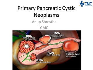
5. • The three most common types of PCNs are
serous cystic neoplasms (SCNs), mucinous
cystic neoplasms (MCNs), and IPMNs,
representing approximately 90% of all PCNs.
• MCNs and IPMNs are the most common and
more importantly have the highest potential
for malignant transformation.
• SCNs occur much less frequently and are
almost always benign.
6. SEROUS CYSTIC NEOPLASMS(SCN)
• described as multilobulated multiloculated cystic
masses with central stellate scars and calcifications
• SCNs are most commonly observed in women (3 : 1
female to male ratio)
There are four subtypes of SCNs:
• serous microcytic adenoma
• serous macrocytic (oligocystic) adenoma
• von Hippel-Lindau (VHL)-associated pancreatic cysts
• serous cystadenocarcinoma.
7. Pathology
• SCNs are characterized by
serous fluid–filled cysts lined
by a single layer of cuboidal
epithelial cells with uniform,
round, darkly stained nuclei
and a glycogen-rich cytoplasm.
• Notably, there is a lack of
atypia, necrosis, and mitotic
features in SCNs.
• On gross examination,
microcystic SCNs have a
characteristic honeycomb
appearance with multiple thin-
walled cysts around a central
scar
8. Von Hippel-Lindau–Associated
Pancreatic Cysts
• VHL disease is an autosomal dominant mutation
of chromosome 3p25 causing multiorgan
pathology. VHLassociated pancreatic cysts show a
loss of heterozygosity on chromosome 3p25 and
mutations in the VHL gene.
• These lesions are indistinguishable from sporadic
serous cystadenomas. In contrast, VHL patients
tend to have multifocal disease rather than a
single lesion. These lesions behave similarly to
non-VHL serous cystadenomas with minimal
malignant potential.
9. Serous Cystadenocarcinoma
• Malignant SCNs are
exceedingly rare
• Serous cystadenocarcinomas
are misdiagnosed as a
malignancy due to the
presence of vascular
impingement on imaging.
• These malignant cysts are
nearly identical to benign
serous cystadenomas and are
distinguished only by the
presence of metastases
10. Imaging
• Three different imaging patterns exist for
SCNs: microcystic, honeycomb, and oligocystic
1.Microcystic
13. MRI
• SCNs on MRI appear as a
“cluster of grapes” on T2-
weighted images with
multiple small cysts and
enhanced septations.
• The presence of bright T1-
weighted cystic fluid suggests
hemorrhagic fluid content.
• The main disadvantage of
MRI is the inability to detect
central calcifications
commonly seen with SCNs
14. EUS-FNA and Cytology
EUS
• SCNs typically appear as
microcystic compartments
with the absence of fluid.
• Some SCNs have
macrocystic components
that can resemble MCNs.
Cytology
• For cytological assessment
and cyst fluid drainage, in
order to distinguish
between serous and
mucinous lesions
• The cytology of SCNs
includes bland cuboidal
cells containing glycogen,
clear cytoplasm without
cellular atypia or necrosis
15. Treatment and Survillence
• Asymptomatic patients with radiological evidence of an
SCN should be followed up for 1 year.
• When the diagnosis of SCN is clear, surgery is
recommended only in patients with symptoms related to
the compression of adjacent organs (ie, bile duct, stomach,
duodenum, portal vein)
• Other indications include cyst size greater than 4 cm and
uncertainty of diagnosis despite appropriate radiologic
assessment
• enucleation of the cyst.
• Avoid Enucleation : is associated with high morbidity
(approximately 40%) due to the development of a
pancreatic fistula
16. MUCINOUS CYSTIC NEOPLASMS
• The average age range of presentation is 40
to 50 years old
• they are found predominantly in
perimenopausal women (20 : 1 female-to-
male ratio).
• The vast majority of MCNs are found in the
body and tail of the pancreas.
• Most MCNs are approximately 6 to 10 cm
at the time of diagnosis but range from 1.5
to 35 cm in diameter.
• On gross examination, they are spherical
and infrequently encapsulated by a calcified
fibrous wall.
• The cyst can be filled with mucin, blood, or
a watery fluid. This fluid tends to be thicker
and more viscous than in SCN, due to the
presence of mucus. MCNs do not
communicate with the pancreatic ductal
system
17. MCN- Histology
• MCNs are lined by tall mucin-
producing columnar cells.
• These epithelial cells that line
the cyst may be papillary or
flat and can show a tendency
toward gastric or intestinal
differentiation.
• Ovarian-like stroma is a
histologic feature often seen
• in MCN.
18. MCN-Malignant Potential
• three distinct categories based on the degree of
cellular atypia: low-, intermediate-, and high-grade.
• The classification is based on the greatest degree, and
not the average, of the epithelial dysplasia.
• Less than 20% of MCNs are associated with invasive
carcinoma and thus should be considered a potential
precursor to pancreatic cancer.
• Extensive sampling of the cyst is recommended, given
the relatively small volume of the invasive component.
It appears that the incidence of malignant
transformation is directly correlated to the overall size
of the cyst and the complexity of the cyst.
19. Imaging-CT
(A) Macrocystic form: note
septum and lack of
surrounding inflammatory
reaction.
(B) Several macrocystic areas
(>2 cm) in midbody of
pancreas.
20. MRI
• well-defined, uniloculated or multiloculated cystic lesion(s)
with enhanced septations and occasionally solid components..
• High signal intensity on T1- and T2-weighted imaging can result
from mucin within the cyst.
• The proximity of the cyst to the ductal system is better
assessed with MRI and can help to differentiate MCNs from
pseudocysts and IPMNs.
21. Cystic Fluid Analysis
• The presence of mucin is highly specific for MCNs
• Most MCNs contain higher concentrations of CEA
compared with their SCN counterparts
(>192ng/mL)
22. Treatment and Survilence
• MCN ≥40 mm should undergo surgical resection.
• Resection is also recommended for MCN which
are symptomatic or have risk factors (ie, mural
nodule) irrespective of their size
• MCN measuring <40 mm without a mural nodule
or symptoms may undergo surveillance with MRI,
EUS, or a combination of both. Surveillance is
recommended every 6 months for the first year,
then annually if no changes are observed
23. INTRADUCTAL PAPILLARY MUCINOUS
NEOPLASMS
IPMNs are defined in the WHO Classification of Tumors of the Digestive
System as an intraductal, grossly visible epithelial neoplasm of mucin-
producing cells. Using imaging and histology, IPMNs can be classified into
three types based on duct involvement:
1. Main-duct IPMN (approximately 25% of IPMNs): Segmental or diffuse
dilation of the main pancreatic duct (>5 mm) in the absence of other
causes of ductal obstruction.
2. Branch-duct IPMN (approximately 57% of IPMNs): Pancreatic cysts (>5
mm) that communicate with the main pancreatic duct.
3. Mixed type IPMN (approximately 18% of IPMNs): Meets criteria for both
main and branch duct.
24. Pathology
• IPMN lesions are
characterized by papillary
projections of columnar-
lined epithelium with
varying degrees of
dysplasia.
• Mucin is typically
abundant both within the
cytoplasm of the lining
epithelial cells as well as
within the acellular fluid
matrix
25. Intraductal Papillary Mucinous Neoplasm–Associated
Malignancy
• Invasive cancer is found in 20% to 50% of
resected IPMN specimens
Factors shown to be associated with the presence
of invasive cancer
• jaundice at presentation
• larger lesions (≥ 30mm)
• mural nodules (≥5 mm)
• main-duct dilation ≥10 mm
• serum cancer antigen (CA) 19-9 levels
26. Risk of high-grade dysplasia or
malignancy according to dilatation of
the main pancreatic duct in IPMN
The European Study Group on Cystic Tumours of the Pancreas. Gut 2018;67:789–804.
27. Genetics of Intraductal Papillary
Mucinous Neoplasm
• Similar to the genetic landscape of pancreatic
cancer, KRAS mutations, loss of p16, and TP53
mutations are frequently observed in IPMN
• SMAD4/ DPC4 expression, which is inactivated
in more than half of patients with pancreatic
adenocarcinoma, is preserved in virtually all
noninvasive IPMNs.
• Another prominent difference is the
mutations of the GNAS gene in IPMN.
28. Clinical presentation
Presentation:
• Abdominal pain.
• Pancreatitis.
• Weight loss.
• Jaundice.
• New onset diabetes.
Investigation
• variable amylase level
• a high CEA level (>200 ng/mL)
• CT Scan characteristics:
• Main pancreatic or duct dilation.
• Involvement of any part of the pancreas
or the whole pancreas.
• Continuity of cyst with ductal system.
• Irregular and poorly demarcated
29. MRCP and EUS
• MRCP may actually be
better than CT for
determining a
communication with the
pancreatic duct and thus
in diagnosing branch-duct
IPMN
• Careful evaluation with
EUS can provide detailed
images of the cyst wall
and internal cyst
architecture. Fine-needle
aspiration and biopsy can
be performed with EUS.
32. Extent of Resection
• Limited to a part of the pancreas and there is no overt
radiographic evidence of malignancy resection of the
IPMN-containing pancreas, with pancreaticoduodenectomy
for the head of the pancreas
• distal pancreatectomy for the body and tail of the
pancreas.
• Patients with MD-IPMN diffusely involving the whole gland
pose a difficult management dilemma.
• In cases in which there is a mural nodule within the MPD
further along the duct, or in patients with an increased risk
for malignancy (ie, patients with familial pancreatic cancer),
a total pancreatectomy can be considered
• MT-IPMN carries a risk of malignant transformation that is
comparable to MD-IPMN, and resection is therefore
advised in patients who are fit for surgery
33. Resection of BD-IPMN
Tanaka M, Fernández-del Castillo C, Adsay V, Chari S, Falconi M, Jang J-Y, et al.
International consensus guidelines 2012 for the management of IPMN and MCN of the
pancreas. Pancreatology. 2012 Jun;12(3):183–97
High-risk stigmata of malignancy
• Obstructive jaundice in a patient with a cystic lesion in the
head of the pancreas
• Enhancing mural nodule ≥ 5 mm
• Main pancreatic duct ≥ 10 mm in size
Worrisome clinical features
• Pancreatitis
Worrisome imaging features
• Cyst ≥ 3 cm
• Enhancing mural nodule < 5 mm
• Thickened/enhancing cyst walls
• Main duct size 5–9 mm
• Nonenhancing mural nodule
• Abrupt change in caliber of pancreatic duct with distal
pancreatic atrophy
• Lymphadenopathy
• Increased serum level of CA-19-9
• Cyst growth rate ≥ 5 mm / 2 years
Resection
without
further
investigation
Should undergo
further
evaluation with
EUS
35. Types of Surgery
• Branch-duct lesions in the head of
the pancreas are resected with
pancreatoduodenectomy
• In the tail, distal pancreatectomy
with or without splenectomy.
• Recently, pancreas-sparing
procedures, such as enucleation
and central pancreatectomy, have
been reported in an effort to
maintain the exocrine and
endocrine pancreatic function
• Total pancreatectomy is reserved
for fit patients with diffuse MD-
IPMN
36. Reference
• European evidence-based guidelines on
pancreatic cystic neoplasms (The European Study
Group on Cystic Tumours of the Pancreas)
• BLUMGART’S Surgery of the Liver, Biliary Tract,
and Pancreas (6th edition)
• Shackelford’s SURGERY Of The ALIMENTARY
TRACT( 8th Edition)
• The many faces of pancreatic serous
cystadenoma: Radiologic and pathologic
Correlation L.C. Chu et al. Diagnostic and
Interventional Imaging (2017) 98, 191—202
walls“I have actually found that the first cystic tumor of pancreas to be described was in a french journal in It was described as a mass in a size of a child’s head composed of very strong fibrous walls.
Incidental finding in 2 % of the abdominal imaging
Klöppel and colleagues classified the lesions
The cystic spaces do not communicate with the pancreatic ductal system. The surrounding stroma usually contains nerves, islets, lymphoid aggregates, and vascular channels
(a.). Arterial phase CECT image shows an infiltrative, heterogeneous, predominantly hypervascular mass involving the pancreatic body (white
arrows) and several hypervascular lesions in the liver. (b.). Portal phase image shows ductal dilation and parenchymal atrophy in the pancreatic tail (black
arrow), as well as peripancreatic lymphadenopathy (black arrowhead) and collateral circulation (black arrow) secondary to splenic vein thrombosis. (c.).
Portal phase image shows splenic vein (white arrow) thrombosis secondary to tumor infiltration by the pancreatic mass (black arrowhead); some of the
liver lesions are shown to have necrotic center (black arrow). (d.). Delayed phase shows contrast washout of the liver lesions (black arrows).
microcystic mass within tail of pancreas with characteristic central stellate scar with calcifications (arrow) and thin internal enhancing septations (arrowhead
Shows lobulated spongiform mass (arrow) with poor visualization of individual microcysts; b: gross pathology photograph illustrate numerous microcysts in the honeycomb pattern
shows a thin-walled cystic masswithin tail of pancreas with minimally lobulated outer margin (arrow); b: gross pathology photograph shows an oligocystic mass wrappingaround a mildly dilated pancreatic duct without direct communication with the pancreatic duct (
MRI is able to detect very small cysts due to superior soft tissue contrast resolution compared with CT.
Top left, T1-weighted image; top right, T2-weighted image; bottom left, T1-weighted gadolinium-enhanced image; bottom right, fat-suppressed T1-weighted gadolinium-enhanced image. The mass is externally lobulated and hypointense on the T1-weighted image and hyperintense on the T2-weighted image, with septal enhancement
The presence of the microcystic appearance usually does not warrant FNA due to the benign nature of disease.
The main limitation of FNA is the nondiagnostic result due to a lack of adequate cellular material, causing a potential false-negative to occur
MCNs are cystic tumors of the pancreas that have a lower incidence than serous cysts or IPMNs.
Almost all MCNs are multiloculated.
The stroma of the ovary is a unique type of connective tissue abundantly supplied with blood vessels, consisting for the most part of spindle-shaped stroma cells. These appear similar to fibroblasts. The stroma also contains ordinary connective tissue such as reticular fibers and collagen. Ovarian stroma differs from typical connective tissue in that it contains a high number of cells. The stoma cells are distributed in such a way that the tissue appears to be whorled. Stromal cells associated with maturing follicles may acquire endocrine function and secrete estrogens. The entire ovarian stroma is highly vascular.
Staining for estrogen and progesterone is positive in most cases. Frequently seen in young women, the mean age at presentation is in the fifth decade
In 2010 the World Health Organization (WHO) classification divided
MRI is able to better identify the contents of the cyst compared with CT imaging
a large oligocystic lesion arising from the pancreatic body/tail (arrowhead = septation, thick arrow = small locule); (B) delayed-phase image shows very faint enhancement of thin walls of septum and locule. Splenic vein occlusion confirmed by the large, tortuous venous collaterals along the tumor margin (long arrow).
Due to the malignant potential of MCNs, it is generally recommended that these lesions be resected
To avoid incomplete treatment of invasive carcinoma, a standard oncologic resection (distal pancreatectomy in 90–95% of MCN) with lymph node dissection and splenectomy is indicated for any MCN with imaging features indicating high-grade dysplasia or cancer (
Some case reports have suggested considerably faster growth of MCN during pregnancy, potentially leading to tumour rupture. Therefore, patients with MCN should be observed closely during pregnancy.
IPMNs are commonly found in the uncinate process
IPMN into the categories of low-grade dysplasia, moderate dysplasia, high-grade dysplasia, and carcinoma
Malignancy arising from IPMN is well documented, and several large series have been reported in the literature (Ajani et al, 2010; D’Angelica et al,
2004; Nara et al, 2009; Salvia et al, 2004; Schnelldorfer et al, 2008; Sohn et al, 2004; Woo et al, 2009).
The presence of a cyst size ≥30 mm, without any other radiological or clinical risk factors, has a positive predictive value for malignancy of between 27% and 33%.Patients with an IPMN measuring ≥30 mm have a 5% risk of developing malignancy, from which they will die within 3 years, whereas the
5 years disease-free survival after resection of IPMN is 96%.
Although IPMN shares many molecular alterations with pancreatic cancer, there are some significant differences with respect to both the incidence of these mutations as well as the presence of novel mutations
p16is a protein that slows cell division by slowing the progression of the cell cycle from the G1 phase to the S phase, thereby acting as a tumor suppressor. The TP53 gene provides instructions for making a protein called tumor protein p53 (or p53). This protein acts as a tumor suppressor, which means that it regulates cell division by keeping cells from growing and dividing (proliferating) too fast or in an uncontrolled way
variable amylase level, reflecting communication with the pancreatic duct, and a high CEA level (>200 ng/mL)
Main-duct IPMN with involvement of the entire main pancreatic duct. Branch-duct IPMN.L
BD-IPMN - note the connection of the lesion via a branch duct to the main duct; the arrowhead points at the BD from which the IPMN arises.
Careful evaluation with EUS can provide detailed images of the cyst wall and internal cyst architecture. Fine-needle aspiration and biopsy can be performed with EUS and may include both biopsy of the cyst and aspiration of cyst fluid for cytology and analysis for tumor markers
EUS showing hyperplastic or a polypoid growth of the epithelial layer (arrows) and hyperechoic ductal margin (arrow heads) of the irregularly dilated main pancreatic duct.
Because these lesions have a high risk of harboring high-grade dysplasia or invasive disease. Even when malignancy or high-grade dysplasia does not exist at the time of presentation, it is believed that most, if not all, MD-IPMNs will progress to malignancy
These recommendations also apply to mixed variants, because there appears to be a similar incidence of high-grade dysplasia or invasive disease whenever the main duct is dilated
When performing partial pancreatectomy for IPMN, intraoperative frozen-section evaluation focused on the identification of high-grade dysplasia or an
occult invasive cancer at the margin should be performed
Ductal dilation throughout the pancreas could be due to MD-IPMN extending throughout the pancreatic duct or due to outflow obstruction caused by malignancy or IPMN in the head of the pancreas.
Revised international consensus guidelines for the management of IPMN and MCN of the pancreas have classified the concerning radiographic features into “worrisome features” and “high-risk stigmata