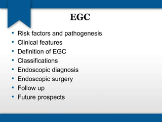
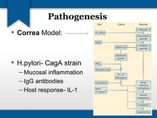
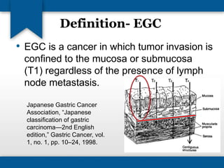
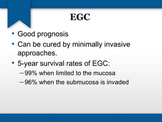
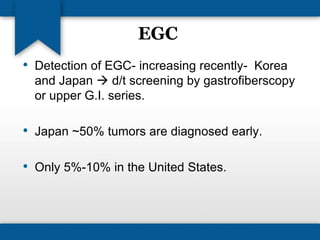
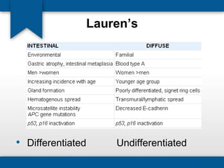
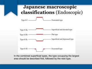
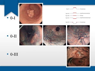
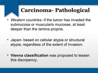
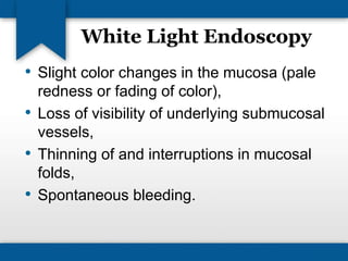
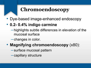
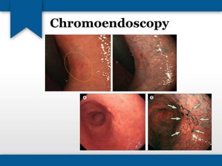
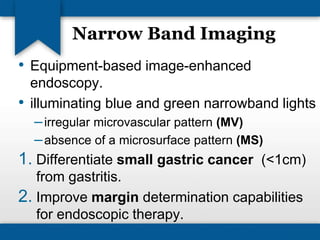
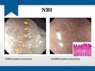
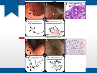
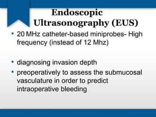
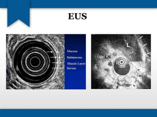
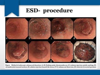
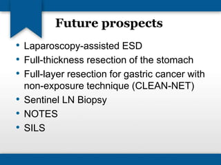
Early Gastric Cancer 
(EGC) 
Dr. Devajyoti Guin 
Postgraduate, General Surgery 
St. John’s Medical College Hospital 
Bangalore, India.
EGC 
• Risk factors and pathogenesis 
• Clinical features 
• Definition of EGC 
• Classifications 
• Endoscopic diagnosis 
• Endoscopic surgery 
• Follow up 
• Future prospects
Introduction 
• Ca Stomach- 2nd most common cause of 
death 
• Males (2:1) 
• Blacks 
• Older age (>7th decade) 
• Shift of site- distal to proximal (cardia)  
smoking, alcohol abuse.
Risk Factors 
Acquired factors 
• Nutritional 
High salt consumption 
High nitrate consumption 
Low dietary vitamin A and C 
Poor food preparation (smoked, salt cured) 
Lack of refrigeration 
Poor drinking water (well water) 
• Occupational 
Rubber workers 
Coal workers 
• Cigarette smoking
Risk Factors 
• Helicobacter pylori, Epstein-Barr virus 
• Radiation exposure 
• Prior gastric surgery- benign gastric ulcer disease (2-6%) 
Genetic factors (1-3%) 
• Type A blood 
• Pernicious anemia 
• Family history 
• Hereditary nonpolyposis colon cancer 
• Li-Fraumeni syndrome
Pathogenesis 
• Correa Model: 
• H.pylori- CagA strain 
–Mucosal inflammation 
– IgG antibodies 
–Host response- IL-1
Precursor lesions 
• Adenomatous gastric polyps 
• Chronic atrophic gastritis 
• Dysplasia 
• Intestinal metaplasia 
• Menetrier's disease (hypoproteinemic 
hypertrophic gastropathy)
Definition- EGC 
• EGC is a cancer in which tumor invasion is 
confined to the mucosa or submucosa 
(T1) regardless of the presence of lymph 
node metastasis. 
Japanese Gastric Cancer 
Association, “Japanese 
classification of gastric 
carcinoma—2nd English 
edition,” Gastric Cancer, vol. 
1, no. 1, pp. 10–24, 1998.
EGC 
• Good prognosis 
• Can be cured by minimally invasive 
approaches. 
• 5-year survival rates of EGC: 
–99% when limited to the mucosa 
–96% when the submucosa is invaded
EGC 
• Detection of EGC- increasing recently- Korea 
and Japan  d/t screening by gastrofiberscopy 
or upper G.I. series. 
• Japan ~50% tumors are diagnosed early. 
• Only 5%-10% in the United States.
Classifications 
• 1926- 
• 1942- Border’s classification- degree of 
cellular differentiation. 
• 1965- Lauren- Intestinal, Diffuse types. 
• 1990- WHO- Adeno Ca., AdenoSq., 
SqCC, Small cell Ca., Undifferentiated Ca.
Lauren’s 
• Differentiated Undifferentiated
Japanese macroscopic 
classifications (Endoscopic) 
In the combined superficial types, the type occupying the largest 
area should be described first, followed by the next type.
• 0-I 
• 0-II 
• 0-III
Carcinoma- Pathological 
• Western countries- if the tumor has invaded the 
submucosa or muscularis mucosae, at least 
deeper than the lamina propria. 
• Japan- based on cellular atypia or structural 
atypia, regardless of the extent of invasion. 
• Vienna classification was proposed to lessen 
this discripency.
Vienna classification 
of gastrointestinal epithelial neoplasia 
Category 1 Negative for neoplasia/dysplasia 
Category 2 Indefinite for neoplasia/dysplasia 
Category 3 Non-invasive low grade neoplasia (low 
grade adenoma/dysplasia) 
Category 4 Non-invasive high grade neoplasia 
4.1 High grade adenoma/dysplasia 
4.2 Non-invasive carcinoma (carcinoma in situ)* 
4.3 Suspicion of invasive carcinoma 
Category 5 Invasive neoplasia 
5.1 Intramucosal carcinoma† 
5.2 Submucosal carcinoma or beyond
Japanese Gastric Cancer 
Association Staging System 
• CECT- the 
modality of 
choice for 
staging of 
gastric cancer. 
• Sensitivity to 
determine 
nodal status - 
50% to 95% 
• Specificity - 
40% to 99%.
LN stations
Endoscopic Diagnosis 
• White light endoscopy, 
• Chromoendoscopy, 
• Narrow band imaging (NBI), 
• Endoscopic ultrasonography (EUS)
White Light Endoscopy 
• Slight color changes in the mucosa (pale 
redness or fading of color), 
• Loss of visibility of underlying submucosal 
vessels, 
• Thinning of and interruptions in mucosal 
folds, 
• Spontaneous bleeding.
Chromoendoscopy 
• Dye-based image-enhanced endoscopy 
• 0.2- 0.4% indigo carmine 
– highlights subtle differences in elevation of the 
mucosal surface 
– changes in color. 
• Magnifying chromoendoscopy (x80): 
– surface mucosal pattern 
– capillary structure
Chromoendoscopy
Narrow Band Imaging 
• Equipment-based image-enhanced 
endoscopy. 
• illuminating blue and green narrowband lights 
– irregular microvascular pattern (MV) 
– absence of a microsurface pattern (MS) 
1. Differentiate small gastric cancer (<1cm) 
from gastritis. 
2. Improve margin determination capabilities 
for endoscopic therapy.
NBI 
Differentiated Carcinoma Undifferentiated Carcinoma
Endoscopic 
Ultrasonography (EUS) 
• 20 MHz catheter-based miniprobes- High 
frequency (instead of 12 Mhz) 
• diagnosing invasion depth 
• preoperatively to assess the submucosal 
vasculature in order to predict 
intraoperative bleeding
EUS
Endoscopic Therapy 
• The frequency of LN metastasis in EGC: 
– 3% for intramucosal carcinoma 
–20% for submucosal carcinoma 
• Indications: 
–Lesions where lymph node metastasis can be 
disregarded
Endoscopic Therapy 
• Strip biopsy method (two-channel 
method)- 1984. 
• EMR (Endoscopic Mucosal Repair) 
–Endoscopic Resection With A Cap-fitted 
Panendoscope (EMRC) 
• ESD (Endoscopic Submucosal Dissection)
Indications 
• a differentiated elevated intramucosal 
cancer <2 cm in size 
• a differentiated depressed intramucosal 
cancer <1 cm in size without ulcer findings
EMR 
• Initially: 
– injecting saline under the lesion thus raising 
the tissue and allowing it to be grasped for 
snaring 
• Later: 
– different injection solutions- hypertonic saline 
with dilute epinephrine, 
–addition of cap-fitted panendoscopes, 
– variceal ligation devices to capture the lesions
EMR
EMR- Strip biopsy 
•
EMR 
Disadvantage: 
• Large tumors (>1.5cm) which cannot be 
resected en bloc are removed piecemeal 
which makes difficult to assess completion 
and curability of the resection by 
histopathology and increases the 
incidence of residual tumor.
ESD 
• dissecting along the submucosal layer directly 
using a high-frequency knife 
• Indications: 
– differentiated intramucosal cancers without ulcer 
findings, irrespective of tumor size, 
– differentiated intramucosal cancers less than 3 cm in 
size with ulcer 
– differentiated minute invasive submucosal SM1 (less 
than 500 μm below the muscularis mucosa) cancers 
less than 3 cm 
– undifferentiated intramucosal cancers less than 2 cm 
in size without ulcer
Rate of LN metastasis
Procedure selection
ESD
ESD- procedure 
•
ESD 
• Disadvantage: 
– increased instances of perforation or bleeding 
• can be treated with endoscopy
Management, Surveillance 
Postendoscopic Resection 
• EMR: annual endoscopic surveillance to ensure 
early detection of metachronous cancer (5.9%) 
• ESD: annual endoscopic surveillance + half-yearly 
abdominal computed tomography or 
endoscopic ultrasonography, for at least 3 years 
in order to detect lymph node or distant 
metastasis.
Procedure selection
Limited surgical resection 
• Gastrotomy with full-thickness mural 
excision (to allow accurate pathologic 
assessment of T status) 
• Aided by intraoperative gastroscopy for 
tumor localization. 
• Formal lymph node dissection is not 
required in these patients.
Gastrectomy 
• Lower, upper or total gastrectomy with D1 
or D2 LN dissection.
Future prospects 
• Laparoscopy-assisted ESD 
• Full-thickness resection of the stomach 
• Full-layer resection for gastric cancer with 
non-exposure technique (CLEAN-NET) 
• Sentinel LN Biopsy 
• NOTES 
• SILS
Bibliography 
• Sabiston Textbook of Surgery- 18th Edition 
• DeVita- Cancer- Principles and Practice of 
Oncology 8th edition 
• Diagnostic and Therapeutic Endoscopy, Volume 
2013, Article ID 241320 
• www.surgicaloncology.net 
• Annals of gastroenterology, vol 25, no. 4, 2012 
• Gastroenterology, 2011 
• BMC Gastroenterology, 2011
Thank You

Early gastric cancer

  • 1.
    Early Gastric Cancer (EGC) Dr. Devajyoti Guin Postgraduate, General Surgery St. John’s Medical College Hospital Bangalore, India.
  • 2.
    EGC • Riskfactors and pathogenesis • Clinical features • Definition of EGC • Classifications • Endoscopic diagnosis • Endoscopic surgery • Follow up • Future prospects
  • 3.
    Introduction • CaStomach- 2nd most common cause of death • Males (2:1) • Blacks • Older age (>7th decade) • Shift of site- distal to proximal (cardia)  smoking, alcohol abuse.
  • 4.
    Risk Factors Acquiredfactors • Nutritional High salt consumption High nitrate consumption Low dietary vitamin A and C Poor food preparation (smoked, salt cured) Lack of refrigeration Poor drinking water (well water) • Occupational Rubber workers Coal workers • Cigarette smoking
  • 5.
    Risk Factors •Helicobacter pylori, Epstein-Barr virus • Radiation exposure • Prior gastric surgery- benign gastric ulcer disease (2-6%) Genetic factors (1-3%) • Type A blood • Pernicious anemia • Family history • Hereditary nonpolyposis colon cancer • Li-Fraumeni syndrome
  • 6.
    Pathogenesis • CorreaModel: • H.pylori- CagA strain –Mucosal inflammation – IgG antibodies –Host response- IL-1
  • 7.
    Precursor lesions •Adenomatous gastric polyps • Chronic atrophic gastritis • Dysplasia • Intestinal metaplasia • Menetrier's disease (hypoproteinemic hypertrophic gastropathy)
  • 8.
    Definition- EGC •EGC is a cancer in which tumor invasion is confined to the mucosa or submucosa (T1) regardless of the presence of lymph node metastasis. Japanese Gastric Cancer Association, “Japanese classification of gastric carcinoma—2nd English edition,” Gastric Cancer, vol. 1, no. 1, pp. 10–24, 1998.
  • 9.
    EGC • Goodprognosis • Can be cured by minimally invasive approaches. • 5-year survival rates of EGC: –99% when limited to the mucosa –96% when the submucosa is invaded
  • 10.
    EGC • Detectionof EGC- increasing recently- Korea and Japan  d/t screening by gastrofiberscopy or upper G.I. series. • Japan ~50% tumors are diagnosed early. • Only 5%-10% in the United States.
  • 11.
    Classifications • 1926- • 1942- Border’s classification- degree of cellular differentiation. • 1965- Lauren- Intestinal, Diffuse types. • 1990- WHO- Adeno Ca., AdenoSq., SqCC, Small cell Ca., Undifferentiated Ca.
  • 12.
  • 13.
    Japanese macroscopic classifications(Endoscopic) In the combined superficial types, the type occupying the largest area should be described first, followed by the next type.
  • 14.
    • 0-I •0-II • 0-III
  • 15.
    Carcinoma- Pathological •Western countries- if the tumor has invaded the submucosa or muscularis mucosae, at least deeper than the lamina propria. • Japan- based on cellular atypia or structural atypia, regardless of the extent of invasion. • Vienna classification was proposed to lessen this discripency.
  • 16.
    Vienna classification ofgastrointestinal epithelial neoplasia Category 1 Negative for neoplasia/dysplasia Category 2 Indefinite for neoplasia/dysplasia Category 3 Non-invasive low grade neoplasia (low grade adenoma/dysplasia) Category 4 Non-invasive high grade neoplasia 4.1 High grade adenoma/dysplasia 4.2 Non-invasive carcinoma (carcinoma in situ)* 4.3 Suspicion of invasive carcinoma Category 5 Invasive neoplasia 5.1 Intramucosal carcinoma† 5.2 Submucosal carcinoma or beyond
  • 17.
    Japanese Gastric Cancer Association Staging System • CECT- the modality of choice for staging of gastric cancer. • Sensitivity to determine nodal status - 50% to 95% • Specificity - 40% to 99%.
  • 18.
  • 19.
    Endoscopic Diagnosis •White light endoscopy, • Chromoendoscopy, • Narrow band imaging (NBI), • Endoscopic ultrasonography (EUS)
  • 20.
    White Light Endoscopy • Slight color changes in the mucosa (pale redness or fading of color), • Loss of visibility of underlying submucosal vessels, • Thinning of and interruptions in mucosal folds, • Spontaneous bleeding.
  • 21.
    Chromoendoscopy • Dye-basedimage-enhanced endoscopy • 0.2- 0.4% indigo carmine – highlights subtle differences in elevation of the mucosal surface – changes in color. • Magnifying chromoendoscopy (x80): – surface mucosal pattern – capillary structure
  • 22.
  • 23.
    Narrow Band Imaging • Equipment-based image-enhanced endoscopy. • illuminating blue and green narrowband lights – irregular microvascular pattern (MV) – absence of a microsurface pattern (MS) 1. Differentiate small gastric cancer (<1cm) from gastritis. 2. Improve margin determination capabilities for endoscopic therapy.
  • 24.
    NBI Differentiated CarcinomaUndifferentiated Carcinoma
  • 26.
    Endoscopic Ultrasonography (EUS) • 20 MHz catheter-based miniprobes- High frequency (instead of 12 Mhz) • diagnosing invasion depth • preoperatively to assess the submucosal vasculature in order to predict intraoperative bleeding
  • 27.
  • 28.
    Endoscopic Therapy •The frequency of LN metastasis in EGC: – 3% for intramucosal carcinoma –20% for submucosal carcinoma • Indications: –Lesions where lymph node metastasis can be disregarded
  • 29.
    Endoscopic Therapy •Strip biopsy method (two-channel method)- 1984. • EMR (Endoscopic Mucosal Repair) –Endoscopic Resection With A Cap-fitted Panendoscope (EMRC) • ESD (Endoscopic Submucosal Dissection)
  • 30.
    Indications • adifferentiated elevated intramucosal cancer <2 cm in size • a differentiated depressed intramucosal cancer <1 cm in size without ulcer findings
  • 31.
    EMR • Initially: – injecting saline under the lesion thus raising the tissue and allowing it to be grasped for snaring • Later: – different injection solutions- hypertonic saline with dilute epinephrine, –addition of cap-fitted panendoscopes, – variceal ligation devices to capture the lesions
  • 32.
  • 33.
  • 34.
    EMR Disadvantage: •Large tumors (>1.5cm) which cannot be resected en bloc are removed piecemeal which makes difficult to assess completion and curability of the resection by histopathology and increases the incidence of residual tumor.
  • 35.
    ESD • dissectingalong the submucosal layer directly using a high-frequency knife • Indications: – differentiated intramucosal cancers without ulcer findings, irrespective of tumor size, – differentiated intramucosal cancers less than 3 cm in size with ulcer – differentiated minute invasive submucosal SM1 (less than 500 μm below the muscularis mucosa) cancers less than 3 cm – undifferentiated intramucosal cancers less than 2 cm in size without ulcer
  • 36.
    Rate of LNmetastasis
  • 37.
  • 38.
  • 39.
  • 40.
    ESD • Disadvantage: – increased instances of perforation or bleeding • can be treated with endoscopy
  • 41.
    Management, Surveillance PostendoscopicResection • EMR: annual endoscopic surveillance to ensure early detection of metachronous cancer (5.9%) • ESD: annual endoscopic surveillance + half-yearly abdominal computed tomography or endoscopic ultrasonography, for at least 3 years in order to detect lymph node or distant metastasis.
  • 42.
  • 43.
    Limited surgical resection • Gastrotomy with full-thickness mural excision (to allow accurate pathologic assessment of T status) • Aided by intraoperative gastroscopy for tumor localization. • Formal lymph node dissection is not required in these patients.
  • 44.
    Gastrectomy • Lower,upper or total gastrectomy with D1 or D2 LN dissection.
  • 45.
    Future prospects •Laparoscopy-assisted ESD • Full-thickness resection of the stomach • Full-layer resection for gastric cancer with non-exposure technique (CLEAN-NET) • Sentinel LN Biopsy • NOTES • SILS
  • 46.
    Bibliography • SabistonTextbook of Surgery- 18th Edition • DeVita- Cancer- Principles and Practice of Oncology 8th edition • Diagnostic and Therapeutic Endoscopy, Volume 2013, Article ID 241320 • www.surgicaloncology.net • Annals of gastroenterology, vol 25, no. 4, 2012 • Gastroenterology, 2011 • BMC Gastroenterology, 2011
  • 47.

Editor's Notes