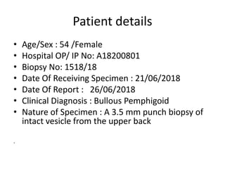
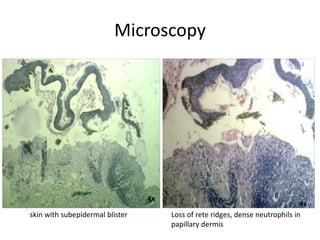
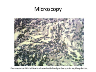
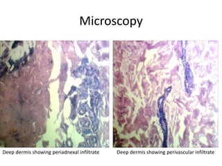
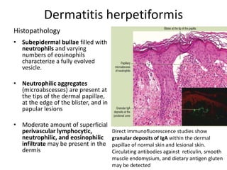
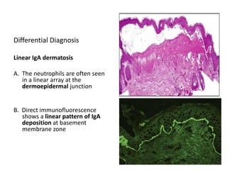
Patient details
• Age/Sex : 54 /Female
• Hospital OP/ IP No: A18200801
• Biopsy No: 1518/18
• Date Of Receiving Specimen : 21/06/2018
• Date Of Report : 26/06/2018
• Clinical Diagnosis : Bullous Pemphigoid
• Nature of Specimen : A 3.5 mm punch biopsy of
intact vesicle from the upper back
.
Gross Examination
Container labelled from upper back
• Received single skin attached soft tissue bit
measuring 0.5 x 0.3cm. All embedded in one block.
Microscopy
skin with subepidermal blister Loss of rete ridges, dense neutrophils in
papillary dermis
4x
4x
Microscopy
subepidermal blister composed of predominantly neutrophils
10x 40x
Microscopy
40x
Dense neutrophilic infiltrate admixed with few lymphocytes in papillary dermis.
Microscopy
Deep dermis showing periadnexal infiltrate Deep dermis showing perivascular infiltrate
Impression
• Biopsy of intact vesicle from the upper back shows
histopathological features consistent with Dermatitis
herpetiformis.
• Advised Immunofluorescence for further evaluation.
Histopathology
• Subepidermal bullae filled with
neutrophils and varying
numbers of eosinophils
characterize a fully evolved
vesicle.
• Neutrophilic aggregates
(microabscesses) are present at
the tips of the dermal papillae,
at the edge of the blister, and in
papular lesions
• Moderate amount of superficial
perivascular lymphocytic,
neutrophilic, and eosinophilic
infiltrate may be present in the
dermis
Dermatitis herpetiformis
Direct immunofluorescence studies show
granular deposits of IgA within the dermal
papillae of normal skin and lesional skin.
Circulating antibodies against reticulin, smooth
muscle endomysium, and dietary antigen gluten
may be detected
Differential Diagnosis
Linear IgA dermatosis
A. The neutrophils are often seen
in a linear array at the
dermoepidermal junction
B. Direct immunofluorescence
shows a linear pattern of IgA
deposition at basement
membrane zone
Differential Diagnosis
Bullous systemic lupus
erythematosus
A. Histologic sections show a
subepidermal bulla with
separation of the epidermis
from the underlying dermis.
B. Direct immunofluorescence
study shows IgG with a strong
linear deposition along the
basement membrane zone.
C. Indirect immunofluorescence study shows IgG at a 1:10 titer binding to the
dermal side of 1M salt-split skin. (Original magnification ×200).
Differential Diagnosis
Bullous Pemphigoid
• Subepidermal vesicle often filled with
eosinophils.
• Superficial perivascular mixed
inflammatory cell infiltrate rich in
eosinophils.
• In the cell-poor variant, only scant
inflammatory cell infiltrate is present.
• Early lesions may present with
spongiosis and infiltrate of eosinophils
(eosinophilic spongiosis)
Histologic section shows subepidermal
blister containing eosinophils and some
neutrophils.
Techniques for Diagnosis
Differential Diagnosis
Bullous Pemphigoid
• Direct immunofluorescence studies
- a linear deposition of C3 and IgG at
the dermoepidermal junction.
• Salt-split skin immunofluorescence
shows that the pemphigoid
antibodies are localized to the roof
of the blister in most cases
Dermatitis herpetiformis

Dermatitis herpetiformis

  • 1.
    Patient details • Age/Sex: 54 /Female • Hospital OP/ IP No: A18200801 • Biopsy No: 1518/18 • Date Of Receiving Specimen : 21/06/2018 • Date Of Report : 26/06/2018 • Clinical Diagnosis : Bullous Pemphigoid • Nature of Specimen : A 3.5 mm punch biopsy of intact vesicle from the upper back .
  • 2.
    Gross Examination Container labelledfrom upper back • Received single skin attached soft tissue bit measuring 0.5 x 0.3cm. All embedded in one block.
  • 3.
    Microscopy skin with subepidermalblister Loss of rete ridges, dense neutrophils in papillary dermis 4x 4x
  • 4.
    Microscopy subepidermal blister composedof predominantly neutrophils 10x 40x
  • 5.
    Microscopy 40x Dense neutrophilic infiltrateadmixed with few lymphocytes in papillary dermis.
  • 6.
    Microscopy Deep dermis showingperiadnexal infiltrate Deep dermis showing perivascular infiltrate
  • 7.
    Impression • Biopsy ofintact vesicle from the upper back shows histopathological features consistent with Dermatitis herpetiformis. • Advised Immunofluorescence for further evaluation.
  • 8.
    Histopathology • Subepidermal bullaefilled with neutrophils and varying numbers of eosinophils characterize a fully evolved vesicle. • Neutrophilic aggregates (microabscesses) are present at the tips of the dermal papillae, at the edge of the blister, and in papular lesions • Moderate amount of superficial perivascular lymphocytic, neutrophilic, and eosinophilic infiltrate may be present in the dermis Dermatitis herpetiformis Direct immunofluorescence studies show granular deposits of IgA within the dermal papillae of normal skin and lesional skin. Circulating antibodies against reticulin, smooth muscle endomysium, and dietary antigen gluten may be detected
  • 9.
    Differential Diagnosis Linear IgAdermatosis A. The neutrophils are often seen in a linear array at the dermoepidermal junction B. Direct immunofluorescence shows a linear pattern of IgA deposition at basement membrane zone
  • 10.
    Differential Diagnosis Bullous systemiclupus erythematosus A. Histologic sections show a subepidermal bulla with separation of the epidermis from the underlying dermis. B. Direct immunofluorescence study shows IgG with a strong linear deposition along the basement membrane zone. C. Indirect immunofluorescence study shows IgG at a 1:10 titer binding to the dermal side of 1M salt-split skin. (Original magnification ×200).
  • 11.
    Differential Diagnosis Bullous Pemphigoid •Subepidermal vesicle often filled with eosinophils. • Superficial perivascular mixed inflammatory cell infiltrate rich in eosinophils. • In the cell-poor variant, only scant inflammatory cell infiltrate is present. • Early lesions may present with spongiosis and infiltrate of eosinophils (eosinophilic spongiosis) Histologic section shows subepidermal blister containing eosinophils and some neutrophils.
  • 12.
    Techniques for Diagnosis DifferentialDiagnosis Bullous Pemphigoid • Direct immunofluorescence studies - a linear deposition of C3 and IgG at the dermoepidermal junction. • Salt-split skin immunofluorescence shows that the pemphigoid antibodies are localized to the roof of the blister in most cases