Download to read offline

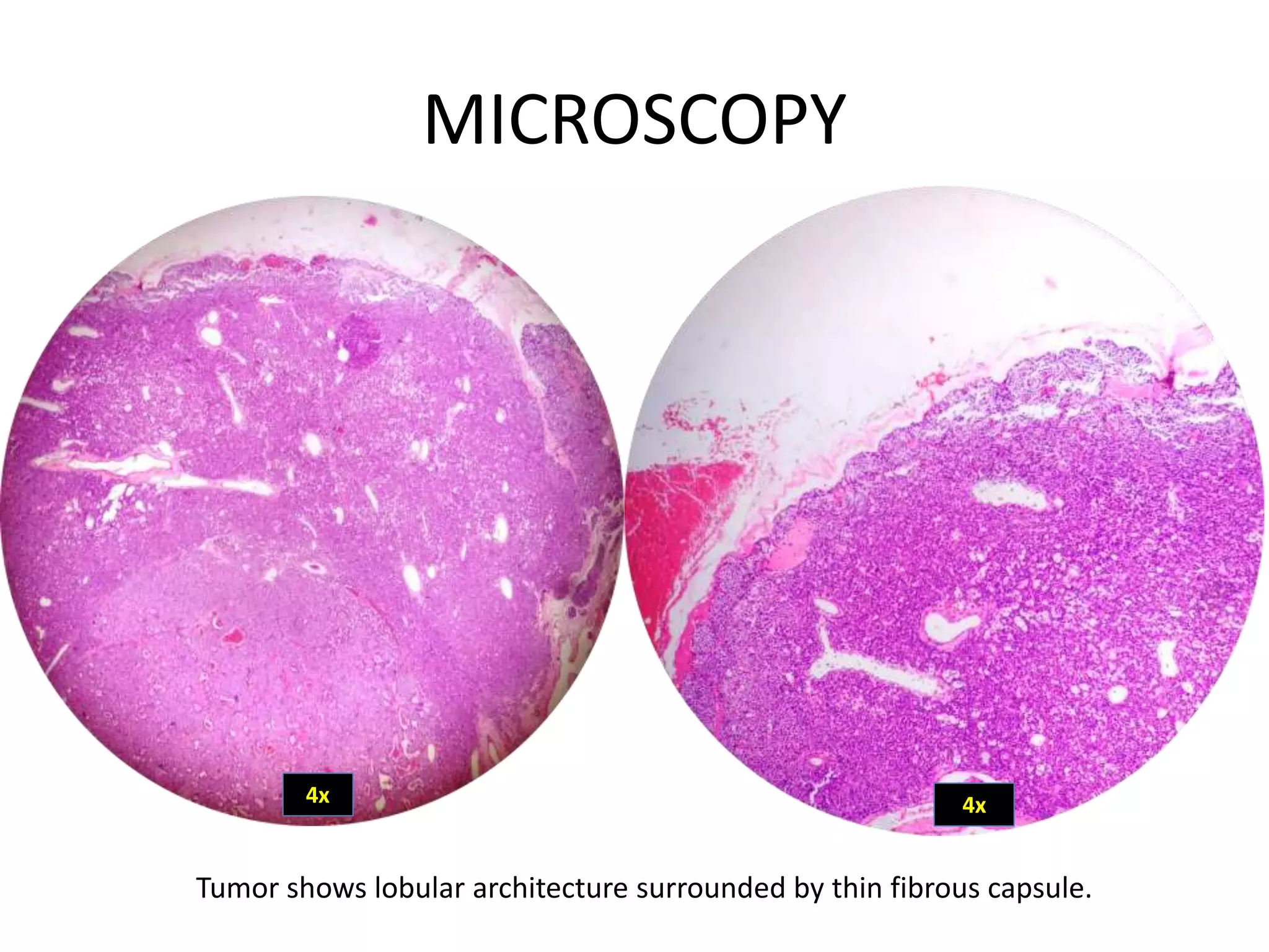
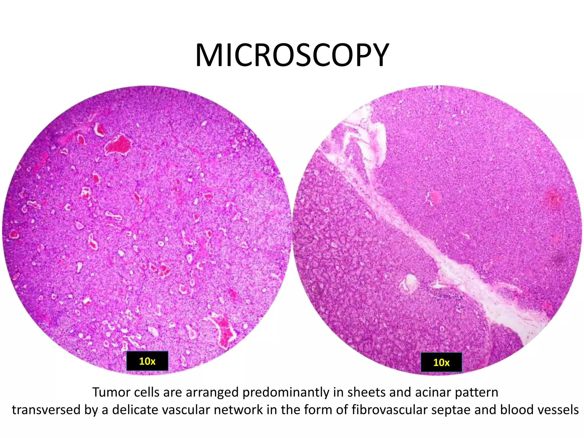
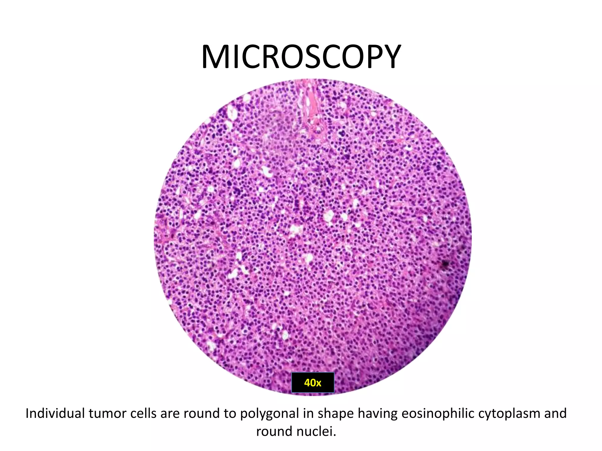

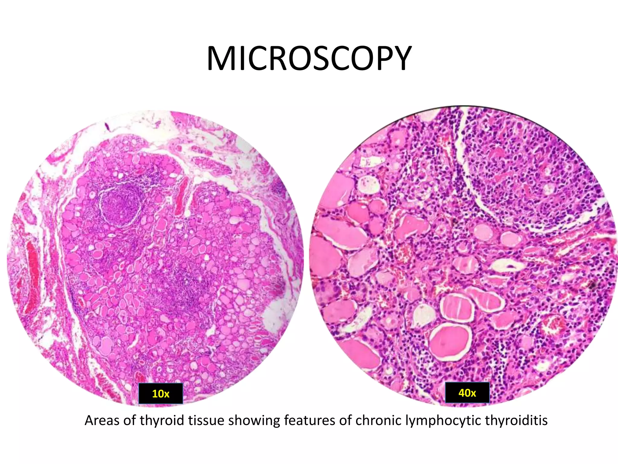
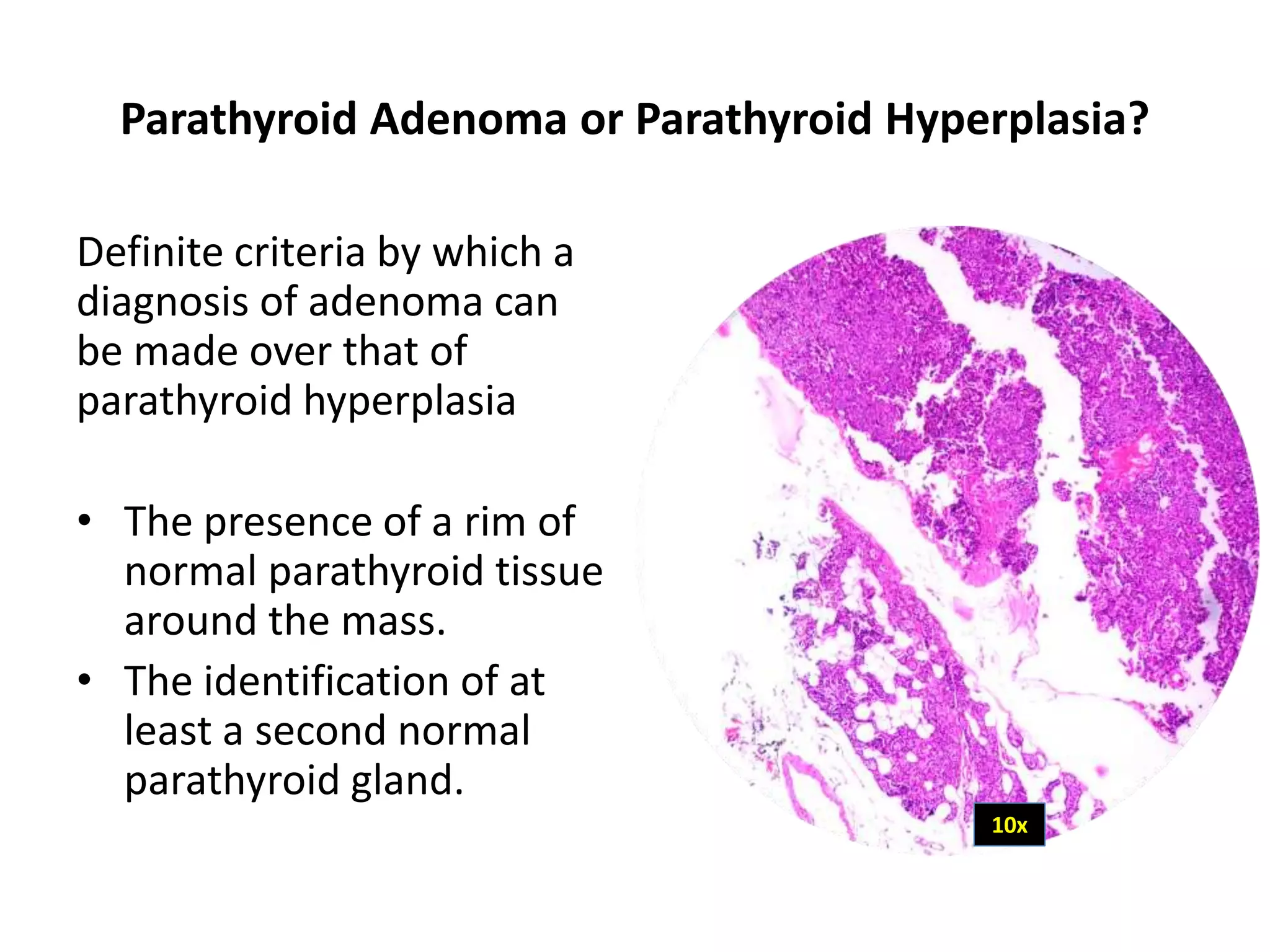


A 46-year-old female underwent biopsy of a right superior parathyroid mass. The biopsy showed a lobular tumor surrounded by a thin fibrous capsule composed of cells arranged in sheets and an acinar pattern. Individual cells had eosinophilic cytoplasm and round nuclei. Areas of normal parathyroid and thyroid tissue were also present. The histopathological features were consistent with a diagnosis of right superior parathyroid adenoma.


