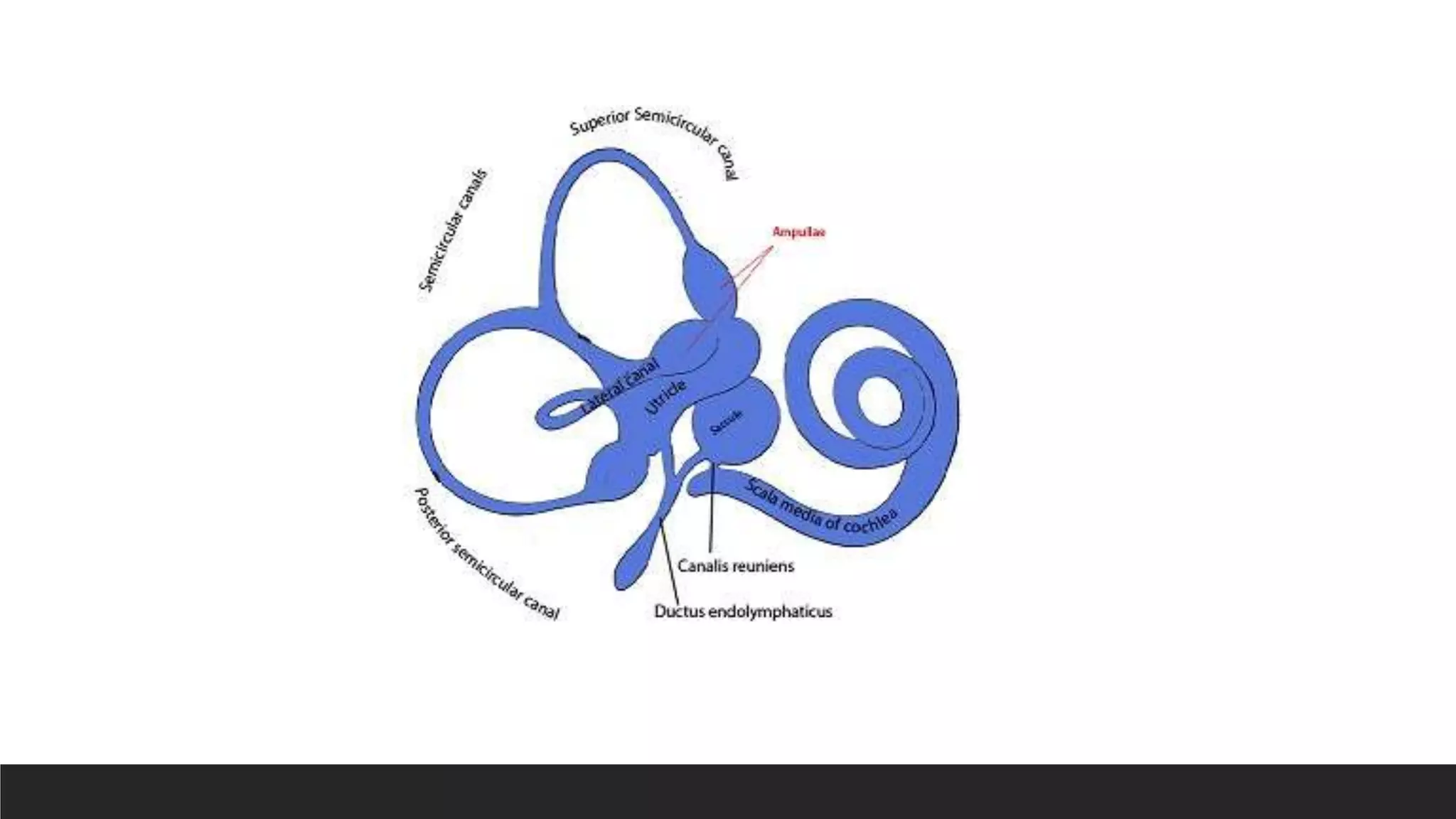
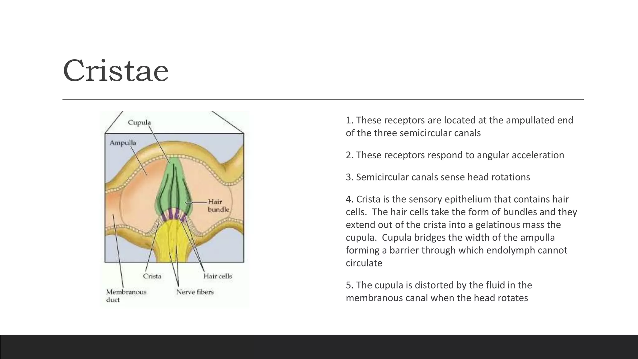
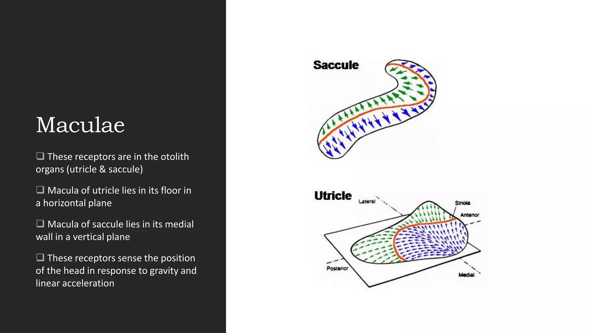
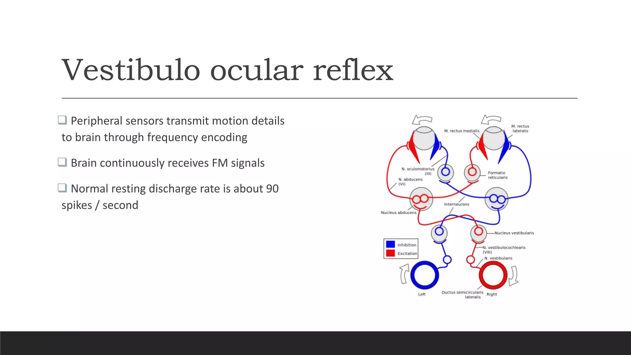
Physiology of
Vestibular
system
PROF BALASUBRAMANIAN THIAGARAJAN
(DRTBALU)
Introduction
All sensory systems have evolved in order to ensure better species survival.
Vestibular organ is the most primitive phylogenetically
Orientation of the individual in relation to environment is the most important sensation for aquatic
organism
Mammalian vestibular system generates information and feeds them to the brain
Functions of vestibular system
 Gaze stabilization ensuring that the field of vision stays focused to the subject of interest
 Body position & locomotion to enable balanced locomotion without compromising body posture
Orientation of the body with respect to gravity
 For readjusting autonomic functions after body reorientation
Vestibular system input system
 Visual
 Vestibular
 Proprioceptive
 Hearing cues (less important)
Brain process these inputs leading to various outcomes like reflexes and other responses
Vestibular system reflexes &
responses
 Vestibulo ocular reflex (VOR). This ensures gaze stabilization
 Vestibulo spinal reflex – Ensures maintenance of upright position
 Vestibulo colic reflex – Ensures head stabilization in space
 Orientation and in higher species, navigation and perception of self position with respect to
surroundings and gravity is mediated by vestibular cortex.
 It influences circadian rhythm
Additional factors playing a role in
vestibular sensation
 Learning
 Memory
 Drugs
 Ageing
 Environmental conditions
Vestibular receptors
1. Cristae – Seen in ampullated ends of semicircular canals
2. Macula – Seen in utricle & saccule
Cristae
1. These receptors are located at the ampullated end
of the three semicircular canals
2. These receptors respond to angular acceleration
3. Semicircular canals sense head rotations
4. Crista is the sensory epithelium that contains hair
cells. The hair cells take the form of bundles and they
extend out of the crista into a gelatinous mass the
cupula. Cupula bridges the width of the ampulla
forming a barrier through which endolymph cannot
circulate
5. The cupula is distorted by the fluid in the
membranous canal when the head rotates
Physiology of Crista
Hair cells of crista are organized with kinocilia
pointing in the same direction.
when the cupula moves in the appropriate
direction, the entire population of hair cells is
depolarized and activity in all of the
innervating axons increases. When the cupula
moves in the opposite direction, the
population is hyperpolarized and neuronal
activity decreases. Deflections orthogonal to
the excitatory-inhibitory direction produce
little or no response.
Contd…
 Passive forms of movements like in aeroplanes, merry-go-rounds, cars, rollercoasters generate more intricate
movements and can readily induce motion sickness since the vestibular apparatus is not adapted to these kinds of
motions.
Three forces act upon the endolymph and cupula in the canal. These include:
1. The inertial force, proportional to the mass of the endolymph and cupula.
2. The elastic restoring force of the cupula that positions the cupula back to the centre position after stimulation.
3. The viscous forces that act upon the fluid when sliding past the internal wall of the tube. This force is dependent on
the speed of relative movement of endolymph with respect to the wall.
Hair cells of crista
Two types:
Type I hair cells: Flask shaped, with a single cup like nerve terminal surrounding the base
Type II hair cells: Cylindrical cells with multiple nerve terminals at the base. From the upper surface
of each cell project a single hair known as the kinocilium and a number of other cilia known as the
stereocilia. The kinocilium is thicker and is located at one edge of the cell. These sensory hair cells
are surrounded by supporting cells that display microvilli on their upper ends
Image of Hair cells
Maculae
 These receptors are in the otolith
organs (utricle & saccule)
 Macula of utricle lies in its floor in
a horizontal plane
 Macula of saccule lies in its medial
wall in a vertical plane
 These receptors sense the position
of the head in response to gravity and
linear acceleration
Otolith organs
Continuous
G
r
a
v
I
T
y
Acceleration
Due to gravity
needs to be
monitored
continuously
Otolith organs sense gravity
These organs
Also detect
linear
accelerations
Einstein's equivalence principle states that no
single physical device can distinguish gravity
from linear acceleration. This really poses a
difficult scenario for the central nervous system
since otoliths cannot differentiate between
linear acceleration and tilt since only the
deflection of teh base of the hair cells are
encoded and sent to the brain. During natural
movements (both active & passive), the otolith
organs sense the sum of all accelerations acting
on the head and interpret these signals to
initiate postural and eye reflexes mediated by
the vestibular nuclei, directing appropriate
signals to the limb, trunk, and neck muscles via
the vestibulospinal tracts or to the eye muscles
via the vestibulo ocular reflex (VOR). The
interplay of several senses at the same time
(visual, vestibular and proprioceptive) enables
the CNS to cope up with the ambiguity of linear
accelerations under normal conditions. However
in darkness when visual stimulus is rather
useless it relies on the vestibular cues.
Ultrastructure of Macula
 It consists of two parts
 Sensory neuroepithelium of type I and type II cells as seen in crista of semicircular canal
 Otolithic membrane made up of gelatinous mass on top of which are seen crystals of calcium
carbonate (otoconia)
 The cilia of the hair cells project into the gelatinous layer
Vestibulo ocular reflex
 Peripheral sensors transmit motion details
to brain through frequency encoding
 Brain continuously receives FM signals
 Normal resting discharge rate is about 90
spikes / second
Features of VOR
1. During head rest, hair cells in both semicircular canals have a resting discharge of 90 spikes / second.
2. Endolymph fluid lags behind within each SSC due to inertia.
3. In right sided head turning, the leading SSC (the right), the stereocilia bends towards the kinocilium.
4. During the postulated right head turn, the discharge rate increases in the leading right ear from 90 to 120 spikes /
second.
5. In the following SSC i.e. the left, the stereocilia bend away from the kinocilium
6. The discharge rate decreases in the following left ear from 90 to 20 spikes / second
7. The vestibular nuclei interprets the difference in the discharge rates between the left and right SSC's as movement
towards the right, and triggers the oculomotor nuclei to drive the eyes to the left to maintain gaze stabilization.
Vestibulo spinal reflex
 The purpose of this reflex is to stabilize the body
 It involves motor output to the muscles below the neck
 Sensory input is from the canals and otolith or both
 When head is tilted to one side both the canals and otoliths are stimulated
 The vestibular nerves and vestibular nucleus are activated
 Impulses are transmitted via the lateral and media vestibulo spinal tracts to the spinal cord
 Extensor activity is induced on the side to which the head is inclined, and flexor activity is induced
on the opposite side
Cervical reflexes
 Cervico ocular reflex – Consists of eye movements driven by neck proprioceptors. This is relevant
when considering recovery from vestibular lesions
 Cervico spinal reflex – Also known as tonic neck reflex. This involves changes in the limb position
driven by neck afferent activity. It interacts with VOR. This involves extension of the limb on the side
to which the chin is pointed and flexion of the limb on the opposite side.
 Cervicocollic reflex – This reflex stabilizes the head on the body. Changes in the neck position,
creates opposition to that stretch by reflexive contraction of neck muscles. This reflex has longer
latency than vestibulocollic reflexes.
Vestibulo sympathetic reflex
 Getting up from recumbent position creates lot of orthostatic stress on the body
 This is due to pooling of blood in the lower parts of the body
 About 800 ml of blood gets pooled in dependent portions of the body
 This reflex increases the heart rate and blood pressure ensuring steady blood supply to the brain
Central projections of vestibular
system
 Movements sensed by vestibular organs on the right and left sides converge into the vestibular
nuclei after passing through the ganglion of scarpa. The semicircular canals and otolith maculae
project to different portions of vestibular nuclei
 Vestibular nuclei triggers other brain centers in order to maintain gaze stabilization as well as body
stabilization
 Superior vestibular nerve gets afferents from horizontal and anterior canals as well as from the
utricular macula and antero superior region of saccular macula
 Inferior vestibular nerve is formed by fibers from the posterior canal and saccular macula
 Caloric tests and rotatory tests evaluate the horizontal canal (superior vestibular nerve) and cVEMP
(collic vestibular evoked myogenic potentials) evaluates the inferior vestibular nerve
Cont..
 Vestibular primary efferents project into the
vestibular nuclear complex in the pontomedullary
region of the brain stem and the cerebellum. Nodulus
and uvula gets the maximum projection
 Superior, lateral, medial and descending vestibular
nuclei
 Afferents from scc’s and otoliths enter vestibular
nuclear complex at the level of lateral vestibular
nucleus and rostral descending vestibular nucleus.
They then divide into ascending and descending
pathways. The ascending pathway projects into the
superior vestibular nerve and further on to the
cerebellum. The descending branch innervates the
central region of the vestibular nuclear complex
Accepted facts…
1. SCC's project to all four vestibular nuclei with the most heavy projection to the Medial vestibular nerve
and Superior vestibular nerve
2. Saccular afferents project strongly to the Dorsal vestibular nerve, the INT8 and the Y-group and weakly
to other vestibular nuclei
3. Utricle mainly projects to the lateral and dorsal portions of the Medial vestibular nucleus, the ventral
and lateral portions of the Superior vestibular nerve and the rostral portion of the Dorsal vestibular nerve.
Afferents from the non vestibular systems like optokinetic system, the neck proprioceptive system and the
cerebellar purkinje cells also project into the vestibular nuclear complex.
Within the vestibular brainstem nuclei there are commissural projections reinforcing the vestibular inputs.
Ewald’s Laws
First law: Stimulation of semicircular canal causes movement of the eyes in the plane of the
stimulated canal.
Second law: In the horizontal canals an ampullopetal endolymphatic movement causes a greater
stimulation than an ampullofugal one
Third law: In the vertical canals the reverse is true.
.
Physiology of vestibular system

Physiology of vestibular system

  • 1.
  • 2.
    Introduction All sensory systemshave evolved in order to ensure better species survival. Vestibular organ is the most primitive phylogenetically Orientation of the individual in relation to environment is the most important sensation for aquatic organism Mammalian vestibular system generates information and feeds them to the brain
  • 3.
    Functions of vestibularsystem  Gaze stabilization ensuring that the field of vision stays focused to the subject of interest  Body position & locomotion to enable balanced locomotion without compromising body posture Orientation of the body with respect to gravity  For readjusting autonomic functions after body reorientation
  • 4.
    Vestibular system inputsystem  Visual  Vestibular  Proprioceptive  Hearing cues (less important) Brain process these inputs leading to various outcomes like reflexes and other responses
  • 5.
    Vestibular system reflexes& responses  Vestibulo ocular reflex (VOR). This ensures gaze stabilization  Vestibulo spinal reflex – Ensures maintenance of upright position  Vestibulo colic reflex – Ensures head stabilization in space  Orientation and in higher species, navigation and perception of self position with respect to surroundings and gravity is mediated by vestibular cortex.  It influences circadian rhythm
  • 6.
    Additional factors playinga role in vestibular sensation  Learning  Memory  Drugs  Ageing  Environmental conditions
  • 8.
    Vestibular receptors 1. Cristae– Seen in ampullated ends of semicircular canals 2. Macula – Seen in utricle & saccule
  • 9.
    Cristae 1. These receptorsare located at the ampullated end of the three semicircular canals 2. These receptors respond to angular acceleration 3. Semicircular canals sense head rotations 4. Crista is the sensory epithelium that contains hair cells. The hair cells take the form of bundles and they extend out of the crista into a gelatinous mass the cupula. Cupula bridges the width of the ampulla forming a barrier through which endolymph cannot circulate 5. The cupula is distorted by the fluid in the membranous canal when the head rotates
  • 10.
    Physiology of Crista Haircells of crista are organized with kinocilia pointing in the same direction. when the cupula moves in the appropriate direction, the entire population of hair cells is depolarized and activity in all of the innervating axons increases. When the cupula moves in the opposite direction, the population is hyperpolarized and neuronal activity decreases. Deflections orthogonal to the excitatory-inhibitory direction produce little or no response.
  • 11.
    Contd…  Passive formsof movements like in aeroplanes, merry-go-rounds, cars, rollercoasters generate more intricate movements and can readily induce motion sickness since the vestibular apparatus is not adapted to these kinds of motions. Three forces act upon the endolymph and cupula in the canal. These include: 1. The inertial force, proportional to the mass of the endolymph and cupula. 2. The elastic restoring force of the cupula that positions the cupula back to the centre position after stimulation. 3. The viscous forces that act upon the fluid when sliding past the internal wall of the tube. This force is dependent on the speed of relative movement of endolymph with respect to the wall.
  • 12.
    Hair cells ofcrista Two types: Type I hair cells: Flask shaped, with a single cup like nerve terminal surrounding the base Type II hair cells: Cylindrical cells with multiple nerve terminals at the base. From the upper surface of each cell project a single hair known as the kinocilium and a number of other cilia known as the stereocilia. The kinocilium is thicker and is located at one edge of the cell. These sensory hair cells are surrounded by supporting cells that display microvilli on their upper ends
  • 13.
  • 14.
    Maculae  These receptorsare in the otolith organs (utricle & saccule)  Macula of utricle lies in its floor in a horizontal plane  Macula of saccule lies in its medial wall in a vertical plane  These receptors sense the position of the head in response to gravity and linear acceleration
  • 15.
    Otolith organs Continuous G r a v I T y Acceleration Due togravity needs to be monitored continuously Otolith organs sense gravity These organs Also detect linear accelerations Einstein's equivalence principle states that no single physical device can distinguish gravity from linear acceleration. This really poses a difficult scenario for the central nervous system since otoliths cannot differentiate between linear acceleration and tilt since only the deflection of teh base of the hair cells are encoded and sent to the brain. During natural movements (both active & passive), the otolith organs sense the sum of all accelerations acting on the head and interpret these signals to initiate postural and eye reflexes mediated by the vestibular nuclei, directing appropriate signals to the limb, trunk, and neck muscles via the vestibulospinal tracts or to the eye muscles via the vestibulo ocular reflex (VOR). The interplay of several senses at the same time (visual, vestibular and proprioceptive) enables the CNS to cope up with the ambiguity of linear accelerations under normal conditions. However in darkness when visual stimulus is rather useless it relies on the vestibular cues.
  • 16.
    Ultrastructure of Macula It consists of two parts  Sensory neuroepithelium of type I and type II cells as seen in crista of semicircular canal  Otolithic membrane made up of gelatinous mass on top of which are seen crystals of calcium carbonate (otoconia)  The cilia of the hair cells project into the gelatinous layer
  • 17.
    Vestibulo ocular reflex Peripheral sensors transmit motion details to brain through frequency encoding  Brain continuously receives FM signals  Normal resting discharge rate is about 90 spikes / second
  • 18.
    Features of VOR 1.During head rest, hair cells in both semicircular canals have a resting discharge of 90 spikes / second. 2. Endolymph fluid lags behind within each SSC due to inertia. 3. In right sided head turning, the leading SSC (the right), the stereocilia bends towards the kinocilium. 4. During the postulated right head turn, the discharge rate increases in the leading right ear from 90 to 120 spikes / second. 5. In the following SSC i.e. the left, the stereocilia bend away from the kinocilium 6. The discharge rate decreases in the following left ear from 90 to 20 spikes / second 7. The vestibular nuclei interprets the difference in the discharge rates between the left and right SSC's as movement towards the right, and triggers the oculomotor nuclei to drive the eyes to the left to maintain gaze stabilization.
  • 19.
    Vestibulo spinal reflex The purpose of this reflex is to stabilize the body  It involves motor output to the muscles below the neck  Sensory input is from the canals and otolith or both  When head is tilted to one side both the canals and otoliths are stimulated  The vestibular nerves and vestibular nucleus are activated  Impulses are transmitted via the lateral and media vestibulo spinal tracts to the spinal cord  Extensor activity is induced on the side to which the head is inclined, and flexor activity is induced on the opposite side
  • 20.
    Cervical reflexes  Cervicoocular reflex – Consists of eye movements driven by neck proprioceptors. This is relevant when considering recovery from vestibular lesions  Cervico spinal reflex – Also known as tonic neck reflex. This involves changes in the limb position driven by neck afferent activity. It interacts with VOR. This involves extension of the limb on the side to which the chin is pointed and flexion of the limb on the opposite side.  Cervicocollic reflex – This reflex stabilizes the head on the body. Changes in the neck position, creates opposition to that stretch by reflexive contraction of neck muscles. This reflex has longer latency than vestibulocollic reflexes.
  • 21.
    Vestibulo sympathetic reflex Getting up from recumbent position creates lot of orthostatic stress on the body  This is due to pooling of blood in the lower parts of the body  About 800 ml of blood gets pooled in dependent portions of the body  This reflex increases the heart rate and blood pressure ensuring steady blood supply to the brain
  • 22.
    Central projections ofvestibular system  Movements sensed by vestibular organs on the right and left sides converge into the vestibular nuclei after passing through the ganglion of scarpa. The semicircular canals and otolith maculae project to different portions of vestibular nuclei  Vestibular nuclei triggers other brain centers in order to maintain gaze stabilization as well as body stabilization  Superior vestibular nerve gets afferents from horizontal and anterior canals as well as from the utricular macula and antero superior region of saccular macula  Inferior vestibular nerve is formed by fibers from the posterior canal and saccular macula  Caloric tests and rotatory tests evaluate the horizontal canal (superior vestibular nerve) and cVEMP (collic vestibular evoked myogenic potentials) evaluates the inferior vestibular nerve
  • 23.
    Cont..  Vestibular primaryefferents project into the vestibular nuclear complex in the pontomedullary region of the brain stem and the cerebellum. Nodulus and uvula gets the maximum projection  Superior, lateral, medial and descending vestibular nuclei  Afferents from scc’s and otoliths enter vestibular nuclear complex at the level of lateral vestibular nucleus and rostral descending vestibular nucleus. They then divide into ascending and descending pathways. The ascending pathway projects into the superior vestibular nerve and further on to the cerebellum. The descending branch innervates the central region of the vestibular nuclear complex
  • 24.
    Accepted facts… 1. SCC'sproject to all four vestibular nuclei with the most heavy projection to the Medial vestibular nerve and Superior vestibular nerve 2. Saccular afferents project strongly to the Dorsal vestibular nerve, the INT8 and the Y-group and weakly to other vestibular nuclei 3. Utricle mainly projects to the lateral and dorsal portions of the Medial vestibular nucleus, the ventral and lateral portions of the Superior vestibular nerve and the rostral portion of the Dorsal vestibular nerve. Afferents from the non vestibular systems like optokinetic system, the neck proprioceptive system and the cerebellar purkinje cells also project into the vestibular nuclear complex. Within the vestibular brainstem nuclei there are commissural projections reinforcing the vestibular inputs.
  • 25.
    Ewald’s Laws First law:Stimulation of semicircular canal causes movement of the eyes in the plane of the stimulated canal. Second law: In the horizontal canals an ampullopetal endolymphatic movement causes a greater stimulation than an ampullofugal one Third law: In the vertical canals the reverse is true. .