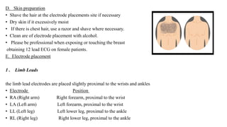
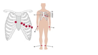
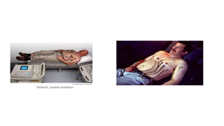
JAMIA MILLIA ISLAMIA
CENTRE FOR PHYSIOTHERAPY AND REHABILITATION SCIENCES
Topic :- Placement of ECG lead: resting
submitted by;- hafiza Sania Urooj
Submitted to :-dr. Jamal moiz
Class:- bpt 4th year
Subject:- physiotherapy in cardiopulmonary rehabilitation
Date of submission :- 19 -02-2021
Introduction
• Electrocardiography (ECG or EKG) : Is the recording of the electrical activity of the heart.
• Traditionally this is in the form of a transthoracic (across the thorax or chest)
interpretation of the electrical activity of the heart over a period of time, as detected
by electrodes attached to the surface of the skin and recorded or displayed by a device
external to the body.
The recording produced by this noninvasive procedure is termed an electrocardiogram
(ECG or EKG).
• An ECG is used to measure the heart‘s electrical conduction system.
• It picks up electrical impulses generated by the polarization and depolarization of cardiac tissue and translates
into a waveform.
• The waveform is then used to measure the rate and regularity of heartbeats, as well as the size and position of
the chambers, the presence of any damage to the heart, and the effects of drugs or devices used to regulate the
heart, such as a pacemaker
General symptoms indicating use of electrocardiography include:
• Symptoms of myocardial infarction
• Symptoms of pulmonary embolism
• Cardiac murmurs
• Syncope or collapse
• Seizures
• Perceived cardiac dysrhythmias
• It is also used to assess patients with systemic disease, as well as monitoring during anesthesia and critically
ill patients.
Contraindications :-
• A. Trauma (must be no delay in transport)
• B. Respiratory Arrest
• C. Cardiac Arrest
12 Lead ECG Placement:-
• The correct positioning of leads is essential to taking an accurate 12 lead resting ECG and incorrect placement of
leads can lead to a false diagnosis of infarction or negative changes on the ECG.
A. Room Preparation
• the patient area and ECG machine are clean and tidy
• the bed is adjustable and large enough to support the patient’s body and limbs 6
• the ECG machine is safe and ready to use. The date and time should be correct on the machine
• there is sufficient stock for performing an ECG including ECG paper, ECG electrodes, towelling, paper, hair
removal device, skin prep and cleaning wipes
• hands are washed 2
• staff are aware of the location of the crash trolley and emergency buttons in case of emergency
B. Equipment within the room should include:
• disposable electrode tabs
• ECG paper
• skin preparation equipment (hair removal device, cleaning wipes, abrasive tape)
• adjustable bed / couch that a patient can lie on comfortably (with all limbs resting on the bed surface) and
can be adjusted to the correct height for the person performing the ECG.
C . Patient Preparation
• Patient position :-
• Confirm patient’s identity
• Explain the procedure in detail
• Place the patient in a supine position or semi-Fowlers position . if the patient
cannot tolerate being flat , you can do the ECG in a more upright position
• Instruct the patients to place their arms side and to relax their shoulders .
• Make sure the patients legs are uncrossed .
• Remove any electrical devices , such as cell phone , aways from the patient as they
may interfere with the machine .
• Every effort should be made to respect the patient and minimize
embarrassment by allowing them to dress and undress in private.
• The female patient should be covered
D. Skin preparation
• Shave the hair at the electrode placements site if necessary
• Dry skin if it excessively moist
• If there is chest hair, use a razor and shave where necessary.
• Clean are of electrode placement with alcohol.
• Please be professional when exposing or touching the breast
obtaining 12 lead ECG on female patients.
E. Electrode placement
1 . Limb Leads
the limb lead electrodes are placed slightly proximal to the wrists and ankles
• Electrode Position
• RA (Right arm) Right forearm, proximal to the wrist
• LA (Left arm) Left forearm, proximal to the wrist
• LL (Left leg) Left lower leg, proximal to the ankle
• RL (Right leg) Right lower leg, proximal to the ankle
2. Precordial (chest) leads
• Electrode Position
• V1 Fourth intercostal space at the right sternal edge
• V2 Fourth intercostal space at the left sternal edge
• V3 Midway between V2 and V4
• V4 Fifth intercostal space in the mid-clavicular line
• V5 Left anterior axillary line at same horizontal level as V4
• V6 Left mid-axillary line at same horizontal level as V4 and V5
• When recording an ECG on female patients, leads V1-V3 should be placed in the standard positions, while it
is recommended that leads V4-V6 chest electrodes are placed beneath the left breast where the breast tissue
meets with the chest .
Documentation
• A patient care report will be completed per policy
• A 12 lead ECG will be turned to the receiving hospital with the patient care report. A copy will also be kept
with the department copy of the patient care report.
Reference
• Goldberger’s Clinical Electrocardiography EIGHTH EDITION
• Amita health st marys kankakees
• Adult resting electrocardiography
• Cash cardio
• Numed health care

Ecg placement resting

  • 1.
    JAMIA MILLIA ISLAMIA CENTREFOR PHYSIOTHERAPY AND REHABILITATION SCIENCES Topic :- Placement of ECG lead: resting submitted by;- hafiza Sania Urooj Submitted to :-dr. Jamal moiz Class:- bpt 4th year Subject:- physiotherapy in cardiopulmonary rehabilitation Date of submission :- 19 -02-2021
  • 2.
    Introduction • Electrocardiography (ECGor EKG) : Is the recording of the electrical activity of the heart. • Traditionally this is in the form of a transthoracic (across the thorax or chest) interpretation of the electrical activity of the heart over a period of time, as detected by electrodes attached to the surface of the skin and recorded or displayed by a device external to the body. The recording produced by this noninvasive procedure is termed an electrocardiogram (ECG or EKG). • An ECG is used to measure the heart‘s electrical conduction system. • It picks up electrical impulses generated by the polarization and depolarization of cardiac tissue and translates into a waveform. • The waveform is then used to measure the rate and regularity of heartbeats, as well as the size and position of the chambers, the presence of any damage to the heart, and the effects of drugs or devices used to regulate the heart, such as a pacemaker
  • 3.
    General symptoms indicatinguse of electrocardiography include: • Symptoms of myocardial infarction • Symptoms of pulmonary embolism • Cardiac murmurs • Syncope or collapse • Seizures • Perceived cardiac dysrhythmias • It is also used to assess patients with systemic disease, as well as monitoring during anesthesia and critically ill patients. Contraindications :- • A. Trauma (must be no delay in transport) • B. Respiratory Arrest • C. Cardiac Arrest
  • 4.
    12 Lead ECGPlacement:- • The correct positioning of leads is essential to taking an accurate 12 lead resting ECG and incorrect placement of leads can lead to a false diagnosis of infarction or negative changes on the ECG. A. Room Preparation • the patient area and ECG machine are clean and tidy • the bed is adjustable and large enough to support the patient’s body and limbs 6 • the ECG machine is safe and ready to use. The date and time should be correct on the machine • there is sufficient stock for performing an ECG including ECG paper, ECG electrodes, towelling, paper, hair removal device, skin prep and cleaning wipes • hands are washed 2 • staff are aware of the location of the crash trolley and emergency buttons in case of emergency B. Equipment within the room should include: • disposable electrode tabs • ECG paper • skin preparation equipment (hair removal device, cleaning wipes, abrasive tape) • adjustable bed / couch that a patient can lie on comfortably (with all limbs resting on the bed surface) and can be adjusted to the correct height for the person performing the ECG.
  • 5.
    C . PatientPreparation • Patient position :- • Confirm patient’s identity • Explain the procedure in detail • Place the patient in a supine position or semi-Fowlers position . if the patient cannot tolerate being flat , you can do the ECG in a more upright position • Instruct the patients to place their arms side and to relax their shoulders . • Make sure the patients legs are uncrossed . • Remove any electrical devices , such as cell phone , aways from the patient as they may interfere with the machine . • Every effort should be made to respect the patient and minimize embarrassment by allowing them to dress and undress in private. • The female patient should be covered
  • 6.
    D. Skin preparation •Shave the hair at the electrode placements site if necessary • Dry skin if it excessively moist • If there is chest hair, use a razor and shave where necessary. • Clean are of electrode placement with alcohol. • Please be professional when exposing or touching the breast obtaining 12 lead ECG on female patients. E. Electrode placement 1 . Limb Leads the limb lead electrodes are placed slightly proximal to the wrists and ankles • Electrode Position • RA (Right arm) Right forearm, proximal to the wrist • LA (Left arm) Left forearm, proximal to the wrist • LL (Left leg) Left lower leg, proximal to the ankle • RL (Right leg) Right lower leg, proximal to the ankle
  • 7.
    2. Precordial (chest)leads • Electrode Position • V1 Fourth intercostal space at the right sternal edge • V2 Fourth intercostal space at the left sternal edge • V3 Midway between V2 and V4 • V4 Fifth intercostal space in the mid-clavicular line • V5 Left anterior axillary line at same horizontal level as V4 • V6 Left mid-axillary line at same horizontal level as V4 and V5 • When recording an ECG on female patients, leads V1-V3 should be placed in the standard positions, while it is recommended that leads V4-V6 chest electrodes are placed beneath the left breast where the breast tissue meets with the chest .
  • 10.
    Documentation • A patientcare report will be completed per policy • A 12 lead ECG will be turned to the receiving hospital with the patient care report. A copy will also be kept with the department copy of the patient care report.
  • 11.
    Reference • Goldberger’s ClinicalElectrocardiography EIGHTH EDITION • Amita health st marys kankakees • Adult resting electrocardiography • Cash cardio • Numed health care